Cartilage and Bone Flashcards
(47 cards)
3 types of cartilage and their locations
Hyaline cartilage: nose, articular joints, intercostal joints, rings of the trachea/lungs/larynx
Fibrocartilage: intervertebral discs & pubic symphysis
Elastic cartilage: external ear and epiglottis
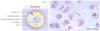
What kind of cartilage is this?
Hyaline cartilage
What kind of cartilage is this?
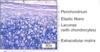
Elastic cartilage
What kind of cartilage is this?
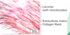
Fibrocartilage
How do chondrocytes receive nutrients?
By diffusion because cartilage is AVASCULAR
Components of cartilage

Chondrocytes
Collagen & elastic fibers
Ground substance (lots of GAGs, proteoglycans)
Matrix is the functional component
Ground substance is ___philic
Basophilic because of its high carbohydrate concentration (lots og GAGs, proteoglycans)
Chondrocytes have well developed ___ and have __.
rERs because they’re constantly secreting proteins
Also have lipids
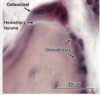
Describe the ring around chondrocytes
Lacunae: cavity in the ECM that chondrocytes sit in
The ring its territorial matrix is slightly darker, but the ones that are farther out between the cells that is barely stained is the interterritorial/interstitial matrix

Type __ collagen is in the territorial matrix

Type II collagen
There are also proteoglycans
Describe the main fibers in hyaline, elastic, and fibrocartilage.
Hyaline = type II collagen
Elastic = elastic fibers (requires special stain)
Fibrocartilage = type I collagen (network) as dense irregular connective tissue

Explain the color differences between perichondrium and cartilage?

There’s a collagen (pink) in both, but there’s so much more ground substance (basophilic) in the cartilage
Two types of chondrogenesis
Appositional growth: at the surface of existing cartilage, perichondrial cells differentiate into chondroblasts
- Growth in girth of cartilage
Interstitial growth: within the cartilage plate, pre-existing chondrocytes are dividing mitotically
- Occurs in the early phases of cartilage formation to lengthen long bones
Describe the composition of the hyaline cartilage matrix
Capsular (pericellular) matrix
Territorial matrix
Interterritorial matrix
–
Collagen type II
Aggrecan (proteoglycan)
Chondronectin (glycoproein)
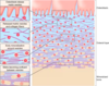
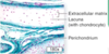
Describe the two layers of perichondrium
Outer fibrous layer: dense connective tissue = type I collagen + fibroblasts
Inner chondrogenic layer: chondroblasts; give rise to new cartilage
From top to bottom, you can see the progenitors > chondroblasts > chondrocytes

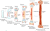
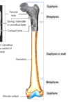
___ cartilage forms the fetal skeleton that will be replaced by bone through endochondral ossification
Hyaline cartilage

What kind of cartilage does not calcify with age?
Elastic cartilage
- Appositional growth
- type II collagen + elastic fibers
- Ears, epiglottis

What kind of cartilage does not have perichondrium? What is this cartilage type mostly made up of and how does it look ona lside?
Fibrocartilage
- Mostly type I collagen, some Type II collagen
- Cells align in an organized fashion to resist compression and shearing forces

What happens if you damage the perichondrium, which is responsible for supplying nutrition to the tissues via diffusion?
Fibroblasts in it will form scar tissue instead of chondrogenic cells
Lamellar/compact/mature bone vs Woven/primary/immature bone
Lamellar/compact/mature bone - regular alignment of collagen fiber
Woven/primary/immature bone - irregular alignment of collagen fiber
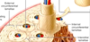
Osteon / Haversian system
the circular unit found within the compact portion of mature bone

Longitudinal Haversian Canal
Vertical blood vessel channels
Transverse / oblique / Volkmann’s canal
Horizontal blood vessel channels
Interstitial lamellae
Outer cicumferential lamellae
Inner circumferential lamellae
Lamellae between osteons
The most external layers of compact bone
The most internal layers of compact bone