Cell (Part 3) Flashcards
(42 cards)
Lysosomes are: (2)
- lysosomes are the end product of _____
- ____ bodies
- lysosomes are the end product of endosomal sorting
- spherical bodies (single membrane)
- several hundred may be present in a single animal cell
Lysosomes digest (3)
Lysosomes digest proteins, lipids, nucleic acids
What do lysosomes have in their membrane? (and what is the effect? where are they commonly found?)
Have proton pumps in their lysosomal membrane
-pump in acidic molecules
- lower pH
- increase activity of lysosomal enzymes that breakdown large molecules (or cells)
- will be able to transport broken down molecules back into the cytosol to be recycled and reused
-lysosomes prevalent in immune cells like macrophages or lymphocytes

Lysosomal storage disorders
- what are they
- 2 types (and subtype of each)
Lysosomal storage disorders: Enzymes for lysosomal degradation are absent or defective
1) Destination label errors (exaple: I cell disease)
2) Enzyme deficiency errors (example: Gaucher’s disease)

Destination label errors
- what is it a type of?
- main example (what happens and what is the net result)
Destination label errors:
-type of lysosomal storage disorder (enzymes for lysosomal degradation are absent or defective
- example: I-cell disease
- the correct enzymes are produced in the ER and trafficked to golgi, but during sorting process, gets wrong the wrong label (i.e. the wrong “molecular address”) so it’s routed away from the lysosome
- net result: the enzymes that are needed to be brought into the lysosomes to degrade all of that material are not present, so end up with building up of material (lysosomal storage) in those disorders.
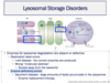
Enzyme deficiency errors
- what type of disorder is it?
- what happens? how can it be fixed?
- main type? what is the net result?
Enzyme deficiency errors:
-type of lysosomal storage disorder (enzymes for lysosomal degradation are absent or defective)
- Enzyme is not made properly earlier in the protein trafficking pathway (and therefore not transferred to the golgi)
- can be fixed with enzyme replacement therapy (because net problem is the absense of the enzyme, if the enzyme was there, it would be packaged and sorted correctly)
- example: Gaucher’s disease - large amounts of lipids (that the lysosome would normally be breaking down) accumulate in the lysosomes

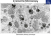
what is shown with arrows?

Lysosomes (in TEM)
- Membrane-bound and electron dense; contain very dark matter = the material that is being digested
- Residual body – lysosome that has completed digestive function.
(You do not have to differentiate between the different lysosomal stages. You do not have to differentiate a residual body from a lysosomes.)
Can visualize lysosomes using a light microscope if:
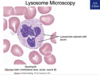
Can visualize lysosomes using a light microscope if you have used a stain specific for that cell’s lysosomes

Four parts of mitochondria in diagram and what they do

-
Matrix: inside
-
has hundreds of enzymes
- contains enzymes needed for the oxidation of pyruvate and fatty acids and for the citric acid cycle
-
has hundreds of enzymes
-
Inner membrane: folded into folds called cristae
- contains proteins that carry out oxidative phosphorylation, including the eletron transport cain and ATP synthase
- contains transport proteins that move selected molecules into and put of the matrix
-
Outer membrane:
-
contains large, channel-forming proteins called porins
- permeable to all molecules of 5000 daltons or less
-
contains large, channel-forming proteins called porins
-
Intermembrane space: space between inner and outer membrane
- contains several enzumes that use the ATP passing out of the matrix to phosphorylate other nucleotides
- also contains proteins that are released during apoptosis
Main purpose of mitochondria:
convert ____ to ____
Convert food to cellular energy
mitochondria are either located/structured:
1) _____ (structure) located in _____ where there is _____
2) form ______ (structure) that are ______ (located)
mitochondria either:
- fixed in one location where there is high energy consumption (cardiac cell, sperm)
- form elongated, dynamic tubular networks that are diffuse throughout the cytoplasm
- continuously break apart by fission and fuse again
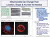
Number of mitochondria depends on _____ and can change with ______
Vary depending on the cell type and can change with energy needs
- Liver cell – 1000-2000 per cell
- Skeletal muscle cells – many many more mitochondria becaue higher energy need
- can divide up to 5-10x if the muscle has been stimulated to contract repeatedly
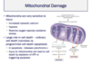
Mitochondria are very sensitive to what types of injuries (3)?
•Mitochondria are very sensitive to injury
- Increased cytosolic calcium
- Hypoxia
- Reactive oxygen species (oxidative stress)

Mitochondria plays a large role in cell death:
-differentiates between 2 types of cell death:
- ordinary cell death = _______
- injury to mitochondria can cause this by ______
- programmed cell death = ______
- injury to mitochondria can trigger this
- _____ (a molecule located in the _____), which is released during this process
Mitochondria plays a large role in cell death:
-differentiates between 2 types of cell death:
- ordinary cell death = necrosis
- injury to mitochondria can trigger this by depletion of ATP
- programmed cell death = apoptosis
- injury to mitochondria can trigger apoptosis
- cytochrome-c (located in the intermembrane space) is released and stiumalates apoptosis
Mitochondria genetic information
- mitochondria are thought to have originated when _____ was engulfed by _____
- mitochondria are virtually cells within a cell, and each has its own _____
- ______-stranded
- ______ molecule (shape)
- codes for ______ units, all of which are components of _____
- mitochondrial DNA (mtDNA) is only inherited through _____
- frequency of mutations increases with _____
- how does this compare to nuclear DNA? why?
Mitochondria genetic information
- mitochondria are thought to have originated when an aerobic bacterium was engulfed by a larger anaerobic eukaryotic cell
- mitochondria are virtually cells within a cell, and each has its own DNA
- double-stranded
-
closed circular molecule (shape)
- codes for 13 polypeptide units, all of which are components of the respiratory chain
- mitochondrial DNA (mtDNA) is only inherited through the maternal lin
- Frequency of mutations increases with age
- has 100-fold higher occurance of deletions and point mutations than in nuclear DNA (because replicates differently than nuclear DNA and does not have the same DNA repair mechanisms)


A = outer mitochondrial membrane
B = inner mirochondrial membrane
C = crista
D = matrix
E = matrix granules (dark granules)
TEM appearance of mitochondrion (can see with light microscope but hard to distinguish)
- Double membrane with the inner membrane folded inward into cristae
- Matrix, enclosed by the inner membrane
- can contain matrix granules (calcium 2+ and other divalent cations) to regulate amount of cations in the cytosol by creating a concentration gradient

A =
B =
C =
D =
Why does this structure appear round?

A = outer mitochondrial membrane
B = inner mitochondrial membrane
C = crista
D = matrix
TEM appearance of mitochondrion (can see with light microscope but hard to distinguish)
- can see double membrane with pieces of the inner membrane extending into the middle - aka folded into cristae
- Matrix, enclosed by the inner membrane
Why are these mitochondria round?
- because of the plane of cut (they are usually longer/more oval-shaped)
- even though they are round, we know they are not a lysosome or endosome or some other round structure because of the double membrane with cristae (which is unique to mitochondria)

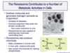
Peroxisomes - 2 main functions
1) _______, producing _____ as a byproduct
* 2 subtypes
2) _______
* 3 subtypes
Peroxisomes contribute to a number of metabolic activities in cells
1) oxidizes molecules - generates hydrogen peroxide as a byproduct
- oxidizes long chain fatty acids into simpler compounds that are more easily metabolized by cells
- oxidizes toxic chemicals, inactivating them
2) produces chemicals
- makes bile acids in liver cells
- contains the enzymes that make phospholipids
- makes cholesterol in animal cells

Peroxisomes role in oxidation:
Peroxysomes oxidize molecules, generating _____ as a byproduct
- Converts _____ into _____ using _____, which they employ for oxidative purposes
- contains _____ to destroy the excess (converts _____ into ____ and ____)
- Used to convert _____ into simpler compounds
- More easily ______
- Peroxisomes are also capable of oxidizing ______ - inactivating them
Peroxysomes oxidize molecules, generating hydrogen peroxide as a byproduct
- Converts molecular oxygen into hydrogen peroxide using oxidases, which they employ for oxidative purposes
- contain catalases to destroy the excess (converts hydrogen peroxide into water and oxygen)
- used to convert long-chain fatty acids into simpler compounds
- More easily metabolized by cells
- Peroxisomes are also capable of oxidizing toxic chemicals - inactivating them
Peroxisomes produce chemicals:
- make _____ in animal cells
- make _____ in liver cells
- also contain ______
•Peroxisomes produce chemicals
- Make cholesterol in animal cells
- In liver cells - produce bile acids.
- Also contain the enzymes for making phospholipids

where are peroxisomes most abundant?
peroxisomes are present in all cells, but in certain kinds of specialized differentiated cells, we may see an abundance of peroxisomes - specifically in metabolically active cells and cells that need to handle a lot of sort of biochemical pathway waste
what are the stars?

* = Lipid droplets
Lipid droplet TEM Appearance:
- Extremely round and not membrane-bound
- Depending on type of lipid, can be electron-dense or electron-lucent
- size is variable (sometimes a lipid droplet could take up broken up into little droplets)

A =
B =
C =
*why is structure B near structure C?
A = mitochondria (double membrane, cristae)
B = lipid droplet (usually a perfect circle, size varies)
C = smooth ER (looks like little noodles)
- Why might this lipid droplet be in close proximity to the smooth endoplasmic reticulum?
- these are often associated with each other because one of theh functions of the smooth ER is phospholipid biosynthesis - they get these lipids from the lipid droplets where they are stored
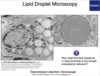
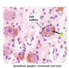
Lipid droplet apperance in light microscope
H&E vs. osmium tetroxide
Lipids in H&E: appear as white circles
- when preparing slides for H&E staining, the lipids are removed with the parafin wax
- so the white circles we see are the space the lipids left behind (so there is nothing left for H&E to stain)
*in image, one cell is outlines and the purple circle is the nucleus
Lipids in osmium tetroxide: appear as black circles
- osmium tetroxide allows lipids to be preserved during staining, so the white spots are now black
So I’ve outlined one cell for you and then the purple circle in the middle is the nucleus. And we see these very circular white shapes. And basically I recall from And basically recall from when you prepare slides for each and each feeding, when you prepare slides for H&E staining, And basically this is the space that the liquid left patents. the space that the liquid left behind. So there’s nothing left for H&E to stain.
Or you can stain them in a way that preserves the lipids and use osmium tetroxide to cover them black, the lipids and use osmium tetroxide to color them black, But in this case again, In this case again, instead of weight, but there are circular. instead of white but they are circular. to identify lipid inclusions.