Cerebrovascular Disease Flashcards

(86 cards)

1
Q

Understand different functions of the brain

A

know the different cortexes

2
Q

Go over the primary motor and sensory cortex

A

review image

3
Q

Blood supply to the brain:

A

review carotid

internal cartoid

ect

4
Q

Know circle of Willis

A

Know branches of circle of Willis

5
Q

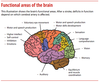
Functional areas of the brain

A
6
Q

Blood supply by major divisions

A
7
Q

Location of MCA

A
8
Q

Location of Lacunar vessels

A
9
Q

Location of watershed infarcts

A

ACA-MCA

MCA-PCA

10
Q

Path of the midbrain

A

Posterior limb is location of corticospinal tracts; may see lacunar infarct here

11
Q

Where is the pyramidal decussation?

A

In the brain stem

12
Q

Review of corticospinal tract

A

note how lateral coticospinal crosses in the decussation (motor for distal muscle)

anterior doesn’t until level of spinal cord (proximal muscles and trunk muscles)

13
Q

Damage here see contralateral paralysis: upper limb and face

Contralateral loss of sensation to upper limb and face

Aphasia if in dominant hemisphere

Hemineglect if lesion affects non-dominant (often right) side

A

MCA

(feeds motor and sensory cortex, Temporal lobe at Wernikes area and frontal lobe at Broca’s area)

14
Q

Contralateral paralysis of Lower limb and Contralatera loss of sensation of lower limb

A

ACA supplies both motor and sensory cortex for lower limb

15
Q

Contralateral hemiparesis/hemiplegia

A

Lenticulostriate artery: common location of lacunar infacts 2nd to unmanaged HTN!

lesion would be in the striatum, internal capsule

16
Q

Contralateral hemiparesis–upper and lower limbs and decreased contralateral proprioception. Ispilateral hypoglossal dysfnx (tongue deviates ipsilaterally)

A

ASA supplies: lateral coticospinal tract and medial lemnisucs as well as caudal medulla (hypoglossal nerve)

***MEDIAL MEDULLAY SYNDROME (often bilateral stroke)

17
Q

Vomit/nystagmus/vertigo with decreased pain and temp sensation from ipsilatareal face and contralateral body

Dysphagia, hoarsness and decreased gag with ipsilateral horner syndrome, ataxia and symetria

A

PICA

***Lateral medullary syndrome or Wallenburg syndrome

(Dont Pick A horse that can’t eat) PICA, horsness, dysphagia

18
Q

Vomit/vertigo/nystagmus

paralysis of face, decreased lacrimiation, salivation, decreased taste from anterior 2/3 of tongue, decreased corneal reflex

FAce: decreased pain and temp

Ipsi decreased hearing and ipsi horner syndrome

ataxia and dymetria

A

AICA; facial nucleas effects specific to AICA lesions

**Lateral pontine syndrome

“FAcial droop means AICA’s pooped”

19
Q

Contralateral hemianopia with macular sparing

A

PCA

supplies occipital cortex and visual cortex

20
Q

Preserved consiousness adn blinking

quadriplegia, loss of voluntary facial, mouth and tounge movements

“Locked-in syndrome”

A

Basilar Artery stroke

supplies: pons/medulla, lower midbrain, corticospinal and bulbar tract, ocular cranial nerve nuclie, paramedian pontine reticular formation

21
Q

CNIII palsy

eye is down and out with ptosis and pupil dilation

A

Pcom

*common site of saccular aneurysm, lesions are usually aneurysms, not strokes

22
Q

Affect of Acom anuerysm

A

visual field defects: can lead to stroke: saccular or berry aneurysm can impinge cranal nerves

23
Q

Hypoxia (deprivation of O2) - in brain occurs by several mechanisms: list 3

A

Low level of oxygen in blood (ex. respiratory arrest, near drowning, severe anemia, carbon monoxide poisoning)

Low blood flow to tissue-ischemia (ex. cardiac arrest, vessel obstruction, increased intracranial pressure)

Oxygen utilization by tissue is impaired (ex.-cyanide poisoning)

24
Q

= low blood flow

causes more damage than hypoxia

A

Ischemia

25
Describe Global Ischemia
**systolic pressure \< 50 mmHg**  Generalized **reduction in cerebral perfusion,** usually due tocardiac arrest, shock, or severe hypotension; outcome dependent upon severity & duration of ischemia
26
Where is damage the worst in Global ischemia?
Brain damage is most severe in watershed/borderzone territories
27
If severe, widespread neuronal death may result in : Persistent vegetative state  Brain death
Case for Global Ischemia
28
infarction from obstruction of local blood supply (stroke)  Results most often from arterial stenosis and/or thrombosis, atheroemboli, or thromboemboli.
Focal Ischemia
29
What are the borderzone areas that are vulnerable in global cerebral ischemia?
Low flow areas between anterior, middle and posterior cerebral arteries
30
Certain brain cells and regions are more susceptible to hypoxia/ischemia than others  Most vulnerable cells (in decreasing order)
– neurons, oligodendrocytes, astrocytes
31
 Most vulnerable regions to hypoxia and ischemia– adults (in decreasing order)
 Hippocampus (CA1 sector – Sommer sector)  Lamina 3 and 5 of cerebral cortex (laminar necrosis)  Purkinje cells in cerebellum
32
What determines the selective vulnerability?
 Variable oxygen/energy requirements of different neurons and neuronal populations  Glutamate receptor densities: Glutamate is **neurotoxic** when present i**n excess,** as occurs in hypoxic/ischemic brain damage
33
Describe what you see on histology in acute hypoxia/ischemia
**“Red is dead”** Pyknotic cell with shrunken & dark nucleus, no nucleolus visible Red cytoplasm (no Nissl substance visible)
34
What area of brain is infarcted and why
Hippocampal infarct: selective vulnerability
35
Laminar necrosis occurs in ischemia, which areas are susceptible?
3, 5 and 6
36
causes widespread neuronal death, irrespective of regional vulnerability
Severe Global ischemia
37
Clincal S/S of severe global ## Footnote  Persistent vegetative state: unconscious , but with retention of sleep-wake cycles, primitive orienting responses, brainstem and diencephalon reflexes  Brain death: diffuse irreversible cortical injury with brainstem injury (absent reflexes and respiratory drive)
38
 Corresponds to brain death  Non-perfused brain  Gross – swollen brain, slit-like ventricles, often herniation
39
Histology of severe global ischemia
Microscopic – pallor, vacuolation of parenchyma, sparse eosinophilic neurons
40
A thrombototic event leading to FOCAL ischemia (infarct, stroke) are often due to: Where do they usually occur?
Atherosclerosis!!!! Origin of MCA Origin/end Basilar Carotid bifurcation
41
What's going on in this internal carotid artery?
atheroma: there can be a liquid core in the plaque or it can become occluded by a thrombus
42
A 73 year old African American male smoker with HTN and DM presented with right sided weakness and aphasia. MRI diffusion weighted image showed:
Left MCA stroke | (was d/t MCA stenosis)
43
Focal ischemia caused by Emboli – Infarcts are more likely\_\_\_\_\_ and are often from a\_\_\_\_\_\_ source and most often affect ______ vessel
hemorrhagic Cardiac MCA
44
Two causes of Emboli leading to focal ischema (infarct) that are from a cardiac source
45
What are common non-cardiac causes of Focal ischemia caused by EMBOLI
Atheroma (often from plaques in carotid arteries) Fat, neoplasm, air
46
Recent hemorrhagic infarct secondary to embolus
occurs because with emboli you can break down embolus and get reperfusion leading to hemorrhage (with throbmus, you just keep scarring over it because it wasn't lodged from somewhere else thus you get white or liquifactive necorsis)
47
85 y/o woman had been noticing palpitations for several weeks. Her primary set her up for a monitor to look for atrial fibrillation but in the meantime she presented with acute left sided visual distortions. An MRI showed:
All lesions are the same age as they are all seen on diffusion weighted imaging. Source is atrial fibriliation as it is dispersed throughout the entirty of brain and all appear same age
48
Third cause of focal ischemia: Due to Hyaline arteriolosclerosis caused by hypertension and diabetes mellitus
Lacunar infarcts/slit hemorrhages
49
Causes of lacunar infarct and LOCATION
 Small strokes (\< 1-1.5 cm) in subcortical brain structures (usually basal ganglia, internal capsule, thalamus, white matter, pons)  May be hemorrhagic
50
45 y/o man with smoking and high cholesterol presented with right face arm and leg plegia. Sensation and language were intact. MRI showed:
lacunar infarct \*\*\*\*severe weakness on whole side of body clear indication for lacunar stroke vision and speech are intact
51
Three common causes of focal ischemia
Thrombis: liquifactive Embolis: hemorrhagic Lacunar: deep and slit like
52
Less common causes of Infarction
 Vasculitis: Non-infectious causes and Infectious causes  Arterial dissection of carotid arteries  Coagulation disorders  Microvasculopathy such as: Arteriosclerotic leukoencephalopathy and CADASIL  Amyloid angiopathy  Drug abuse
53
- -Involves multiple small/medium sized meningeal & parenchymal vessels - -Characterized by chronic inflammation, **fibrinoid necrosis, multinucleated giant cells** (often, not always), & wall destruction
Primary angitis (see fibriod necrosis with vessel wall inflammation)
54
Morphology of infarct Acute: Subacute Chronic:
 Acute: up to 48 hours: **soft, swollen**; gray-white distinction blurred  Subacute: up to 2-3 weeks: **liquefactive necrosis**  Chronic: several months: **cavitated**, all dead tissue removed
55
Describe histology of acute infarct
 Acute:  8-12 hours: red neurons, pallor (with preservation of small area around vessel)  Up to 48 hours: neutrophils (not always)
56
Subacute and chronic presentaiton of brain infarct histology
 Subacute: 48 hrs. to 3 weeks: macrophages, necrotic tissue, reactive astrocytes, vascular proliferation  Chronic: several months: cavity with glial scar
57
What will we see on CT vs MRI in acute hypoxic ischemia?
CT looks normal, even conventional MRI looks normal Get a diffusion weighted MRI; you can see evidence of ischemia in the white areas; there is BRIGHTNESS in areas lacking oxygen or filled with water= edema
58
takes days to weeks: see liquefactive necrosis, many macrophages, reactive astrocytes with vascular proliferation and necrotic tissue
Subacute (organizing) infarct
59
Describe what you see on CT in subacute infarct What about MRI?
CT: see hypodense area where all water is: often tehre is midline shift On MRI: we have diffuse edema and swelling
60
Describe what you see grossly in chronic infarct What about on imaging?
See cavity with fibrillayr astrocytic scar
61
 Small areas of infarction – multiple lacunar infarcts  **Diffuse white matte**r disease – Binzwanger disease and CADASIL  Strategic infarcts – occur in areas **important for cognition/memory**
Pattner of damage in VASCULAR dementia
62
Causes of Cerebral venous thrombosis
Infection, injury, neoplasm, surgery, Pregnancy, oral contraceptives, hematologic abnormalities, dehydration, and malignancy
63
What brain structures are susceptible to vascular dementia?
 Hippocampus, dorsomedial thalamus, frontal cortex, cingulate cortex
64
 Causes hemorrhagic infarcts  Usually superior sagittal sinus or lateral sinuses which results in parasagittal hemorrhagic infarcts
Cerebral venous thrombosis
65
Superior sagittal sinus thrombosis and para-sagittal hemorrhagic infarct
66
 28 year old woman presents with severe headache.  Pressure behind left eye x 1 week, acute intensification of headache while brushing teeth in morning, developed throbbing sensation.  PMHx: none. Healthy recreational distance runner.  Exam: Afebrile, Alert, uncomfortable with severe headache, non-focal neurologic exam.  CT normal  LP normal
Cerebral venous thrombosis: this is before and after being placed on anti-coags; pt was severely dehydrated
67
\_\_\_\_\_\_\_\_\_\_– most common cause of primary ICH  Peak occurrence in 60s  Abrupt onset of severe neurologic dysfunction when hematoma is large  Putamen, thalamus, pons, cerebellum  Hyaline arteriolosclerosis
Hypertension
68
Most common cause of Intracerebral hemorrhage Location where we see this in the brain
Hypertension see in the Putamen, thalamus, pons and cerebellum
69
Less common causes of intracerebral hemorrhage
Amyloid angiopathy and vascular malformations most often from Hypertension
70
means bleeding. Any time a blood vessel is broken and bleeding, whether internally or externally
Hemorrhage
71
Describe a hematoma
a describes a collection of blood in the body’s tissues. A bruise or contusion is also a hematoma. In head injuries, internal bleeding is often described based on how deep it is in relation to the three layers of the meninges. The meninges is the membrane surrounding the brain. Its layers from outside to inside are the: Dura mater Arachnoid mater Pia mater
72
Descibe a Charcot Bouchard microaneurysm
Type of hyaline ateriolosclerosis from uncontrolled HTN
73
How does hyaline ateriolosclerosis cause intracerebral hemorrhage
often secondary to HTN; will weaken the arteriole and presdispose it to rupture
74
WTF happened to this person?
Massive hypertensive hemorrhage: in the deep grey matter "ganglionic"
75
 58 y/o man presented with right sided sensory loss and elevated blood pressure. CT scan showed a left thalamic intracerebral hemorrhage.How do we tx pt?
Need to lower BP of pth then deal with excisting coagulapathies resulted in tiny hematoma
76
Arteriovenous malformation-most common vascular malformation Male or female predominance? What age does it present? Location?
 M\>F  Presentation between 10-30 years  Most often in distribution of MCA
77
Whats going on in this fucked up image?
Arteriovenous malformation: see Tangled network of vessels with arteriovenous shunt
78
How do pts with arteriovenous malformations present?
Can present with bleeding, but often with seizures, headaches, or focal deficits
79
Vascular malformation:  Cerebellum, pons, white matter  No intervening brain tissue  Evidence of prior bleeding
Cavernous angioma
80
Cause of Lobar hemorrhage: not located deep; on the hemisphere
 Neoplasms  Drug abuse  Vasculitis  Hemorrhagic diathesis  Amyloid angiopathy
81
Describe where you would see amyloid angiopathy and what it looks like histologically
Located more in hempispheres: NOT deep congo red stain bc of amyloid and would see beta stain (below) with immunostain
82
89 y/o woman presented with aphasia. CT showed left temporal ICH. MRI showed old bleeds c/w amyloid angiopathy.
Not best image: point is you see old and new infarcts in pt
83
Cause of subarachnoid hemorrhage
84
 “The worst headache I’ve ever had!”  Increased risk with hypertension, smoking, AVM, etc.  Increasing risk of rupture as size increases
Berry or Saccular aneurysms: leads to subarachnoid hemorrhage
85
What is the pathophysiology of Berry anuerysms? Where do they typically occur?
 Not present at birth, but **defect in media** is congenital and the aneurysm develops over time  Occur typically at branch points, 90% in the anterior circulation
86
 59 y/o woman with severe headache followed by unsteady gait and then coma.  PMHx: 40 pack-year smoking, HTN  BP 187/106  Exam: Intubated, opens eyes to pain, pupils equally reactive, moving all 4 extremities spontaneously. Does not follow commands
Not loss of structure in the brain on CT we have ventricles full of blood: in the subarachnoid space get sever reactive edema \*\*Subarachnoid hemorrhage