Cervical fx Flashcards

Atlas fx - C1 Axis fx- odontoid peg traumatic spondylolithesis C2-hangman fx cervical facet dislocations/fx cervical spine fx (56 cards)

1
Q

What is the epidemiolgy of atlas fx?

A
  • 7% of all cervical fx
  • risk of Neurologic injury= LOW
  • commonly missed due to inadequate imaging of occiptocervical junction
2
Q

What is the pathophysiology of atlas Fx?

A
  • Hyperextension
  • lateral compression
  • axial compression
3
Q

Name any associated conditions with atlas fx?

A
  • Spine fractures
    • 50% associated spinal injury
    • 40% assoc AXIS fracture
4
Q

What is the prognosis of atlas fx?

A
  • Stabilty dependent on degree of injury and healing potential of TRANSVERSE ligament
5
Q

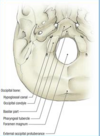
Describe the anatomy of Atlas bone?

A
  • C1 is a ring containing 2 articular lateral masses
  • lacks vertebral body or spinous process
  • forms form 3 ossification centres
  • incomplete formation of post arch is relatively common anatomic variant- doesn’t represent traumatic injury
  • occipital-cervical junction & atlantoaxial junction are coupled
  • intrinsic ligaments provide most stability
    • transverse ligament
    • paired alar ligaments
    • apical ligament
    • tectorial membrane- connects posterior bocy of axis to anterior foramen magnum and is the cephalad continuation of PLL
6
Q

What is the classification of atlas fractures?

A
  • Type 1
    • Isolated ANT or POST ARCH Fx
  • Type 2
    • Jefferson Burst Fx
    • Bilateral ANT & POST Arch FX
    • Stability determined by transverse ligament
  • Type 3
    • ​Unilateral Lateral Mass Fx
    • stability determined by integrity of transverse ligament
7
Q

What is the classification of transverse ligament injuries?

A
  • Type 1 - Intrasubstance tear
  • Type 2 - Bony avulsion
8
Q

What imaging aids dx of atlas fx?

A
  • Lateral xray
    • Atlanto-dens interval
      • <3mm normal adult ( <5mm child)
      • 3-5mm= injury transverse ligament
      • >5mm = injury to transverse lig, alar and tectorium membrane
  • Open mouth odontoid view
    • to identify atlas fracture
    • sum of lateral mass displacement
    • if >7mm = transverse lig rupture assured- unstable
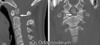
CT

  • delinate fracture pattern & assoc injuries

MRI

  • More sensitive at detecting injury to transverse lig
9
Q

What are the tx for atlas fx?

A

Non operative

  • Hard cervical orthosis vs halo immobilisation 6-12 wks
    • for Stable Type 1- intact TL
    • Stable Jefferson fx- intact TL
    • Stable type 3- intact TL

Operative

  • Posterior C1-2 Fusion vs Occipitocervical Fusion
    • ​for Unstable Type 2
    • unstable Type 3
  • posterior C1-2 fusion preserves motion cf occiptocervical fusion
  • C1-2 transarticular screw placement or *C1 lateral mass to C2 pedicle screw- *see pic
  • Occiptocervical fusion used when unable to get adequate puchase of C1
10
Q

What are the complications of atlas fx?

A
  • Delayed c spine clearance
    • higher rates of complications in pts with delayed c spine clearance so important to clear expeditiously
11
Q

Define an odontoid fracture?

A
  • a fracture of the dens of the AXIS C2
12
Q

What is the epidemiology of Odontoid fracture?

A
  • Incidence
    • most common fracture of the axis
    • accounts for 10-15% of all cervical fx
    • occurs bimodal distribution
      • elderly
        • missed, caused by simple falls
        • assoc increased morbidity/mortality
      • Young pts
        • blunt trauma to head-> cervical hyperextension/flexion
13
Q

What is the pathophysiology of odontoid fractures?

A
  • Displacement maybe Anterior ( hyperflexion) or Posterior (hyperext)
  • Anterior displacement=
    • TL failure
    • Atlanto-axial instability
  • Posterior displacement
    • direct impact from ant arch during hypextension
  • *A fx thru the base of the odontoid process severly compromises the stability of the upper cervical spine*
14
Q

Name any associated conditions with odontoid fx?

A
  • Os odontoideum
    • Appears like a type 2 odontoid fx on xray
    • previously thought to be due to failure of fusion at the base of the odontoid
    • may represent the residules of old traumatic process
    • tx is obervation
15
Q

Describe the anatomy of axis?

A
  • axis has odontoid process
    • develops from 5 ossification centres
    • subdental synchondrosis is an intial cartilaginous junction between dens & vertebral body that does not fuse until 6 years of age
    • secondary ossification centres appear 3ys fuses to dens at 12
  • Axis Kinematics
    • C1-C2 atlantoaxial articulation
      • Diathrodal joint which provides
      • 50 degrees of cervical rotation
      • 10 degrees of flexion/extension
      • 0 lateral bend
    • C2-3 joint
      • 50 degrees of rotation
      • 50 degrees of flex/ext
      • 60 degrees lat bend
  • Ligamentous stability
    • transverse ligament
    • Apical ligament
    • alar ligament
  • Blood supply
    • Wateshed exists between apex and base of odontoid
    • apex supplied branches internal carotid A
    • base supplied branches vertebral A
    • limited blood supply affect healing type 2 odontoid fx
16
Q

Describe the classification of axix fractures?

A
  • Anderson and D’Alonzo
  • Type 1 = Oblique Avulsion fx, tip odontoid
    • avulsion by alar ligament
  • Type 2= Fx thru WAIST
    • high non union rate- watershed blood supply
  • Type 3 = fx extends into cancellous body C2
    • involves variable portion of C2/3 joint
17
Q

What are the symptoms and sign of axis fracture?

A

Symptoms

  • Neck pain worse with motion
  • dysphagia maybe present when assoc large retropharyngeal haematoma

Signs

  • Myelopathy
  • v rare as large x ssection of c spine here
18
Q

What imaging is important in axis fx?

A

Xrays

  • Ap, Lateral. open mouth odontoid peg view
  • flexion-extension: c spine instability in type 1
    • ADI ( atlantodens- interval) >10mm
    • <13mm Space Available for the cord

CT

  • delinate fractures and assess stability

MRI

  • If neurology present

Ct angio

  • To determine locality of vertebral artery prior to post instrumentation
19
Q

What is the tx of axis fx?

A
  • OS Odontoideum = Observe
  • Type 1 avulsion = Hard Cervical Orthosis
  • Type 2 Young pt
    • Halo vest immobilisation 6-12 wks if no risk factors for non union
    • Surgery if risk of Non union
  • Type 2 Elderly
    • Hard Cervical orthosis 6-12wks- if not surgical fit
    • Surgery if surgically fit
  • Type 3
    • Hard Cervical Orthosis 6-12 wks
    • no evidence to support halo over orthosis!!
    • elderly pt poorly tolerate halo-> aspiration, penumonia, death
20
Q

Describe the techniques of surgery to Axis fx?

A
  • Posterior C1-2 fusion
    • for Type 2 fx w risk fx of nonunion
    • type 2/3 fx non unions
    • posterior c1-2 transarticular screw - see pic- avoid in pt w aberrant vertebral artery
    • or post C1 lateral mass and c2 pedicle
    • loss of 50% neck motion
  • Anterior Odontoid osteosynthesis
    • iin type 2 fx with risk nu &
    • acceptable alignment/minimal displacement
    • obliq fx pattern perpendicular to screw trajection
    • pt body habitus allows screw trajection
    • assoc higher failure rates than post fusion
  • transoral odontoidectomy
    • in severe post displacment & cord compression/neurological deficits
21
Q

Decribe the technique for anterior odontoid screw osteosynthesis?

A
  • anterior apporach cervical spine
  • single screw adequate
  • assoc with higher failure rate cf post fusion
  • preserves atlanto axial motion
22
Q

What are the complcaitions of axis fx?

A
  • Non union
    • increased in type 2
    • risk factors include
      • >5mm posterior displacement
      • >1mm fracture displacement
      • fx comminution
      • angulation >10o
      • age >50 years
      • delay in tx > 4 days
      • posterior redisplacement >2mm
23
Q

What is a hangman’s fx?

A
  • Traumatic anterior spondylolitheis if AXIS due to BILATERAL fx of PARS INTERARTICULARIS
24
Q

What is the mechanism of a hangman’s fx?

A
  • Hyperextension
    • leads to fx of pars interarticularis
  • Secondary Flexion
    • Tears PLL and disc
    • leads to Subluxation
  • 30% have concomitant c spine fx
25
What are the symptoms and signs of trumatic spondylothiesis of axis
Symptoms * Neck Pain Signs * Pts are Neurologically Intact
26
What imaging is helpful in dx of traumatic cervical spondylolithesis of axis?
* xrays * flexion/extension shows subluxation * CT * delinate Fracture pattern * MRI * suspicious of vascular injru to vertebral artery
27
What is the classification system for traumatic cervical spondylolithesis of axis?
* **Levine and Edwards**
28
Can you describe and the tx for type I traumatic cervical spondylolithesis of axis?
* * no angulation * C2/3 remains intact * stable fx pattern tx * Rigid cervical collar 4-6 wks
29
Can you describe and the tx for type 2 traumatic cervical spondylolithesis of axis?
* **\>3mm horizontal displacement** * **Significant angulation** * **vertical fracture line** * C/3 disc& PLL interupted * Unstable fx pattern Tx * If \<5mm displacment then reduce with traction then HALO immobilisation for 6-12 wks * if \>5mm displacement ?surgery/ prolonged traction * normally autofuse depsite displacement
30
Can you describe and the tx for type 2a traumatic cervical spondylolithesis of axis?
* No horizontal displacment * **horizontal fracture line** * sig angulation TX * _Avoid TRACTION_ * Reduction with hyperextension then halo immobilisaiton 6-12 weeks
31
Can you describe and the tx for type 3 traumatic cervical spondylolithesis of axis?
* Type 1 with assoc bilateral C2/3 facet disslocation * rare injury tx * Surgical reduction of facet dislocation * then Stabilisation * anterior C2-3 interbody fusion * posterior c1-3 fusion * Bilateral C2 pars screw osteosynthesis
32
What is a cervical disc dislocation/fx?
* Spectrum of osteoligamentous pathology includes * **Unilateral facet dislocation** * most freq missed c spine injury * -\>25% subluxation on xray * assoc w monoradiculopathy- improves w traction * **Bilateral Facet dislocation** * -\>50% subluxation on xray * assoc sig spinal cord injury * **Facet fractures** * more freq involves superior facet * maybe unilateral/ bilateral
33
What is the epidemiology of cervical facet dislocations/fx?
* **75%** of all facet dislocations occur within the subaxial spine **C3-C7** * 17% of all injuries are fx of C7 or dislocations of C7-T1 junction
34
What is the mechanism of cervical facet dislocations?
* Flexion & distraction +/- element of rotation * so in a facet dislocation the posterior structures (interspinous ligament, facet capsule, liagmentum flavum, posterior annulus) are likely disrupted,
35
What is the signs of cervical facet dislocation?
Signs * **Monoradiculopathy** * pt w unilateral dislocation * C5/6 unilater * C6 radiculopathy * Weakness wrist extension * numbness in thumb * C6/7 unilat * C7 radiculopathy * weakness in triceps/wrist flexion * numbness to index/middle finger * **Spinal cord injury** * Seen in _bilateral facet dislocations_ * symptoms worsen with increasing subluxation
36
What imaging is useful in dx of cervical facet dislocation/fx?
* Xrays * lateral * unilateral facet dislocation- 25% subluxation(pic) * bilateral facet dislocation- 50% subluxation * loss of disc height ? retropulsed disc in canal * CT * essential for more detailed bony anatomy * MRI * when **acute facet dislocation in pt w altered mental state** * **failed closed reduction before open reduction to look for _disc herniation_** * _if find ant disc herniation need to open anterior first_ * **any neurological deterioration**
37
what is the tx for cervical facet dislocation/fx?
Non operative * **Cervical Orthosis/ external immobilisation 6-12 wks** * for facet fractures wout sig subluxation/dislocation/kyphosis Surgery 1. **Immediate closed reduction , then MRI then surgical stabilisation** * for bilat facet disclocation w deficits in awake & cop pt * unilat facet dislocation w deficits in awke & coop pt * _never closed reducition on pt w altered mental state_ * **Always do MRI b4 surgery** so check no disc * PSF/ ACDF in absence of disc * ACDF if disc herniation * 26% pts will fail closed reduction & require open **2.****MRI then open reduction + surgical stabilisation** * for facet dislocations when pt changed mental state * or failed closed reductions * _if anterior disc need to go in anteriorly_
38
Can you how you do a closed reduction of cervical facet dislocation?
* Adequate aneathesia * sedation * supervision of respiratory function * serial cross table laterals- do in theatre * gradually increase axial traction with addition of weight ( can give \>50lbs) * a compotnet of cervical flexion can aid reduction * perform serial neuro exams and xray after each weight * abort if neurological exam worsens & obtain urgent MRI
39
Describe the type of cervical vertebral body fractures?
By fracture Pattern * **Compression** * **Burst fracture** * **flexion teardrop fx** * **extension teardrop fx** **Allen and ferguson classification** of subaxial spinal injuries by mechanism used only for research
40
What compression cervical fractures characterised by?
* **Compressive failure of Anterior vertebral body** _without disruption of posterior body cortex_ and without retropulsion into canal * often assoc with posterior ligamentous injury
41
What Burst cervical fx characterised by? What are they associated with?
* **fracture extension thru posterior cortex with retropulsion into spinal canal** * often assoc with _posterior ligamentous injury_ * often assoc with _complete/incomplete spinal injury_ * frequently _unstable_ * usually requires **Surgery**
42
What flexion teardrop cervical fractures characterised by?
* **fx of anterior inferior portion of vertebra** * **post portion of vertebra RETROPULSED POST** * often assoc w post ligament injury * assoc with SPINAL cord injury * normally unstable * Requires Surgery
43
What extension teradrop avulsion cervical fractures characterised by?
* small fleck of bone avulsed of anterior endplate * usually at c2 * must differentiate from true tear drop fx * mechanism= extension * stable injury pattern * Not assoc with Spinal cord injury * TX= cervical collar 6-12 wks
44
What are the tx options for cervical spine fractures?
Non operative * **collar immobilisation 6-12 wks** * stable mild compression fx * ant teardrop avulsion fx * **ext halo immobilisation** * only if stable fx pattern Surgery * **Anterior decompression, corpectomy, strut graft and fusion with instrumentation** * burst fx w cord compression * unstable tear drop * compression fx with angulation 110  or 25% loss in height * **Posterior decompression, fusion w instrumentation** * sig injury to post elements * ant decomp not required
45
Describe the 2 types of occiptiocervical dislocation?
* **Traumatic occipitocervical dislocation** * _severe injury, pt rarely survives_ * most pt die of brainstem destruction * 19% of fatal cervical injuries * os those survive high neurological injury * mechanism- _translation/distraction_ * _​_**Acquired occipitocervical instability** * **​in Down's syndrome**
46
Can you describe the classification of occiptiocervical instability?
* type 1 - anterior * type 2- longitudinal dislocation * type 3 - posterior
47
What do the radiographs show of occipitocervical instability?
* Low sensitivity in detecting injury * **Powers ratio** * used to detect occipitocervical instability * Powers ratio = C-D/A-B * C-D = _distance from basion to post arch_ * B-A= **distance from ant arch to opisthion** * **ratio normal =1** * **if \>1 = anterior dislocation** * **if \<1** * post ​atlanto-occipital dislocation * odontoid fx * ring of atlas fx
48
What is th tx of occiptiocervical instability?
* Non op- **don't use traction**= 10% risk of neurological deterioration * Operative * **Occipitocervical fusion** * mot cases require stabilisation * modular occiptial plates * position 8mm unicortical screw 2cm lateral and 2cm inferior of **external occiptal protruberance**-5cm lateral is the thickest portion of occiput- see pic * don't put screw just below external occiptal protuberance as **major dural venous sinuses** here and risk of penetration
49
What is the epidemiology of occipital condyle fx?
* Involve the craniocervical junction * approx 1-3% population * often missed due to low sensitivity of plain xrays * dx on CT
50
What is the mechanism of occiptial condyle fx? Name any assoc injuries?
* High energy- non pentrating to head/neck * fx patterns dependent on directional forces assoc injuries * c spine fx * polytrauma * intrcranial bleeding * brainstem and vascular lesion * elevated ICP
51
What is the prognosis of occiptial condyle fx?
* High mortality rate 11% - associated injuries
52
Describe the anatomy of occipital condyles?
* Paired prominences of occipital bone * form lateral aspects of foramen magnum * forms the occiptioatlantoaxial complex * 6 main synovial articulations * ligamentus structure * Transverse L * apical ligament * paired alar ligaments * tectorial membrane * Proximity to * Medulla oblongata * vertebral arteries * Lower cranial nerves CN IX-XII
53
Describe the classification of occipital condyle fractures?
* **_Anderson and Montesano_** * Type 1 * impaction type due to compression between atlanto-odontoid joint * stable as minimal fragment displacment into foramen magnum * Type 2 * basilar skull fx extends into 1/2 occipit condyles * direct blow ot skull * stable as alar lig and tentorium membrane intact * Type 3 * avulsion fx of condyle in region of alar lig * due to forced rotation and lat bending * unstable due to craniocervical disruption
54
What are the signs and symptom of occiptial condyle fracture?
Symptoms * high cervical pain * reduced head rom * torticollis * lower cranial n deficit * motor paresis signs * CN IX, X, XI affected
55
what imaging useful in occipital condyle fx?
* Xrays * Ap, lateral ope mouth ap view * CT * method of choice * MRI * soft tissue craniocervical trauma
56
What is the tx of occiptial condyle fx?
Non op * **Analgesia, cervical orthosis( semi-rigid/rigid)** * type 1 & 2 * type 3 without instability _Operative_ * Type 3 with overt instability * neural compression from displaced fx * assoc occipital- atlanto-axial injuries * **C0-C2/3 occipitocervical arthrodesis using semi rigid segmental fixation or post decompression and instrumented fusion** * may need bone graft/removal of bony fragments