What are the histologic findings of a seborrheic keratosis that is “irritated”? (2)
- Squamous eddy formation (eddy = whirlpool)
- Spindling of cells
Seborrheic keratoses may have what common genetic mutations? (2)
- BCL-2 (marker of resistance to programmed cell death)
- Fibroblast growth factor receptor 3 (FGFR-3, a tyrosine kinase receptor)
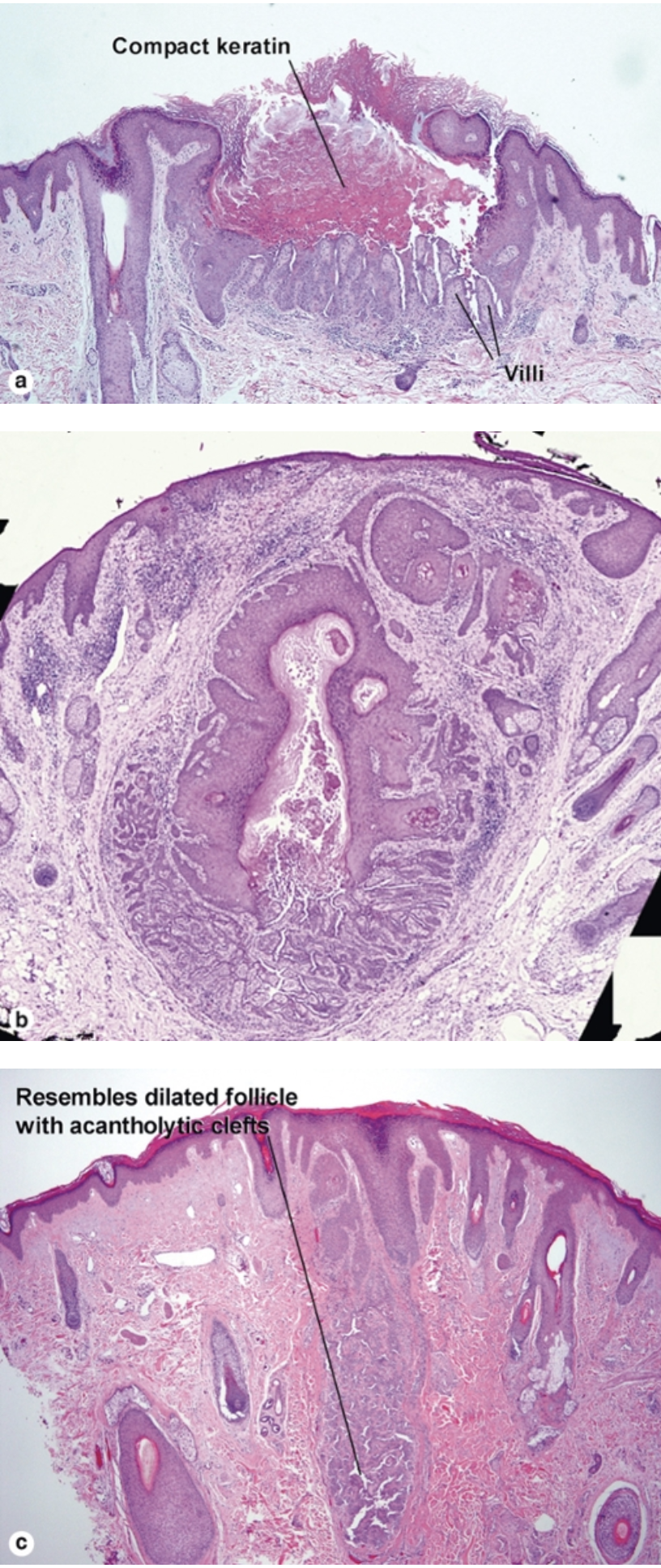
What are the histologic findings of a warty dyskeratoma? (at least 3)
- Corps ronds (stain pale pink to red with wide, clear halo around nucleus)
- Grains (flattened, basophilic, dyskeratotic cells)
- Often resemble a dilated hair follicle with acantholytic clefts
Seborrheic keratoses are acanthomas of keratinocytes that are about the size of normal keratinocytes at what location?
Acrosyringeal keratinocytes (i.e., cells that compose the intraepidermal portion of the eccrine duct)
Keratinocytes of seborrheic keratoses are typically SMALLER than normal keratinocytes. If the cells are larger, then it is termed a large cell acanthoma.
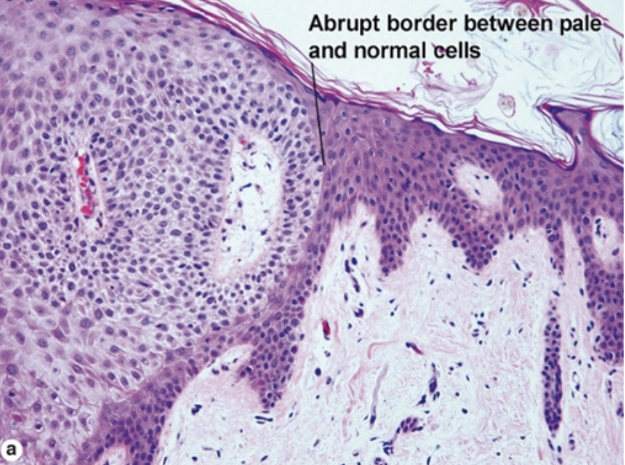
What are histologic features of a clear cell acanthoma? (3)
What else would be on the differential?
- Abrupt transition from normal epidermis to glycogenated epidermis
- Parakeratosis with neutrophils overlying the abnormal epidermis
- Neutrophil fragments in epidermis itself (“karyorrhectic debris”)
- dDx includes psoriasis but look for abrupt cut-off and glycogenated keratinocytes, and Bowen’s disease
What may develop if HPV infects an eccrine duct or hair follicle?
- A cystic papilloma (also known as a verrucous cyst)
- Look for hypergranulosis in the dells
- Papillomatosis
- Compact red stratum corneum with round cookie cutter holes
What are the two types of cyst that can have a “shark tooth cuticle”?
- Steatocystoma (has vellus hairs)
- Dermoid cyst (has terminal hairs)
What is the other name for a steatocystoma?
Simple sebaceous duct cyst
What diagnoses feature acantholytic dyskeratosis?
- Darier disease
- Acantholytic acanthoma
- Grover disease
- Warty dyskeratoma
What is an important histologic finding in a branchial cleft cyst?
Lymphoid aggregates
What are two histologic findings of a verrucous cyst/cystic papilloma?
Coarse hypergranulosis and round parakeratosis
Stratum corneum with cookie cutter holes
What are the histologic findings of a median raphe cyst? (3)
What is the common location?
What is the analogue in women, and where does this occur?
- Variable lining that may be ciliated, cuboidal, or simple epithelium
- Debris-filled cyst
- Surrounding skin has findings of genital skin (delicate collagen, random smooth muscle, many small nerves, and prominent vascularity)
- Occurs in men between urethral meatus and anus
- Analogue in women is the cutaneous ciliated cyst, which occurs on the leg
True or false:
Other skin cancers, such as Bowen disease or melanoma, can arise from a seborrheic keratosis.
True
What are histologic findings of a pilar cyst?
- Epidermal lining without a granular layer
- Uniform, red, compact keratin inside the cyst
What are findings of a dermoid cyst?
What is the most common location for a dermoid cyst?
- Eccrine glands and terminal hair follicles embedded in cyst wall
- Lamellar keratin inside cyst
- Most common location: lateral brow
What are the histologic findings of porokeratosis ptychotropica? (2)
- Unique form of porokeratosis, typically on the buttocks
- Multiple cornoid lamellae
- Psoriasiform acanthosis
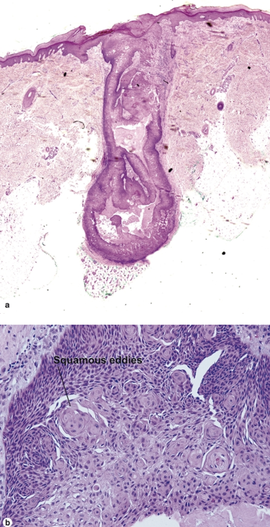
What can an inverted follicular keratosis be likened to, according to Elston?
- “An irritated seborrheic keratosis in a massively dilated hair follicle”
- An endophytic acanthoma with squamous eddies
What are the histologic findings of an epidermolytic acanthoma?
Where are 90% of these located?
- Crateriform acanthoma
- Expanded granular layer shot full of holes
- Compact horn (stratum corneum)
- 90% located in genitalia
- Not HPV-associated
What are the histologic findings of inflammatory linear verrucous epidermal nevus (ILVEN)?
- Variable acanthosis
- Stratum corneum with alternating orthokeratosis (having a granular layer) and parakeratosis (having NO granular layer)
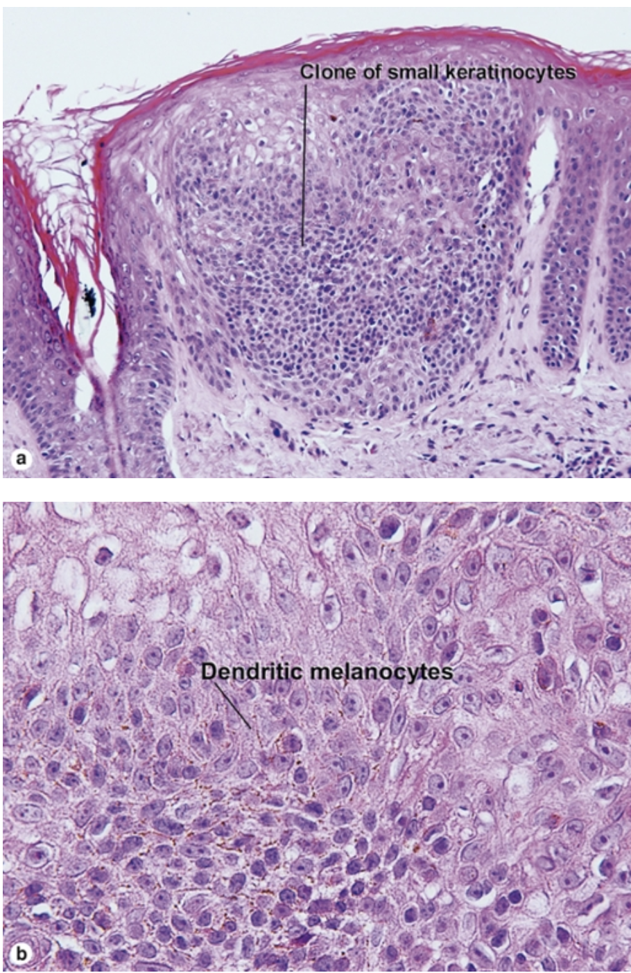
What are the features of a melanoacanthoma?
- Acathoma of both small keratinocytes and pigmented dendritic melanocytes
- Most pigment is within dendrites
- Overlying stratum corneum is almost always compact eosinophilic and parakeratotic
What are the features of a clear cell acanthoma (pale cell acanthoma)?
- Discrete acanthoma with overlying parakeratosis
- Distinct transition between normal keratinocytes and clearer cells in stratum spinosum that lack phosphorylase and thus have accumulated glycogen stores
- Peppered with neutrophils
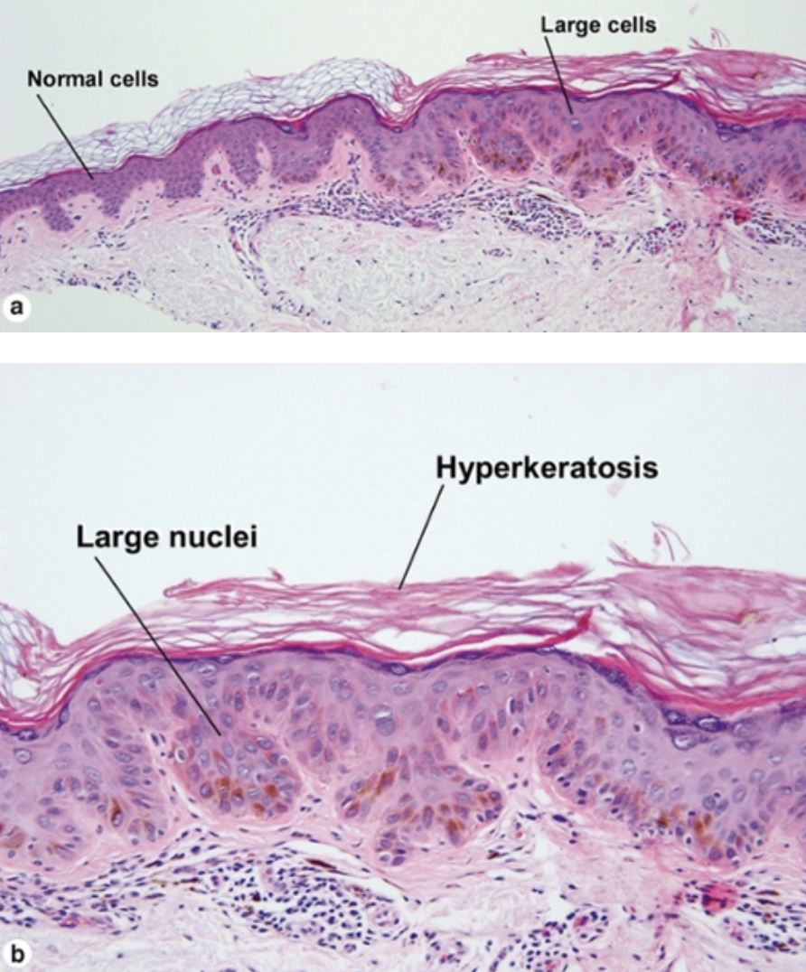
What are the features of a large cell acanthoma?
- Discrete acanthoma composed of cells with large nuclei typically twice the size of nuclei in the surrounding epidermis
- Overlying lamellar hyperkeratosis common
- May be pigmented
- Some clinicians prefer to destroy any remaining lesion with cryotherapy given possibility this may be early Bowen disease
What are the features of inverted follicular keratosis (IFK)?
- Endophytic lesion resembling an expanded hair follicle
- Squamous eddies
- Unrelated to HPV infection
- Multiple lesions → Cowden syndrome
What are the features of a warty dyskeratoma?
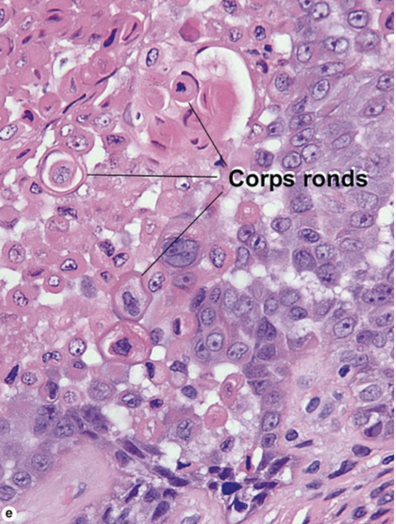
- Endophytic growth
- Acantholytic dyskeratosis
- Overlying parakeratotic crust
- Corps ronds → round, dyskeratotic cells that stain pale pink to red with wide, clear halos around the nucleus (see initial photo)
- Grains → flattened, basophilic (blue or purple) dyskeratotic cells