clinical and biochemical features of metabolic bone disease Flashcards

(72 cards)

1
Q

what is a metabolic bone disease

A

a group of diseases that causes a change in bone density and bone strength

by indreaing resorption or decreasing bone formation or altering bone structure

it may be associated with disturbances in mineral mechanism

2
Q

what are the 5 common metabolic bone disorders *

A

primary hyperthyroidism

rickets/osteomalacia

osteoporosis

pagets disease

renal osteodystrophy

3
Q

what are metabolic symptoms of bone diseases

A

hyper/ocalcaemia

hyper/ophosphtaemia

4
Q

what are the bone symptoms of metabolic bone disorders

A

deformity

fracture

5
Q

what is hydroxyapatite made of

A

calcium and phosphorus

6
Q

how is cancellous bone metabolically active

A

remodelling - 5% body is remodelling at any time

whole skeleton remodels over 7years

acts as a ca reserve - there is a balance with blood and bone

7
Q

what makes a bone strong

A

mass - genetic

material properties - collagen cross-linking, woven/lamella, mineralisation (young bone less mineralised so ductile, older is more brittle), microcracks

microarchitecture - trabecular thickness, trabecular connectivity, cortical porosity (holes in corticies - when teenager, grow fast and fracture)

macroarchitecture - hip axis length and diameter

8
Q

summarise the age related changes in bone mass *

A

build bone up until 20s,

stable until 40

lose

accelaratd loss in women in menopause - lose 30% of bone mass

both men and women have a slow phase

9
Q

how can exercise change bone mass

A

change in dimention - add perisoteum

change in shape - lay down bone specific to force

10
Q

describe bone remodelling during the growth of the tibea

A

lay bone down in anterior and posterior compartments - specific to minimise the mass of bone that is layed down

therefore there is increased bending strength anterio-posteriorly, than medio-laterally

bone is layed down in the periosteum region and resorbed from the endosteum

11
Q

descrieb teh sexual dismorphism in bone growth

A

men develop bigger bones with a bigger cavity

women’s bones have thicker endosteum so are stronger, as they get older - endocortical bone resorption and periosteal bone formation occurs - never matches mens

12
Q

descrieb cortical bone microfractures *

A

the bone has a structure to absorb energy - cortex has osteons and alternating density of lamellae

irreversible plastic deformation does occur - causing microfractures that dissipate the energy - they crack the matrix - usually limited to the interstitial bone between osteons - if they accumulate the bone strength will be comprimised

this is detected so has to be repaired

they are repaired through bone remodelling

each oseton represents a previous remodelling event

13
Q

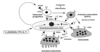
describe the bone remodelling cycle *

A

activation phase - osteocytes detect a microcrack - the crack crosses canaliculi so severing the osteocyte’s dendritic processes

this causes osteocytic apoptosis

this acts as a signal to the connected surface lining cells which are of osteoblast lineage

the lining cells and osteoblasts release factors that attract cells from the blood and marrow into the remodelling compartment, these cells are monocytes and

resorption phase - osteoclasts are generated locally from the recruited cells - they resorb the matrix and the offending microcrack

reversal phase - switch from resorption to formation, osteoclasts are apoptosed

formation - then osteoblasts deposit new lamellae bone

the osteoblasts that are trapped in the matrix become osteocytes, others die or form new osteoblast lining

osteoblasts last months, clasts last weeks so have to keep recruiting clasts

this is happening all over the skeleton

14
Q

what are the biochemical investigations in bine disease *

A

serum

  • bone profile - ca, corrected ca, phosphate, alkaline phosphate, mg

renal func

  • creatinine
  • PTH
  • 25-hydroxy vut D

urine

  • ca/phos
  • NTX (bone resorptoion marker)
15
Q

summarise Ca balance

A

some absorption from GIT but not very efficient

reabsorb some from kidney - but excrete some, this cannot be helped

there is a big influx in and out of bone - cancellous bone has a huge supply

16
Q

how do you correct serum ca measurements

A

total ca is 2.15-2.56mmol/L

46% of this is protein bound

47% is fress ionised

7% is complexed

measure ionised in casualty

acid base balance affects ca - in alkalosis ca bind to protein = ca drop = tinglking feeling

venous stasis might falsely elevate levels

corrected ca = [ca] +0.02(45-[albumin])

if albumin is high, true ca is low - corrected ca compensates for the protein level

17
Q

explain how PTH regulates ca levels *

A

has the predominant role in min by min ca reg

if ca drops PTH increases in minutes

immediately causes bone resorption by stimulating osteoclasts - releases ca and phos

acts on kidney - increase ca resorption in DCT, increase phos excretion by inhibiting NAP co-transporter in PCT (phosphaturic hormone), increases 1a hydroxylation so increases vit D activation = increase in intestinal ca and phos absorption

18
Q

clinical relevance of PTH response system

A

84aa peptide but N1-34 is active - this is used clinically

mg dependant - alcoholics have low mg = low ca

PTH half life - 8mins - so can be measured intraoperatively

PTH receptor activated by PTHrP during breast feeding - releases ca from bone, PTHrP also produced by tumours so hypocalcaemia might be a presenting feature of a tumour

19
Q

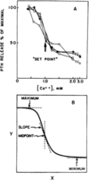
describe the relationship between PTH and ca *

A

a steep inverse sigmoidal function relates PTH and Ca

minimum - even at high levels there is always some PTH production

set-point - point of half max suppression of PTH, steep part of slope, small perturbation causes large change in PTH

20
Q

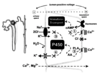
describe the mechanism by which PTH drives ca reabsorption *

A

PTH increases the number and activity of TRPV5/6 (ca channels)on the lumen - increase ca reabsroption

also increases the active transport of ca into the blood out of the cells

21
Q

describe how PTH causes bone resorption through the RANK system *

A

increases RANKL production = increase in osteoclast differentaition

22
Q

how common is primary hyperparathyroidism *

A

occurs in 50s

female:male 3:1

2% people develop it post-menopausally

23
Q

what are the causes of primary hyperparathyroidism *

A

mainly parathyroid adenoma - benign

parathyroid hyperplasia - genetic condition when under 40

parathyroid carcinoma <1%

rare familial syndromes - MEN1, MEN2a HPT-JT

24
Q

how do you diagnose primary hyperparathyroidism *

A

an elevated total/ionised ca with PTH levels frankly elevated or in the upper half of normal range (this is inappropriately high)

corrected ca >2.6mmol/l with PTH>3.9pmol/l (normal range 1-6.8)

25
clinical features of primary hyperparathyroidism \*
thirst polyuria tiredness/fatigue muscle weakness fractures due to secondary bone resorption 'stones, abdominal moans, psychic groans' * renal colic, nephrocalcinosis, chronic renal failure * dyspepsia (excess acid production), pancreatitis, constipation, nausea, anorexia * impaired concentration, drowsy, coma
26
describe how high serum ca can cause diuresis \*
high Ca is sensed on teh basolateral membrane, this inhibits the Na Cl K transporter ion the apical membrane - stop resorption of these = loss of water this can become med emergancy if Ca \>3mmol/L - act like a loop diuretic
27
effect of PTH on renal stone risk \*
elevated PTH causes an increased renal stone risk caused before diagnosis after surgery, incidince of stones goes almost back to normal
28
effect of high PTH on bone resorption \*
increases cortical boen turnover acute/pulsed PTH is anabolic - downregulates sclerostin in osteocytes chronic increase = catabolic occurs more in cortical bone than cancellous
29
effect of chronically elevated PTH on fracture risk \*
increases - 3x higher
30
what are the biochemical findings in primary hyperparathyroidism&
increased serum ca - by absorption from bone and gut decreased serum phos - renal excretion in PCT PTH in upper half of normal, or elevated increase urine ca - huge filtered load mean surpass max renal resorption creatine might be elevated - renal damage
31
describe production of vut D \*
7-dehydrocholesterol from skin turned to cholecalciferol by UV - difficult in UK oily fish give you cholecalciferol in liver cholecalciferol is converted to 25-cholecalciferol in kidney converted to calcitriol under PTH and 1a hydroxylase in kidney 25-cholecalciferol is also converted to 24,25 D, an inactive metabolite vitamin D binding protein has a half life of 3 days and is filtered by the kidney
32
actions of vit D \*
vit d acts on intracellular receptors to alter the rate of transcriptionof certain genes in the GIT - increase ca transport proteins called calbindin-D proteins = increased uptake of ca ca and phos absorption in SI ca and phos release from bone, differentiation agent for osteoclast precursers ca resorption and phos excretion from kidney
33
how does vit d increase gut ca absorption\*
it activates TRPV6 and calbindin in duodenum passive - paracellular transport we need AT to get enough ca to account for the 200mg/d obligatory losses - this active transport is vit d dependant
34
effect of vit d on bone \*
synerges with PTH acting on osteoblasts to increase formation of osteoclasts through RANKL increase osteoblast differentiation and bone formation - by increasing Ca levels it keeps PTH levels low therefore protects bone structure - this is a secondary action
35
effect of vit D in kidney \*
fascilitates PTH action to increase ca resorption in distal tubule - including TRPV5 and calbindin
36
what is the definition of vit d def
vit d deficiency is 25 OH D \<50nmol/l vit d insufficiency is 40-72.5nmol/l this is because this is the level that PTH production plateus, ie less than this and the PTH production is increased to compensate
37
what is rickets \*
inadequate vit d activity leads to defective mineralisation of the cartilaginous growth plate - before a low ca
38
symptoms of rickets \*
lack of play because of prox myopathy and axial bone pain and tenderness
39
signs of rickets \*
age dependant derformity myopathy hypotonia short stature tenderness on percussion
40
causes of rickets \*
dietry lack sunlight GI - small bowel malabsorption/bypass, panc insufficiency (alcoholism), liver/bilary disturbance, drugs - phenytoin, phenobarbitone (increase p450 cytochrome activity that increases vit d) renal - chronic renal failure rare hereditory - type 1 is deficiency of 1a hydroxylase, type 2 is defective VDR for calcitriol
41
biochemistry for rickets \*
ca - normal or low phos normal or low (normal if PTH risen to compensate) alk phos - high 25 oh vit d - low urine: phos - high, glycosuria, aminoaciduria, high pH, proteinuria - urine readings are rare
42
descrieb the action of FGF-23 \*
it is made by osteoblasts in the long bone when they sense phosphorus NPT2a and NPT2c receptors reabsorb phos - if stimulated by FGF-23 or PTH these receptors are internalised = waste phosphate FGF-23 also inhibuts activation of vitd by 1a hydroxylase acts as a back up to PTH - have to get rid of ca phos otherwise it goes into tissues
43
describe osteomalacia and phosphate \*
can also get renal phos loss when ca and vut d levels are usually normal _kidney forced to lose phosphate_ isolated hypophosphtaemia - X linked hypophosphtaemic rickets - mutations in PHEX = high levels of FGF-23 = toddlers with leg deformity, enthesopathy, dentin anomolies autosomal dom hypophosphtaemic rickets (ADRR) - variable age of onset, may improve, cleavage site for FGF-23 mutated so high FGF-2 osteogenic osteomalacia - in mesenchymal tumours, produce FGF-23 - causes phosphaturia and inhibit 1a hydroxylase _kidney proximal tubule damaged_ waste glucose, aa and protein and lose phos causes phosphaturia and stops 1ahydroxylation of vit D faconi syndrome is most common - multiple myeloma, heavy metal poisening (lead/mercury), drugs - tenofovir, gentamycin, congenital disease (wilson's, glycogen storage disease)`
44
what is seen in the bone in osteoporosis \*
weak laterally loss of trabeculae lost connectivity cannot reform the connections
45
causes of high turnover osteoporosis \*
oestrogen def - primarily in post-menopausal women hyperparathyroidism hyperthyroidism hypogonadism - premature ovarian insufficiency cyclosporine heparin
46
causes of low turnover osteoporosis \*
liver disease - primary bilary cirrhosis heparin age \>50 chronic kidney disease
47
cause of osteoporosis with increased resorption and decreased formation \*
glucocorticoids
48
how does oestrogen deficiency cause menopausal bone loss \*
increases the number of remodelling units causes increased bone resorption compared to formation - enhanced osteoclast survival and activity remodelling errors - deeper and more resorption pits = trabecular perforation and cortical excess excavation decreased osteocyte sensing - when young bone senses when exercise - osteocytes die in menopause - cant tell that you are exercising - cant keep bone density up there is a disproportionate loss of cancellous bone in women
49
biochemistry in osteoporosis \*
is used to exclude other casues serum biochem should be normal if primary check for vit d def check for secondary endocrine causes * primary hyperparathyroidism - PTH high * primary hyperthyroidism - free t3 high, TSH suppressed * hypogonadism - testosterone low exlude multiple myeloma by electrophoresis may have high urine ca
50
what is the best predictor of fracture risk \*
BMD - represents 70% of fracture risk
51
explain DEXA \*
measures transmission through the body of x-rays of 2 different proton energies enables densities of bone mineral and soft tissue to be inferred
52
how do you calculate a T score \*
measured BMD - young adult mean BMD divide by young adult SD
53
how does fracture risk change with BMD \*
1 SD reduction = 2.5 increase in risk of fracture
54
why do you ise central measurements for osteoporosis DEXA \*
vertebral * commonest fracture * increasing incidence after age 60 * measure of cancellous bone * metabolic bone - quickest response to treatment hip * 2nd commonest site * increase incidence after age 70 * has costs and mortality fracture risk assessment tool FRAX uses hip BMD
55
summarise bone markers as use for diagnosis \*
in most bone diseases teh bone cycle is disrupted markers indicate boen activity they re dynamic markers of formation and resorption
56
markers of bone formation \*
2 a1 and 1 a2 chain of type 1 collagen are produced by osteoblasts extension peptides are cut off - these can be measured in blood P1NP (procollagen type 1 N-terminal propeptide) - this is a procollagen molecule that is released from the N terminal when type 1 collagen is layed down -- 3 hydroxylysine molecules on adjacent tropocollagen fibrils condense to form pyridinium ring linkage - cross link C and N terminal can measure this in urine - indicator of bone resorption
57
markers of bone resorption \*
tehy are used to monitor treatment to anti-resorbitive drugs - BMD change 18months markers fall in 4-6weeks expect a 50% drop of urine NTx by 3months do it because the drugs are unreliable and poorly absorbed
58
what is the problem with cross links \*
we grow during night and remodel so have to measure them in the afternoon reproducibility - CV 20% positive association with age need to correct for Cr diurnal variation in urine markers
59
what is the clinical use of bone formation markers \*
only one in common use is alkaline phosphtase used in diagnosis and monitoring of pagets, osteomalacia, boney metastases (prostate monitoring of PSA) now P1NP is being used as a predictor of response to anabolic treatments - it is a procollagen fragment released from the N terminal when collagen is layed down - PTH treatment rises to peak in 3 months - predicts response
60
describe BSAP \*
role - essential for mineralisation, regulates concentrations of phosphocompounds uses - consistent within individual, half life is 40hours increased in pagets, osteomalacia, bone met, hyperparathyroidism, hyperthyroidism - anything causing increased turnover - bone compensates by increasing mineralisation
61
change in alkaline phosphtase with age \*
higher when growing- need to use age specific range increases \>50yrs especially in post-menopausal women - not used as a marker in osteoporosis
62
describe chronic kidney disease bone mineral disorder
systemic disease skeletal remodelling caused by CKD leads to heterotopic calcification, especially vascular CKD impairs skeletal anabolism, decrease osteoblast function and bone formation rates
63
describe renal osteodystrophy \*
it is a bone disease that results from poor mineralisation due to chronic renal failure increase serum phosphate, reduction in calcitriol if GFR\<60 get secondary hyperparathyroidism in attempt to actiavte vit D this is unsuccessful so hypocalcaemia develops later parathyroids are autonomous causing hypercalcaemia
64
describe the glandular progression of secondary hyperparathyroidism \*
parathyroid hyperplasia develops alongside renal failure the increasing demand on parathyroid glands drives cell proliferation initially the glands respond by increasing proprtion of secretatory chief cells within the gland an dthen increasing the total number of cells = hyperplasia in diffuse hyperplasia, cell growth is polyclonal but is accompanied by down regulation of CaR and VDR as CKD progresses to stage 5 hyperplasia increases and monoclonal abnormalities lead to nodular hyperoplasia of the glands with less CaR and VDRs - they're less responsive to serum ca levels
65
summarise the pathophysiology of CKD-MB
the hyperphosphtaemia binds to ca in circ = metastatic calcification = die of MI or stroke
66
pathology of paget's \*
accelarated rates of bone turnover, abnormoal bone architecture that may lead to deformity may be because of a virus, genetic associations have been identified excessive cellularity and vascularity huge and highly nucleated osteoclasts resultant bone is woven 3 phases 1. osteolytic phase - huge increase in osteolytic activity 2. osteoclast/osteoblast phase - disordered activity as blasts try to fill erosions = woven bone 3. osterosclerotic phase - new sclerotic bone is produced
67
clinical features of paget's \*
may be asymptomatic or cause pain enlargement of the effected bones pathological fractures deafness from nerve compression of bone overgrowth in the skull
68
investigations for paget's \*
serum ca and phos is normal alkaline phosphtase and serum ca are high hydroxyproline present in the urine
69
complication of paget's
secondary osteoarthritis high oputput HF because new vessels form shunts bone sarcoma can occur
70
pathology of renal osteodystrophy \*
caused by: * inadequate renal tissue for making vit D = osteomalacia * high serum phos (in CRF) = hypocalcaemia, precipitates hyperparathyroidism with subsequent bone resorption to release ca stores * haemodialysis - inhibition of calcification of bone matrix = osteomalacia * steroid treatments = osteoporosis/avascular necrosis
71
clinical features of renal osteodystrophy \*
low PTH = low bone turnover in severe CRF - body cant compensate by getting rid of phos = increased phos levels = decreased ca levels = increase in PTH = increased bone resorption high PTH causes fibrosis of the bone marrow - this is osteitis fibrosis cystica failing kidneys cannot activate vit d - ca cannot be resorbed from the GI tract - worsening hypocalc osteosclerosis occurs and chronically there are metastatic calcifications in the skin, eyes, joints and arterial walls parathyroid glands become autonomous
72
pathology of osteomalacia \*
failure of bone mineralisation = softer and wider channels of matrix unmineralisation and underdeveloped epiphyseal cartilage calcification lead to endochondrial bone that is deranged and overgrown presents with muscle and bone pain, looser zones, prox myopathy = waddling rickets = bowing of legs bone biopsy is definitive