Cornea Degeneration and Ectasia Flashcards

(71 cards)

1
Q

which layer of the cornea is capable of regeneration? which ones are not?

A

epithelium is the only layer to undergo mitotic division (Bowman’s, stroma, Descemet’s and endothelium do not regenerate)

2
Q

what are the cell junctions present in the corneal epithelium?

A

desmosomes and gap junctions and connect to Bowman’s layer via hemidesmosomes

3
Q

what type of pumps are in the corneal endothelium and what is the purpose?

A

Na-K-ATPase pumps to keep the stroma from having too much fluid

4
Q

what is a degneration?

A

a process in which normal elements of corneal tissue are converted (age-related or metabolic diseases) - can be benign or detrimental to normal function

5
Q

what are 7 corneal degenerations that are non-sight threatening?

A

crocodile shagreen, arcus, limbal girdle of vogt, farinata, terriens marginal degeneration, moorens ulcer and amyloid

6
Q

what are 2 non-sight threatening corneal degenerations that have the possibility of turning to sight threatening?

A

mooren’s ulcer and amyloid

7
Q

what are 3 exmples of degenerations that are opacificiations and sight threatening?

A

salzmanns nodular, spheroidal and band keratopathy

8
Q

what is crocodile shagreen?

A

age related, benign, common condition, easily seen with slit lamp = plaques of fibrous tissue

9
Q

where is crocodile shagreen located anteriorly and posteriorly?

A

anterior = bowman’s layer

posterior = posterior corneal stroma and descemet’s

10
Q

what are the symptoms and treatment for crocodile shagreen?

A

no symptoms and no treatment required

11
Q

what is arcus?

A

lipid/cholesterol deposits in Bowman’s (not common under age 40)

12
Q

why does arcus have a lucid interval between the limbus?

A

the lipid deposition ends at bowman’s - it has an abrupt ending

13
Q

what does type 1 limble girdle of vogt look like?

A

has a lucid interval - deposition ends at bowman’s swiss cheese holes and sharp edges centrally early form of band keratopathy

14
Q

what does type 2 limble girdle of vogt look like?

A

goes to limbus - elastoid degeneration of sub-epithelial collagen extensions centrally

15
Q

what is farinata?

A

white dust-like particles, pre-descemet’s and occurs with aging (may resemble pigment dispersion syndrome)

16
Q

what is the leading line of a pterygium called?

A

stocker line (iron line)

17
Q

what is a hudson-stahli line?

A

occurs in the interpalpebral zone from tear stagnation (iron deposits in tear film) = typically after chronic inflammatory condition

18
Q

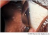
what is Terrien’s marginal degeneration?

A

thinning of the cornea (starts superiorly then circumferential), asymptomatic, bilateral and epithelium stays intact, fine line of lipid deposit, superficial vascularization, males >> females and 40+

19
Q

what is a differential diagnosis for Terrien’s marginal degeneration?

A

Mooren’s ulcer

20
Q

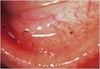
what symptoms will patients have with Mooren’s ulcer?

A

non-infectious (unknown etiology - autoimmune likely), painful, red, photophobia

21
Q

where does Mooren’s ulcer begin?

A

near limbus, typically progressive (circumferentially and centrally) = thinning, stromal melting, potentially perforation (epithelium is not intact)

22
Q

what is Mooren’s ulcer type 1?

A

typically seen in older patients, unilateral and better responses to treatment

23
Q

what is Mooren’s ulcer type 2?

A

seen in younger (african descent) 20-30 y/o, bilateral and poor response to treatment (rare)

24
Q

how do you differentiate between Mooren’s and Terrien’s?

A

Terriens has intact epithelium, no NaFl staining, rarely painful/inflammatory, and rarely perforates

25
what type of systemic work up is needed to Mooren's ulcer?
vasculitis or collagen vascular disease (autoimmune diseases)
26
what is the treatment for Mooren's ulcer?
mostly supportive = topical steroids, conjunctival resection/radiation, bandage CL, topical cyclosporine or systemic immunosupression (perforation = cyanoacrylateor lamellar keratoplasty)
27
what is polymorphic amloid degeneration?
occurs deep stroma, bilateral and appears similar to lattice degeneration - mostly benign
28
what is spheroidal degeneration?
common from UV exposure (actinic), usually interpalpebral, golden brown deposits
29
what is Salzmann's nodular degeneration?
elevated white masses on cornea - tend to be peripheral (milky/glossy), can have a red/irritated eye
30
what causes salzmann's nodular degeneration and who typically gets it?
females, \> 50 y/o, chronic ocular surface disease/inflammation (especially viral) - hyaline plaques replace bowman's
31
what are the symptoms and treatment for salzmann's nodular degeneration?
symptoms = dry eye symptoms, VA if central treatment = lubricants, steroid, bandage CL (PK if severe)
32
what is band keratopathy?
interpalpebral CA++ deposits in bowman's with clear zone separating the limbus - swiss cheese appearance
33
why does band keratopathy have swiss cheese appearance?
clear areas and small circular areas where nerve endings perforate bowman's (similar to Vogt 1)
34
what causes band keratopathy?
inflammatory disease (mercury), systemic conditions that cause increased Ca, chronic ocular pathology and degenerative conditions or idiopathic
35
what is the treatment/plan for band keratopathy?
monitor, ocular lubricants for mild cases, refer for hypercalcemic work-up, chelation using 2% EDTA for severe cases, PTK
36
what are 3 corneal ectasias?
keratoconus, keratoglobus, pellucid marginal degeneration
37
when does keratoconus typically occur and what causes it?
bilateral, usually after puberty (rarely congenital), progressive then stabilizes, males = females, \>asians, may have an inheritance pattern
38
what is the pathology for keratoconus?
irregular epithelium, breaks in bowman's, fibrosis beneath epithelium, stromal scarring, corneal thinning
39
what is the pathophysiology for keratoconus?
epithelial lysosomal enzyme expression increase, reduction inhibition of proteolytic enzymes, abnormal corneal collagen, lamellae, keratocyte populations
40
what is charleaux's sign?
an irregular red reflex from retinoscopy or retro-illumination in keratoconus
41
what is Rizutti's sign?
a triangle of light seen on the distal iris in keratoconus
42
what is munson's sign?
when a patient with keratoconus looks down it forms a "V" shape
43
what is vogt striae and fleischer ring?
lines seen in cornea with keratoconus (fleischer ring = iron deposits around base of cone)
44
what is the treatment for keratoconus?
optical correction, specialty RGP/hybrid CL, PK, no refractive surgery, intacts, collagen crosslinking
45
what are corneal hydrops?
spontaneous rupture/break in descemet's - can result in flattening of cornea, clears on its own - may scar
46
what is posterior keratoconus?
posterior diffuse or localized curvature (normal anterior surface), unilateral, non-progressive, female \> male
47
what is keratoglobus?
bilateral, congenital or acquired, diffuse corneal thinning \> peripheral (1/3 to 1/5 normal thickness)
48
what does pellucid marginal degeneration look like?
the thinned area is confined to corneal side of inferior limbus (4:00-8:00), pot belly cornea, ATR astigmatism (kissing dove pattern)
49
what are dermoids?
collection of ectodermal tissues: sweat glands, hair follicles, sebaceous glands
50
what is a lipodermoid?
benign fatty tumor beneath conjunctiva laterally
51
what are trisomy 21, 17-18, and 13?
21 = downs syndrome 17-18 = edwards syndrome 13 = bartholin-patau syndrome
52
what happens with a vitamin A deficiency?
bitot spot, impaired goblet cell function, keratinization
53
what disease is caused by sphingolipidoses disorder?
Fabry's disease
54
what is fabry's disease?
x-linked recessive, posterior spoke like deposits in lens along sutures = verticillata
55
what is cystinosis and alkaptonuria caused by?
diseases of protein and amino acid metabolism
56
what does cystinosis look like?
cystine crystal deposits throughout cornea
57
what type of deposits does ciprofloxacin give?
chalky white deposits where epithelium is absent
58
where do mercury deposits occur? what causes them?
(orange-brown) in bowman's typically from preservatives
59
where do silver deposits occur?
argyrosis in descemet's
60
what type of deposits occur from epinephrine drops?
adrenochrome
61
what type of deposits do the drug thorazine cause? and where do they occur?
stellate sub-capsular throughout the stroma
62
what causes wilson's disease?
defect in copper metabolism - Cu+ deposit in descemet's peripherally with no clear interval
63
what is this picture of?
corneal hydrops
64
what is this picture of?
band keratopathy
65
what is this picture of?
crocodile shagreen
66
what is this picture?
furrow degeneration
67
what is this topography of?
keratoconus
68
what is this topography of?
pellucid marginal degeneration
69
what is this picture of?
Salzmann's nodular degeneration
70
what is this picture of?
vogt striae in keratoconus
71
what is this picture of?
limbal girdle of vogt type 1