DCM Flashcards

1
Q

Causes of DIFFUSE abdominal pain (7)

A
  1. Acute Pancreatitis
  2. Early Appendicitis
  3. Diabetic Ketoacidosis
  4. Gastroenteritis
  5. Intestinal Obstruction
  6. Mesenteric Ischemia
  7. Peritonitis
2
Q

Causes of URQ Abdominal Pain

A
  1. Biliary Tract Disease
  2. Perforated Peptic Ulcer
3
Q

Causes of ULQ Abdominal Pain

A

Gastric & Spleen disorders

4
Q

Causes of LRQ abdominal pain

A
  • Appendicitis
  • Chron’s Disease
  • Meckel’s Diverticulum
5
Q

Causes of LLQ abdominal pain

A

Diverticular disease

6
Q

Causes of LOWER abdominal pain

A
  • PID
  • Abscess
  • Ruptured AAA
  • Ectopic Pregnancy
  • Torsion of ovarian cyst or testis
  • Ovulation
7
Q

Non-surgical/Extra-peritoneal Pain

A
  • Acute MI
  • Pericarditis
  • Sickle Cell Crisis
8
Q

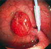
Acute Cholecystitis:

    • symptoms
  • investigations
    • treatment
A

= obstruction of cystic duct, most often due to gallstones

Sx: Acute RUQ or epigastric pain
- Choledocholiathiasis presents with CHARCOT’S TRIAD (pain+jaundice+fever)

Dx: US, CT, HIDA

Tx: ERCP! or cholecystectomy

9
Q

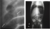
Perforated Peptic Ulcer:

  • symptoms
  • diagnosis
  • treatment
A

Sx: Acute & SEVERE abdominal pain, peritonitis, hemodynamic instability

Dx: CHEST X-RAY shows FREE GAS UNDER DIAPHRAGM

Tx: resuscitation & surgery

10
Q

Acute Pancreatitis:

  • symptoms
  • diagnosis
  • treatment
A

= auto-digestion of pancreas seen in GALLSTONE DISEASE & ALCOHOLISM

Sx: Epigastric abd pain RADIATING TO BACK, worse in SUPINE (will be leaning forward), and after eating

    • Grey Turner’s Sign (bruised flanks)
    • Cullen’s sign (superficial edema + bruising around umbilicus)
    • abdominal distension & epigastric tenderness
    • decreased bowel sounds

Dx: Serum amylase & lipase, LFTs, CT!! (most accurate for Dx & ID), US, ERCP

11
Q

Diverticular Disease

  • symptoms
  • diagnosis
  • treatment
A

= Increased intraluminal P in colon –> inner colonic layer bulges out => false diverticuli

Sx: vague LLQ pain, bloating, diarrhea

Dx: Barium enema (NOT in ACUTE Diverticulitis), CT abdomen & pelvis with oral & IV contrast

Tx: IV abx, IV fluids

12
Q

Complications of Diverticulosis

A

Painless rectal bleeding

13
Q

Complications & Management of Diverticulitis

A

Bowel Obstruction, Pericolic abscess, perforation & peritonitis, fistula formation

Management: CT-guided surgical drainage of abscess, resection of fistulas
***DON’T DO ENEMA OR COLONOSCOPY– could perforate!

14
Q

Acute abdomen.. can’t rule out appendicitis.

A

TAKE IT OUT

15
Q

What are the types of Jaundice?

A
  • Prehepatic– mainly hemolytic
  • Hepatic – hepatocellular or intrahepatic obstruction
  • Post-hepatic – obstruction/pressure of bile duct
  • Cholestatic – intra-/extra-hepatic stasis of bile
  • Physiological
  • Hemolytic disease of newborn
16
Q

Signs of Pre-hepatic Jaundice

A

Due to hemolysis.

Patient is Pale (anemia) and lemon yellow (UCB)
Splenomegaly
High reticulocytes, ↓ Hb

17
Q

Causes & Signs of Post-hepatic Jaundice

A

Due to obstruction/pressure of bile duct (biliary atresia, BILE DUCT STONE (MCC), Head of Pancreas CA, UC, 1* biliary cirrhosis)

or absent bile pigments in gut –> STEATORRHEA - Fat soluble vit defx

18
Q

Gilbert Syndrome: - Etiology - Clinical

A

AD mutation of promotor of UGT1A1 (Bilirubin UDP Glucuronosyl Transferase) –> decr hepatic bilirubin uptake —–> unconjugated hyperbilirubinemia

7% pop, not severe– no tx

19
Q

Dubin-Johnson Syndrome: - etiology - clinical

A
  • *Faulty excretory fx** of hepatocytes due to pt mutation in gene for organic anion transporter
  • -> ↑ CONJ Bilirubin

Gall bladder not visualized on cholecystography;
Bx reveals CENTRILOBULAR BROWN/BLACK PIGMENT

Great prognosis

20
Q

G6PD Deficiency dx

A

G6PD level assessed WEEKS AFTER crisis

21
Q

Hereditary Spherocytosis: - Etiology - Dx - Tx

A

AD abnormality of SPECTRIN or other mem. protein –> SPHEROCYTES (incr cell fragility –> hemolysis –> jaundice)

Dx: RBC fragility test

Tx: Splenectomy after 6y

22
Q

ALT:AST ratio in ALCOHOLIC HEPATITIS

A

AST:ALT > 2

23
Q

Where is ALT found?

A

Hepatocytes— more sensitive than AST in liver damage

24
Q

Next step if ALP is found elevated?

A

Assess GGT– if also elevated, consider Hepatobiliary/bone/placenta/intestinal path

25
Q

ALP levels in cholestasis?

A

>10x elevated due to extra-hepatic biliary tract obstruction

26
Q

What is the best indicator of EXCESSIVE ALCOHOL CONSUMPTION?

A

GGT (Gamma Glutamyl Transpeptidase)

27
Q

Significance of

  1. Mild ↑ of AST & ALT (low 100s)
  2. Moderate ↑ of AST & ALT (high 100s to 1000s)
  3. Severe ↑↑ of AST & ALT (>10k)
  4. AST & ALT normal or ↓
  5. ↑↑ ALP & GGT, ↑ AST & ALT
  6. ↑↑ AST & ALT, ↑ ALP
A
  1. Chronic viral or acute alcoholic hep
  2. Acute viral hep
  3. Extensive hepatic necrosis (ischemia, acetaminophen tox, severe viral hep)
  4. Cirrhosis, metastatic liver dis (↓ # normal hepatocytes)
  5. Cholestasis
  6. Hepatocellular Path
28
Q

Non-Alcoholic Fatty Liver Disease - Etiology

A

= STEATOHEPATITIS & CIRRHOSIS due to INSULIN RESISTANCE associated with obesity

–greater BMI = more liver damage

29
Q

Carcinoma of Head of Pancreas: - S/S - Labs

A
  • Jaundice, dark urine, pale stoole, DISTENDED, NON-TENDER GALLBLADDER
  • ↑ ALP, ↑ CB
  • Courvoisier’s Law: Enlarged, NON-tender gallbladder caused by MALIGNANCY, not gallstones
30
Q

High levels of HBeAg indicate what?

A

Infectivity.

Usually present @6w-3m

31
Q

Anti-HbcAg is indicative of?

A

past infection

32
Q

ONLY Anti-HbS detected is indicative of?

A

vaccine

33
Q

After exposure, HBsAg is present for ____, or ____ in carriers/chronic infx

A

1-6mo exposure, >6mo chronic

34
Q

What is the demographics of an Ulcerative Colitis pt?

A

Caucasian
Jewish
NON-Smoker!! (quitting smoking may worsen)
Young

35
Q

What is backwash ileitis?

A

When ulcerative colitis extends proximally and reaches the terminal ileum, but NO ULCERS are present there

36
Q

What are the features of UC?

A
  • Only colon is affected (rectum always)
  • Mainly involves mucosa
  • Confluent
  • Resection is CURATIVE
  • Cancer is more common
37
Q

What are the features of CD?

A
  • ANY part of GIT is affected– skip lesions present
  • Involves full bowel wall (deep ulcers)
  • Stricture & Fistulae common
  • Perianal diseases are common
38
Q

Ulcerative Colitis: S/S

A
  • Bloody diarrhea +/- mucus
  • Lower abd cramps
  • Fecal urgency (tenesmus)
  • Anemia
  • ↓ albumin
  • Negative stool culture
  • Bright red blood on DRE
39
Q

Disease stages of Ulcerative Colitis

A
  • Severe Disease
    • anorexia, malnutrition, fever
    • hypovolemia (>6 bloody BM/day)
    • ↑ ESR & CRP
    • ↓ Hb & Albumin
  • Fulminant Disease
    • Rapid progression of severe toxicity over 1-2w
    • Requires transfustion
  • Toxic Megacolon
    • Colonic dilation of >6cm on radiograph
    • Heightened risk of perforation
40
Q

Pharmacological treatment options for Ulverative Colitis & Chron’s Disease?

A
  • Supplementation
  • Mesalazine (anti-inflamm)
  • Corticosteroids
  • Cyclosporine & Infliximab in severe cases
    *
41
Q

IBS treatment:

A
  • Diet change (avoid insoluble fiber in
  • Anti-diarrheals (Loperamide)
  • Dicyclomin (anti-spasmodic)
  • Amitriptyline (tricyclic AD)
42
Q

Diagnosis of IBS

A

Generally young women (20s) (rule out cancer in older pts)

at least 6 months of recurrent abdominal pain for 3days/month in last 3 months associated with 2+ of:

  1. improvement with defecation
  2. onset assoc w/change in frequency of stool
  3. Onset assoc w/change in appearance of stool
43
Q

What is:

  1. Hematemesis
  2. Melena
  3. Hematemesis AND Melena?!?!?!
  4. Hematochezia
  5. Occult Blood Test
A
  1. Blood in vomit
  2. Black tarry stools due to blood
    → intestinal enzymes & bacteria acting on blood (>60mL for 10-14h)
  3. Both → Upper GI hemorrhage
  4. Red blood from rectum (>1000mL blood loss)
  5. Occult blood test: (+) = >1mL, <60mL blood
44
Q

Where is the bleeding?

  1. Bright red blood streaks on stool
  2. Bright red within stool
  3. Maroon
  4. Melena
A
  1. Rectum, anal canal, lower sigmoid
  2. Left colon
  3. Right colon / Small intestine
  4. Upper GI
45
Q

Features of Acute Severe Bleeding

A
  • SOB, dizziness, fainting
  • Crampy abdominal pain, diarrhea
  • Anemia 1-3 days later

>500mL loss → systemic signs
20%-40% → orthostatic hypotension
>40% → shock → flat JVP, supine hyptoension

46
Q

Causes of Severe GI Bleed

A
  • Rupture of esophageal varices
  • Peptic ulcer disease (→ torrential hemorrhage)
  • Diverticulitis
  • Amylodysplasia
  • Ischemic colitis → ↓ blood to splenic flexure → mucus membrane sloughs off
  • Meckel’s Diverticulum (40-50% cases, Patent artery = source of bleed)
47
Q

Features of Chronic Bleeding

A
  • Weakness, fainting
  • Fatigue, lethargy
  • SOB
  • Anemia
    If Hb is low, do CBC–
    • NO ↑ Reticulocytes if bone marrow depletion or Fe defx anemia.
48
Q

Causes of Upper GI Hemorrhage

A

Common:

  • Peptic ulcer
  • Erosive gastritis, esophagitis
  • Esophageal variceal rupture
  • Mallory-weiss

Less common:

  • esophageal / gastric cancer
  • Duodenal, diverticular, hemobilia (mix bile & blood due to injury to liver)
  • Arteroenteric fistula (communication bt/w aorta & enterics after removal of AAA)
49
Q

Posterior duodenal ulcers can perforate to pancreas and erode __________ artery?

A

Gastroduodenal

( Anterior ulcers perforate more frequently )

50
Q

Blood vessel visible in gastric ulcer

A

EMERGENCY SURGERY!!!

may rebleed in 1-2y

Upper GI endoscopy is both diagnostic & therapeutic

51
Q

Benign Gastric Antral Ulcer

A
  1. Small
  2. Mucosa surrounding base– NOT swollen
  3. Rugae extend to BASE
  4. REPEAT BIOPSIES! to confirm not bleeding & benign, every 6mo
52
Q

Malignant Gastric Ulcer

A

Difficult to differentiate from benign

Biopsy edge of non-healing ulcer

Most are near gastroesophageal junction

53
Q

Esophageal Varices

A
  • Pressure buildup of L Gastric vein
  • In submucosal layer of esophagus

→ torrential hemorrhage if rupture

54
Q

Ruptured Esophageal Varices

A
  • Stop bleeding in ER!
  • Urgent endoscopy– confirm Dx then transfuse or ressucitate pt
  • Intubate to protect airway
55
Q

Management of Ruptured Varices

A

IV Octreotide / terlipress, balloon tamponade if bleeding cont.

If rebleed - repeat tx; if not - B-blocker to prevent

56
Q

Mallory Weiss

A

Alcoholic, middle-aged men

INITIAL vomitus has NO blood
2-3 vomits later = bloody

Gastric mucosa moves into LES via a-peristalsis during vomiting → ischemia → bleeding!!

Tx: ressuscitate, stop vomit, endoscopy to confirm

57
Q

Gastric Cancer

A
  • Growth in pre-pyloric region
  • non-specific sx early on
  • later, when symptomatic, it has probably metastasized and has a poor prognosis :(
    • indegestion, burning sensation
    • anorexia
    • severe bleed @ late stage

Tx: radical surgery, Chemo, radio adjuvant

58
Q

Gastric Cancer– late symptoms

  1. Kruckenberg
  2. Blumer’s Shelf
  3. Sister Joseph’s Nodule
  4. Virchow’s Node
  5. Trosseau’s Sign
A
  1. Spread to ovary
  2. Can palpate mass in rectouterine/rectovesicular pouch
  3. Tumor spread to umbilicus
  4. Supraclavicular LN
  5. Venous thrombosis & hypercoaguabiltiy where clots form, resolve, & form elsewhere (fleeting thrombophlebitis)
59
Q

Lower GI Bleed causes

A
  • MCC: Piles/Hemmorhoids
  • Hematochezia
  • Fecal occult blood
  • Anemia (of chronic dz)
  • Hemorrhoids: dilation & tortuosity of anal canal veins
  • Infectious diarrhea
  • Anal fissure/fistula
  • Proctitis secondary to radiation
  • Colon cancer
  • Angiodysplasia
  • Meckel’s
  • Diverticular disease
60
Q

Anal fissure

A

MC position @ 6:00 (40%)

painful- don’t wanna poo – severe constipation

Tx: relax sphincter spasm (Ca2+ blocker, B-blocker)
*heal spontaneously

61
Q

Anal Fistula

A

Abnormal communication between hollow organ/skin, or Hollow organ/hollow organ

MC = subcutaneous type (good prognosis)

Bad prognosis: intersphincteric, transphincteric, suprasphincteric, extrasphincteric :(

62
Q

Colonoscopy

A

Both diagnostic & therapeutic

Colonoscopic snare is used to remove colonic polyps

63
Q
  1. Angiodysplasia
  2. Gold standard for detection of colorectal cancer
A
  1. Due to AV malformation– causes torrential bleeds!!
  2. Do total colonoscopy to detect colorectal cancer
64
Q

Management of Lower GI Bleed

A
  • Stabilize
  • Rule out Upper GI bleed (esophagogastroduodenoscopy)
  • Lower GI Dx of choice - selective mesenteric angiography
    • Can use to embolize
  • Surgery
65
Q

Colon Cancer

A

@ LEFT colon (more common)

  • Collicky pain (constant pain = advanced dz)
  • Change in bowel habits
  • Rectal bleeds, diarrhea
  • Palpable lump @ sigmoid colon

@ RIGHT colon

  • anemia, weakness, abdominal lump (LRQ)
  • *obstruction unlikely
66
Q
A

Apple Core appearance on barium enema = carcinoma of sigmoid colon

**most common site of colon malignancy is the rectosigmoid junction– more exposed to carcinogens**

67
Q

Innervation of : Longitudinal muscle | Circular muscle | Submucosa

A

Vagus n.

Auerbach / Myenteric Plexus

Meisssner’s Plexus

68
Q

Stages of swallowing

A
  1. Oral phase (voluntary)
  2. Pharyngeal phase (involuntary)
  3. Esophageal phase (involuntary)
69
Q

Motor Dysphagia

A
  • Neuromscular abnormalities
  • Sjögren’s
  • Sensory loss
  • Striated muscle disorder (MG, ALS, polio..)
70
Q

Mechanical Esophageal Dysphagia

A
  • Damage to esophagus proper (rings & webs, tumor, compression)
  • Large bolus, epiglottitis, pharyngitis
  • Plummer-Vinson Syndrome (esophageal webs + Fe defx anemia in post-menopausal women)
  • Schatzki’s Rings (narrow of lower esophagus)
  • Strictures & tumors
71
Q

Dysphagia + fever, foul breath, sore throat = ?

A

Retropharyngeal Abscess

72
Q

Dysphagia + chest pain radiating thru to back = ?

A

Esophageal cancer

-risk factors: family hx, alcohol/smoking

73
Q
A

Esophageal stenosis

  • barium trickling down after narrowed area
74
Q
A

bird’s beak appearance of the gastroesophageal junction and total aperistalsis of the esophageal body consistent with the diagnosis of achalasia.

75
Q
A

This is the esophagogram from a patient who experienced progressive solid-food dysphagia for several months. He also experienced a 20-pound weight loss during that same period.

Esophagogram showed a malignant neoplasm involving the esophagus.

76
Q
A

high-grade, smooth-walled distal esophageal stricture that resulted from reflux esophagitis

heartburn + dysphagia. The dysphagia is for solids and not liquids, indicating the presence of a lumen-narrowing lesion.

  • NO anorexia or weight loss and slow rate of progression of dysphagia – benign peptic process.
  • Presence of anorexia and weight loss and rapid progression of dysphagia – malignant and most likely an adenocarcinoma arising in a Barrett’s esophagus
77
Q
A

Scleroderma

78
Q

GERD Pathway

A
  • Initial esophageal lesion
  • –> Scar
  • –> Incompetent LES
  • –> Reflux - recurrent injury ——-> Scar (restart cycle) OR
    • Stricture, Pan, Obstruction, Perforation
    • Barrett’s Esophagus
      • -> Cancer
79
Q

Screening of Barrett’s pts

A

Patients with Barrett’s esophagus should get a surveillance endoscopy every 3 years to check for adenocarcenoma

80
Q
A
81
Q

Respiratory Symptoms of Tracheoesophageal Fistula

A

**Asymptomatic at birth**

  • Excessive drooling
  • Aspiration – distress, atelectasis, pneumonia
  • Cyanosis 2ry to coughing/choking
82
Q

Tracheoesophageal Fistual: gastric presentation

A
  • Gastric distention since respiratory status is compromised because inhaled air goes into the abdomen, causing pulmonary compression (–> cause of death)
83
Q

Tracheoesophageal Fistula: presentation

A
  • @ first few hours of life: hypersecretion, choking, cyanosis, respiratory distress
  • TEF @ distal esophagus (esophageal atresia): NO gaseous distention!
84
Q

Tracheoesophageal Fistula: Investigation

A
  • Catheter in nose– see how far it goes.
    • inject dye and take X-ray– is there a blind pouch?
    • (Follow w/ aspiration of medium so it doesn’t get into lungs)
  • Abdominal Distention
  • Bronchoscopy (abnormal communication) + esophaguscopy (blind puch)
  • Echo for heart defect (TOF, VSD)
    • Look @ aortic arch: if on right, then consider L thoracotomy
85
Q

Tracheoesophageal Fistula: associated abnormalities

A

Vertebral
Anal
Cardiac
TEF
Renal
Limp

**polyhydramnios in prenatal ultrasound

86
Q

Tracheoesophageal Fistula: treatment

A
  • IV abx, parenteral nutrition
  • Reconstruction of air & food passage
87
Q

Pyloric Stenosis

A
  • Congenital hypertrophy of muscles
    2* to healing duodenal ulcer
    Gastric malignancy
  • Distended stomach, projectile vomit (non billious)
  • NOT present @ birth– usually @ 2-8w
    >3mo is unlikely
  • Olive-shaped lump in upper abdomen after feeding
88
Q

Pyloric Stenosis: investigations

A

ULTRASOUND!!!! + Hx & Physical

89
Q

Pyloric Stenosis: Treatment

A
  • Correct fluid, electrolyte abnormality
  • Hypochloremic metabolic acidosis & dehydration control
  • NO oral feeding
  • Pylorotomy
90
Q

Familial Adenomatous Polyposis

A
  • AD mutation APC
  • Need <100 polyps
  • Adenoma by 15y
  • 100% will get adenocarcinoma by 30y
  • May have rectal bleeding, anemia

*R/O Upper GI probs since assoc w/ gastric, pancreatic cancer @ young age, plus extra-intestinal manifestations

91
Q

FAP: Tx

A

Removal of Polyp-bearing area of colon

GOLD STANDARD: Remove entire rectum & colon!!!! (can be dangerous)

92
Q

Peutz-Jegher’s Syndrome

A
  • AD
  • Polyps in small intestine
  • Hemorrhage, intestinal obstruction, intussception (emergency!)
  • FRECKLES ON LIP/GINGIVA – fades w/age
  • Associated with pancreas, lung, breast, ovarian cancer
  • Malignancy unlikely to derive directly from PJS
93
Q

Necrotizing Enterocolitis

A

One of MC surgical emergencies in neonates

Necrosis of colon & intestine in neonate (<1mo) - immature immunity

Infections cause ulceration & necrosis

  • -> enterocolitis, hemorrhage, perforation
  • -> peritoneal contamination w/ fecal biota –> sepsis –> death :(
94
Q

Necrotizing Enterocolitis: Clinical Features

A
  • GAS UNDER DIAPHRAGM due to perforation
  • Poor feeding, bilious vomit, abd distension, blood in stool
  • Diarrhea after milk ingestion due to disaccharide defx
  • Sepsis
  • Tender abdomen, periumbilical darkening/erythema
95
Q

What is Pneumatosis intestinalis?

Necrotizing Enterocolitis: tx

A
  1. Pneumatosis intestinalis = gas in wall of intestine 2* to necrotizing enterocolitis
  2. Tx: Surgery, NOTHING BY MOUTH (parenteral nutrition), IV Abx, aspirate stomach
96
Q

Meckel’s Diverticulum

A

TRUE diverticulum

Common (2%), asymptomatic

Vitelline duct does not obliterate!

RULE OF 2s

  • 2% have it
  • 2” in length
  • 2 ft prox of ileocecal junction
  • 1/2 present before 2y
  • 1/2 made up of 2 epithelia (1/2 gastric)
97
Q

Meckel’s Diverticulum: Clinical Signs

A
  • Hemorrhage
  • Intussception
  • Inflammation
  • Chronic Peptic Ulcer
  • Intestinal Obstruction

MCC rectal bleeding in children– maroon hematochezia

98
Q

Meckel’s Diverticulum:

Diagnosis
Treatment
Management

A

Good prognosis

Diagnosis:

  • DRE with maroon blood W/IN stool
  • Radioactive Technicium Scan (accurate)

Treatment:

  • Symptomatic – RESECT
  • Silent & found incidentally– leave it UNLESS *narrow mouth, *thick-walled
99
Q

Intussusception

A
  • MC @ 6-8mo
  • Telescopic invagination of 1 part of gut to distal part
  • Elderly: occurs 2* to colonic polps
  • Due to change in food habit –> infx –> swollen Peyer’s patches –> bridge for intussusception
  • Swollen lymphatic vessels follow due to compression
  • decreased blood flow to affected intestine
100
Q

Intussusception: presentation

A
  • MC @ 6-8mo; MCC intestinal obstruction between 3-6y
  • Colicky abdominal pain
  • Red-currant Jelly stool (blood + mucus- no fecal matter)
  • Bilious vomit
  • Episodes of screaming & drawing up legs
  • Sausage-shaped mass; empty R iliac fossa
101
Q

Intussusception: Treatment

A
  • IV fluid + resusscitation
  • NOTHING by mouth
  • IV abx if infx or gangrene
  • DO NOT OPERATE
    • BARIUM ENEMA: diagnosis & therapy
      • pressure pushes up intussusception & frees it

Only do surgery IF: barium enema doesn’t work, recurrent, secondary to malignancy, unstable pt

102
Q

Cystic Fibrosis:

First sign
Complications
Tx
Management

A
  • First sign: meconium ileus- normally, pancreatic enzymes keep meconium liquid–
    • in CF, it is dry and causes intestinal obstruction @ terminal ileum
  • Complication: perforation, volvolus (axial rotation)
  • Tx: Hyperosmolar gastrogratin enema- absorbs water from wall -> relief of obstruction
  • Management: Supplement Vit KADE, feed via ng tube or gastrostomy tube
103
Q

Celiac Disease

A
  • infants & elderly
  • T-cell mediated Hypersensitive reaction to whet protein (anti-gliadin, anit-endomysial abs)
  • Death of epithelial cells in upper intestine – Malabsorption

Present with: abd pain, bloating, steatorrhea, weight loss

104
Q

Celiac Disease: Investigation

A

Endoscopic small bowel biopsy

  • Atrophy of villi
  • Intraepithelial lymphocytes
  • migration of activated lymphocytes to tip of intestinal villi
105
Q

Celiac Disease: Tx

A
  • Avoid proteins pt is allergic to (prolamins- glaidin & gultenins)
  • Steroids (if that doesn’t work, use steroid + immunosuppressant)
  • Complications: anemia, lactose intolerance, GI lymphoma, etc)
106
Q

Hereditary Spherocytosis

A
  • Weak RBC membrane due to decreased spectrin
  • Na+ influx
  • Swollen cell = spherocyte
    • Fragile cell membrane
    • Macrophages in spleen destroy them –> excessive hemolysis
107
Q

Hereditary Spherocytosis:

presentation, diagnosis, management

A

Presentation:

  • Anemia
  • Gallstones, Jaundice
  • Splenomegaly
  • Unconjugated Billirubinemia

Diagnosis: RBC fragility test, Flow Cytometry w/ EMA

Management: Asymptomatic - leave alone; Symptomatic - splenectomy

108
Q
A
109
Q
A
110
Q

Patient with TRAUMA and 5L of 200 mOsm/kg urine per day and undetectable ADH

Dx?

A

Central Diabetes Insipidus-- post-traumatic polyuria (water diuresis)

111
Q

Middle-aged woman with breast cancer. Polyuria. What do you think of?

A

Central Diabetes Insipidus

Breast cancer commonly metastasizes to pituitary, causing Centra DI

112
Q

Patient with Water Diuresis:

What test needs to be run to distinguish between Central and Nephrogenic Diabetes Insipidus?

A

Water Deprivation Test

increased osmolality = CDI
no change = NDI

113
Q

Central Diabetes Isipidus:

S/S

Causes

A
  1. S/S:
    - Polyuria + polydypsia
    - Abrupt onset
    - Near normal or mildly elevated Serum Na+
  2. Causes
    1. 75% idipathic
    2. Neurosurgical
    3. Head trauma
    4. Ischemic encephalopathy or hypoxia
    5. Neoplasm
    6. Misc. (pregnancy)
114
Q

Nephrogenic Diabetes Insipidus:

S/S

Causes

A
  1. S/S: Gradual onset of polyura + polydypsia
  2. Causes:
    • Acquired: Chronic Renal dis, electrolyte disorders, drugs (lithium)
    • Congenital/Hereditary: Polycystic Kidney, Medullary Cystic Dis
115
Q

Causes of Water Diuresis?

A

Lack of urine concentration from…

  • Pituitary diabetes insipidus (ADH deficiency)– central diabetes insipidus
  • Primary polydypsia
  • Neprogenic Diabetes Insipidus (renal resistance to vasopressin)
116
Q

What is Diabetes Insipidus?

A

Excessive water loss & inability of kidney to concentrate urine due to

  • ADH deficiency (central DI)
  • Renal resistance to ADH (nephrogenic DI)
117
Q

Primary polydypsia

S/S

Causes

Labs

A
  • Polyuria + polydypsia
  • Causes
    • Psych disorder (irregular water consumption)
    • Hypothalamic disorder (sarcoidosis)
  • Labs:
    • Na+: normal or mildly decreased
      • rare caes of lethal hyponatremia
    • After WTD- urine osmolality = 600-800 mOsmol
    • After dDAVP inhalation: <10% increase of urine osmolality
118
Q
A

Blue: normal

Green: Primary Polydypsia

Orange: Partial Central Diabetes Insipidus

Light Blue: Complete Central Diabetes Insipidus

Purple: Nephrogenic Diabetes Insipidus

119
Q

Patient who is a smoker, small cell carcinoma, gained weight.

Highly concentrated urine, low volume.

A

SIADH - paraneoplastic

120
Q

What are the serum & urine lab values of SIADH?

A
  • Serum Na+: very low
  • Serum osmolality: low (normal = 285-295 mOsm/kg)
  • Urine output: low (oliguria)
  • Urine Osmolality: HIGH
121
Q
A