Deep Face II Flashcards
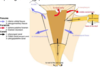
pterygopalatine fossa
small pyramid shaped space that is found inferior to the apex of the orbit.
Borders of the pterygopalatine fossa
Anterior = maxillary tuberosity; Posterior = pterygoid process of sphenoid (lateral plate); Medial = perpendicular plate of palatine bone; Lateral = opens into infratemporal fossa via pterygomaxillary fissure; Roof = (incomplete) greater wing of sphenoid; Floor = pyramidal process of palatine bone

Openings of the pterygopalatine fossa
Superior: opens into inferior orbital fissure
Inferior: closed except for palatine foramen
Openings of pterygopalatine fossa (labeled)
Contents of the pterygopalatine fossa
maxillary N (CN V2); pterygopalatine ganlgion; 3rd part of maxillary artery
Nerves of the pterygopalatine fossa (labeled)

Route of the maxillary nerve w/i the fossa

Parasympathetics of the pterygopalatine fossa
from the facial nerve via the greater petrosal nerve. the greater petrosal joins the deep petrosal to for the nerve of the pterygoid canal. The parasympathetic fibers from the greater petrosal n. supply the pterygopalatine ganlgion.

Sympathetics of the pterygopalatine fossa
The deep petrosal n. arises from the internal carotid plexus and conveys postsynaptic sympathetic fibers which join branches of the maxillary n. via the pterygopalatine ganlgion (but don’t synapse there). Presynaptic parasympathetic fibers are from the superior cervical ganglion.
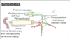
What are the 2 bold arrows?

- maxillary n. (CN V2)
- pterygopalatine fossa
What are the 3 bold arrows?

- maxillary n.
- nerve of pterygoid canal
- pterygopalatine ganglion
arteries of pterygopalatine fossa are ALL FROM _______?
Maxillary Artery
arteries of ptergyopalatine fossa (labeled)

Bony parts of the nose
nasal bones; frontal process of maxilla; nasal part of frontal bone and nasal spine; bony part of nasal septum

Cartilagenous parts of the nose
2 lateral cartilages; 2 alar cartilages; septal cartilages (nasal septum)

Nasal cavity
Except for the vestibule of the nose (considered an external portion), all of the nasal are lined with nasal mucosa. Nasal mucosa is firmly connected to the periostium of the bony parts of the nasal cavity and the perichondrium of the cartilagenous nasal components. The areas lined with nasal mucosa constitute the respiratory area (inferior 2/3) and the olfactory area (superior 1/3).
Nasal cavity olfactory area
has specialized nasal muscosa that the contains the peripheral nerve ending from the olfactory n. (CN I) for special sense olfaction

Nasal cavity divided into 4 passages
- spheno-ethmoidal recess (opening of sphenoid sinus) 2. superior nasal meatus (openings of ethmoidal sinuses) 3. middle nasal meatus (opening of frontal sinus) • maxillary sinus also opens into middle nasal meatus in posterior part of semilunar hiatus at the maxillary ostium (below ethmoid bulla). 4. inferior nasal meatus (opening of nasolacrimal duct)
4 nasal cavity passages labeled

what is the area where all 5 arteries come together in a capillary bed that can bleed profusely?
kiesselbach area

Arterial supply to the nasal cavity
Most of the blood is supplied to the lateral and medial walls of the nasal cavity from branches of the maxillary artery: sphenopalatine a.; anterior & posterior ethmoidal aa.; greater palatine a.
But also from the facial artery: • superior labial a. • lateral nasal branches
Nerve supply to the posterior 2/3 of the nasal cavity is from?
V2 via the nasopalatine n (nasal septum) and greater palatine n. (lateral wall)
nerve supply to the anteriosuperior nasal mucosa of the septum and lateral walls is from?
V1 via the anterior ethmoidal nerves
Paranasal sinuses
air filled extensions of the respiratory portion of the nasal cavity; found in frontal, ethmoid sphenoid and maxillary bones of the face; lined w/ nasal mucosa; drain into the nasal cavity via openings in the spheno-ethmoidal recess, and superior, middle and inferior meatuses.


