Department Images 98 - 151 (Dustin) Flashcards
(53 cards)
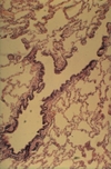
What tissue is this slide from?
(really difficult without seeing more of the lumen)

Esophagus
contains inner circular and outer tongitudinal muscles layers with skeletal muscle interspersed throughout
epithelium is stratified squamous non-keratinized

What tissue is this slide from?

Stomach Cardia
Near the esophageal-gastric junction, the bottom part of the image has strat squam non-keratinizing epithelium

Which tissue is this slide taken from?

Fundus of Stomach
Has many different kinds of cells in the gastric pits: know mucus neck cells, parietal cells, chief cells
One protruding fold of cells is called a plica villosa, surrounded on either side by gastric pits

What tissue is this from?
What stain? What is the purpose of this stain?

Fundus of Stomach
PAS-Congo-Hematoxylin stain
Makes mucus (both surface and neck) cells purple as they are PAS positive
Parietal cells turn orange
Chief cells become basophilic / grey

What cells here are orange?
Which ones are red?

Orange = Parietal Cells
Red = Mucus Neck Cells

What are the orange cells?
What are the greyer/ basophilic cells more at the top of the image?
What are the redder cells at the bottom of the image?

Orange = Parietal Cells
Greyer/basophilic = Chief Cells
Redder = Mucus Neck Cells
All in the gastric glands of stomach fundus

What tissue is this slide from?

Pylorus of Stomach
has branched tubular glands, almost entirely mucus secreting cells, stained poorly

What tissue is this slide from?
What major structures do you see?

Pylorus of Stomach
See Gastric Pits, and below are Pyloric Glands - not to be confused with Brunner’s glands of the duodenum which are in the tela submucosa

What is the large fold you see curving rightward in the lower half of the image?
What tissue is this slide from?

Plica Circularis aka Fold of Kerckring
Slide is from Jejunum
(no Peyer’s Patches or Brunner’s Glands)

What do you call the protruding fold of cells that makes up most of the image?

Intestinal Villi

What tissue is this slide from?
What stain?
(not a typical slide)

Jejunum
Indian Ink
(book says it demonstrates the blood vessels of the villus)
What are the cells with reddish granules at the base of the crypt?
Where do these exist in the GI tract?

Paneth Cells
Exist only in small intestine crypts of Lieberkuhn

What do you call the lighter thing sandwiched between the two darker eosinophilic structures?

Myenteric Plexus (of Auerbach)
Takes place between the inner circular and outer longitudinal smooth muscle layers of the GI tract

What tissue is this slide from?

Duodenum
Have typical small intestine structures + Brunner’s Glands in the tela submucosa

What type of glands are at the bottom of the image?
What tissue is this slide from?

Brunner’s Submucosal Glands
From the Duodenum

What tissue is this slide from?
What are the large purple areas in the middle?

Ileum
Purple areas = Peyer’s Patches

What tissue is this slide from?

Colon
Crypts of Lieberkuhn with a bunch of goblet cells and no villi

What tissue is this slide from?

Appendix
Has secondary follicles, NOT Peyer’s Patches
Many goblet cells, few/short crypts, no teniae

What tissue is this image from?
What stain?
What is the majority of the blue stuff in the image?

Pig Liver (Azan)
Blue = interlobular connective tissue that clearly separates the hepatic lobules, and in some corners between lobules you see the (Glisson’s) portal triad of interlobular bile duct, vein, and artery

What 3 important vessels do you see here?
What is the combination of them called?
What stain?

Interlobular Bile Duct: Simple cuboidal-columnar epithelium
Interlobular Artery/arteriole: thick-walled, small diameter.
Interlobular Vein/venule: largest diameter vessel here, typical thin wall
All 3 together = portal triad
Azan Stain

What organ is this from?
What stain?
What does this stain show?

Liver, India Ink Injection
Shows vascular pattern / Kupffer cells

What liver structure is this image focused on?

Portal Triad again
Really only the interlobular veins/venules are clear

What 3 important vessels do you see here?

Portal Triad again
This time all 3 are very clearly here

What type of cells are here?

Hepatocytes
Make up liver sinusoids, lined with Kupffer Cells which are generally not visible with an H-E stain like this