Derm HX/PE/DX/Therapy Introduction Flashcards

(51 cards)

1
Q

What are the three key factors that can be used to DX a skin condition?

A

1. The degree of pruritus

2. The distribution of the pruritus

3. The nature and distribution of any primary eruption.

2
Q

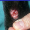
Primary skin lesions

A

Macule

Papule

Nodule

Plaque

Tumor

Pustule

Wheal

Vesicle

Bulla

3
Q

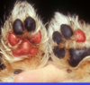
secondary skin lesions

A

Scale

Crust

Scar

Ulcer

Excoriation

Lichenification

Hyperpigmentation

Hyperkeratosis

Epidermal collarette

4
Q

Large vesicle; an intraepidermal or subepidermal accumulation of serous fluid

A

bulla

5
Q

Accumulation of keratin and dried sebum in a hair follicle

A

comedones (comedo)

6
Q

Dried exudate for secretion ± epithelial or bacterial debris

A

crust

7
Q

Inflammation of the skin.

Abnormal condition or disease of the skin

A

Dermatitis*

Dermatosis*

8
Q

Epidermis (name the layers)

A

Outermost, nonvascular layer of the skin.

Made up from outward-in of 5 layers: 1) Stratum corneum, 2) S. lucidum, 3) S. granulosum, 4) S. spinosum, 5) S. basale.

9
Q

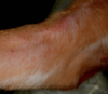
Redness produced by capillary congestion

A

erythema

10
Q

Superficial erosion or ulcer; usually implies scratch­ing or abrasion

A

excoriation

11
Q

Inflammation of hair follicles and associated adnexae

A

folliculitis

12
Q

Thickening of the stratum corneum due to an increased number of keratinized cells. Maybe from increased production or decreased loss. Orthokeratosis is a form wherein in the nucleus is lost in normal fashion (vs. parakeratosis).

A

Hyperkeratosis

13
Q

Cell of the epidermis

A

keratinocyte

14
Q

Thickening of skin with exaggeration of normal markings. Consists of acanthosis, hyperkeratosis and dermal thickening.

A

Lichenification

15
Q

Circumscribed, flat change in skin color. May be pale, hyper­pigmented or erythematous.

A

macule

16
Q

A large papule; a circum­scribed lesion raised above the level of the epidermis. Often extends into dermis.

A

nodule

17
Q

Circumscribed elevation of skin less than 1 cm in diameter.

A

papule

18
Q

Raised flat‑topped lesion

A

plaque

19
Q

Circumscribed epidermal or dermal accumulation of purulent exudate.

A

pustule

20
Q

Purulent dermatitis

A

pyoderma

21
Q

Flake of abnormal or compacted epithelial cells.

22
Q

A functional disturbance of sebaceous glands or of lipid metabolism of the epidermis. Accompanied by abnormal keratinization

23
Q

Layer of epi­dermis com­posed of flat­tened cells with pyk­notic nuclei and keratin gran­ules

A

s. granulosum

24
Q

Swelling or enlargement. Usually, but not always, neoplastic

25
Loss of substance on a cutaneous surface exposing inner layers or tissues. May imply full thickness loss of epidermis.
ulcer
26
A circumscribed elevation of epidermis caused by accumulation of clear fluid within or beneath the epidermis.
vesicle
27
Sharply circumscribed skin elevation produced by edema of the superficial dermis. This is commonly known as a hive.
wheal
28
What are these stains most useful for evaluating:
29
A macule that is \>1cm= a ...
patch
30
Name the lesion?
macule (aka target lesion)
31
Name the lesion?
papule
32
Name the lesion?
plaque
33
Name the lesion?
a pustule
34
DDX for follicular papular/pustule eruption
dermatophyte bacteria demodex
35
DDX for non-follicular pustule/papule?
36
Name the lesion? Caused by?
vesicle contact dermatitis & autoimmune skin dz
37
Name the lesion? Causes?
bulla burns, contact dermatitis, drug rxn, autoimmune dz
38
Name the lesion?
nodule
39
Name the lesion type?
tumor | (may or may not be neoplastic)
40
Lesion type? Test to make sure it is this type of lesion?
wheal (edema under skin) Diascopy - swelling should go down and blanch (if erythematous) when you press on the lesion if it is d/t vascular fluid leakage
41
A dog presents with the following? Primary or secondary lesion? If secondary, what was the primary lesion? Name of lesion?
Secondary- epidermal collarette Primary lesion was a pustule
42
Name the lesion? Primary or secondary? If secondary, what is the primary lesion?
Secondary: scale Primary: large scales= pustule small scales= dandruff
43
lesion? 1 or 2?
crust seondary
44
A lesion associated with hypopigmentation, alopecia, and/or a thinner dermis.
scar
45
Lesion? 1 or 2?
ulcer; secondary
46
Lesions? 1 or 2?
excoriations (implies scratching or abrasions)
47
Lesion? 1 or 2?
lichenification; secondary
48
Lesion? 1 or 2?
hyperkeratosis secondary
49
Lesion? 1 or 2? What structure in the skin is most often affected and why do they loose pigment?
depigmentation basement membrane that contains melanocytes is attacked via an inflammatory or neoplastic process that affects the melanocytes
50
Type of diagnostic test? What can be observed along the hair shaft?
direct microscopic exam of hair arthrospores
51
What is a cytology useful for?
identify secondary infections