Dermatologic disorders Flashcards

(135 cards)

1
Q

The most common form of cutaneous T-cell lymphoma. It generally affects the skin, but may progress internally over time. Symptoms include rash, tumors, skin lesions, and itchy skin.

A

Mycosis fungoides

2
Q

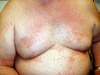
Involves the skin in about 25% of patients. The most common lesions are erythema nodosum, plaques, maculopapular eruptions, subcutaneous nodules, and lupus pernio. Treatment is not required, since the lesions usually resolve spontaneously in two to four weeks.

A

Sarcoidosis

3
Q

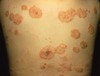
A chronic mucocutaneous disease that affects the skin, tongue, and oral mucosa. The disease presents itself in the form of papules, lesions, or rashes. It is sometimes associated with oxidative stress, certain medications and diseases (HCV), however the underlying pathology is currently unknown.

A

Lichen planus

4
Q

A chronic disease caused by the bacteria Mycobacterium leprae and Mycobacterium lepromatosis.

A

Leprosy

5
Q

It is primarily a granulomatous disease of the peripheral nerves and mucosa of the upper respiratory tract; skin lesions are the primary external sign. Left untreated, it can be progressive, causing permanent damage to the skin, nerves, limbs and eyes.

A

Leprosy

6
Q

Onset within the 1st year of life in 60% of cases. 25% will develop it between 1-5 yrs old. Childhood illness clears up in up to 40% of patients. Pruritic, erythematous, oozing rash with vesicles and edema that often involves the face and flexor surfaces.

A

Atopic dermatitis. If it does not clear up it is called chronic atopic dermatitis

7
Q

It is a type I hypersensitivity reaction (Th2 cells) associated with asthma and allergic rhinitis

A

(Chronic) atopic dermatitis

8
Q

Three types of fungus that commonly causes skin disease (Dermatophyte infection) in animals and humans

A

Microsporum (skin and hair), Epidermophyton (skin and nails) and Trichophyton (skin, hair, and nails)

9
Q

Caused by Trichophyton (most common cause in U.S.) and Microsporum dermatophytes. Seen in children 3-14 yrs/old. 3 clinical patterns: ectothrix, endothrix (more likely to cause hair to break), and favus

A

Tinea Capitis

10
Q

Causes tinea versicolor. It’s a lipophilic fungus that can cause disseminated infection in patients receiving IV lipid preparations. “Spaghetti and meatball” appearance on skin scraping.

A

malassezia furfur

11
Q

Caused by a group of soil-inhabiting fungi and produce warty, nodular, cauliflower-like lesions. The lesions are painless and usually occur on the feet but hands can be involved. No bone or muscle invasion and no fistula formation. It occurs in the tropics and is characterized by sclerotic bodies in biopsy.

A

Chromoblastomycosis

12
Q

Localized, indolent, deforming lesions of the foot and hand. Lesions are characterized by abscesses and draining sinuses; triad: abscesses, welling, grains (granule) exudation; bone and muscle involvement is common; grow slowly and usually painless. Lesions can be caused by bacteria or fungi (worse).

A

Mycetoma

13
Q

Bacterial Mycetoma is caused by

A

Actinomyces israelli, and Nocardia species

14
Q

Fungal (Eumycotic) mycetoma is caused by

A

pseudallescheria boydii

15
Q

Caused by Sporothrix schenckii a saprophyte in soil and vegetation.

A

Sporotrichosis

16
Q

Infection associated with traumatic implantation from rose thorns, sphagnum moss, and straw.

A

Sporothrix schenckii causing Sporotrichosis

17
Q

It is dimorphic: “cigar-shaped” yeast at 37 degrees c, and mycelia with “rosettes” conidia at 23 degrees c. It is easily cultured. It causes a disease that can be lymphocutaneous: ulceration at the site of inoculation, spread along lymphatic tract, with draining sinuses, or Disseminated (only in immunocompromised host): CNS, lungs, etc).

A

Sporothrix schenckii

18
Q

lymphocutaneous Sporotrichosis is treated with

A

Potassium iodide

19
Q

An adverse drug reaction of the skin

A

Drug eruption

20
Q

Benign neoplasm of melanocytes

A

Epidermal nevus

21
Q

Most common mole in children

A

Junctional nevus

22
Q

Most common mole in adults

A

Intradermal nevus

23
Q

Characterized by a flat macule or raised papule with symmetry, sharp borders, evenly distributed color, and a small diameter (

A

Nevus

24
Q

An inflammatory skin disease with erythema and scaling that affects nearly the entire cutaneous surface

A

Exfoliative dermatitis (Erythroderma)

25
The most common cause of erythroderma is
Exacerbation of an underlying skin disease (psoriasis, contact dermatitis, seborrheic dermatitis, lichen planus, pityriasis rubra pilaris or a drug reaction)
26
The conjunctivae and oral mucosa, along with the epidermis (skin) become erythematous (red and inflamed). Edema is often seen in the hands and feet.
Kawasaki's disease
27
Pruritic, planar, polygonal, purple papules often with reticular white lines on the surface (Wickham striae) that commonly involve the wrists, elbows, and oral mucosa. Characterized histologically by a saw-tooth appearance at the dermal epidermal junction. The etiology is unknown but it is associated with hepatitis C infection
Lichen planus
28
A skin disorder characterized by chronic itching and scratching (self-perpetuating scratch-itch cycle). The constant scratching causes thick, leathery, brownish skin.
Lichen simplex chronicus
29
Characterized by a bilateral malar erythema (also known as the "butterfly rash") and lesions that tend to be transient, follow sun exposure
Acute cutaneous lupus erythematosus
30
A clinically distinct subset of cases of lupus erythematosus that is most often present in white women aged 15 to 40, consisting of skin lesions that are scaly and evolve as polycyclic annular lesions or psoriasiform plaques.
Subacute cutaneous lupus erythematosus (SCLE)
31
Chronic skin condition of sores with inflammation and scarring favoring the face, ears, and scalp and at times on other body areas. These lesions develop as a red, inflamed patch with a scaling and crusty appearance. The center areas may appear lighter in color with a rim darker than the normal skin.
Chronic (Discoid) lupus erythematosus (DLE)
32
Characterized by large and irregular (malignant) cells forming in the skin of the nipple, indicating underlying breast cancer.
Paget's disease
33
A benign skin rash that may inflict substantial discomfort in certain cases. Classically, it begins with a single "herald patch" lesion, followed in 1 or 2 weeks by a generalized body rash lasting about 6 weeks
Pityriasis rosea
34
Self limited disease lasting approximately 6 weeks. Age group: 10-43 yrs. More common in the spring and autumn. Rarely recurs suggesting an infectious cause
Pityriasis rosea
35
A group of chronic disorders characterized by reddish orange, scaling plaques and keratotic follicular papules.
Pityriasis rubra pilaris (PRP)
36
Symptoms may include reddish-orange patches on the skin, severe flaking (pityriasis), uncomfortable itching, thickening of the skin on the feet and hands, and thickened bumps around hair follicles. For some, early symptoms may also include generalized swelling of the legs, feet and other parts of the body.
Pityriasis rubra pilaris (PRP)
37
Well-circumscribed, salmon-colored plaques with silvery scale that usually occurs on extensor surfaces and scalp. Pitting of the nails may also be present.
Psoriasis
38
Due to excessive keratinocyte proliferation possibly with an autoimmune ( Th1 T-cell) etiology associated with HLA-C.
Psoriasis
39
Characterized histologically by parakeratosis (retention of nuclei by the stratum corneum), elongation of the dermal papillae which can cause pinpoint bleeds beneath the silvery scale.
Psoriasis
40
Psoriasis is treated with
Corticosteroids and immune-modulating therapy (to combat autoimmune reaction), as well as with UV-A light plus psoralen to damage keratinocytes
41
An inflammatory skin disorder affecting the scalp, face, and torso. Typically presents with scaly, flaky, itchy, and red skin. It particularly affects the sebaceous-gland-rich areas of skin. In adolescents and adults, it usually presents as scalp scaling similar to dandruff or as mild to marked erythema of the nasolabial fold.
Seborrheic dermatitis
42
A benign squamous proliferation that commonly occurs in the elderly and presents as raised, discolored plaques on the extremities or face and often has a coin-like, waxy, 'stuck-on' appearance.
Seborrheic keratosis
43
It is characterized by keratin pseudocysts on histology. If many suddenly arise (Leser-Trelat sign), it suggests underlying carcinoma of the GI tract.
Seborrheic keratosis
44
A rash of oval or irregularly-shaped spots often merging to form a larger patch, occasional fine scaling, mildly itchy, with a sharp border that occurs on the trunk and proximal extremities.
Tinea versicolor
45
It is usually caused by the Malassezia globosa fungus (Malassezia furfur is responsible for a small number of cases). These yeasts are normally found on the human skin and only become troublesome under certain circumstances, such as a warm and humid environments.
Tinea versicolor
46
Flesh-colored papules with a rough surface that is due to HPV infection of keratinocytes characterized by koilocytic change commonly on the hands and feet.
Verruca (Wart)
47
A form of eczema that is characterized by changes that occur when skin becomes abnormally dry, itchy, and cracked. Lower legs tend to be especially affected, although it can appear in the underarm area as well.
Asteotic Eczema
48
The most common form of ichthyosis, affecting around 1 in 250 people. A skin disorder causing dry, scaly skin.
Ichthyosis Vulgaris
49
It is usually an autosomal dominant inherited disease (often associated with filaggrin), although a rare non-heritable version exists. The symptoms of the inherited form are not usually present at birth but generally develop between 3 months and 5 years of age. The symptoms will often improve with age, although they may grow more severe again in old age
Ichthyosis Vulgaris
50
Present with a variety of infectious disorders, often occurring in response to an inflammatory tinea of the feet, resulting in an eczematous dermatitis. The inflammation is in the absence of a locally visible cause.
Id Reaction
51
Characterized by round or oval-shaped itchy lesions. It is recurrent and chronic, and may appear at any age, although it is most common in people in their 60's. Does not appear to be a genetic condition, is not related to food allergies, and is not contagious.
Nummular Eczema
52
Characterized by itchy, coin-shaped patches that can affect any part of the body, but the legs and buttocks are the most common areas. Flare-ups are associated with dry skin, so the winter season is a particularly bad time for those with this condition.
Nummular Eczema
53
A rare, severe infection associated with an immense number of mites, with marked scales and crusts, usually accompanied by lymphadenopathy and eosinophilia. Infection that can be confused for psoriasis
Norwegian Scabies
54
A severe, autosomal recessive form of ichthyosis associated with mutations in the SPINK5 gene.
Netherton's Syndrome
55
Characterized by chronic skin inflammation, universal pruritus (itch), severe dehydration and stunted growth. Patients with this disorder tend to have a hair shaft defect (trichorrhexis invaginata), also known as "bamboo hair".
Netherton's Syndrome
56
An autoimmune blistering disease of the skin and mucous membranes with characteristic lesions that are scaly, crusted erosions, often on an erythematous base. No clinically apparent mucosal involvement is present even with widespread disease.
Pemphigus Foliaceus
57
Presents as a pruritic, erythematous, oozing rash with vesicles and edema that arises upon exposure to allergens or irritants. It is a type IV hypersensitivity reaction
Contact Dermatitis
58
Comedones (Whiteheads and blackheads), Pustules (pimples), and nodules due to chronic inflammation of hair follicles and sebaceous glands.
Acne Vulgaris
59
Acne Vulgaris occurs as a result of hormone-related increases in sebum and excess keratin blocking follicles that become infected with what
Propionibacterium acnes
60
Treatment for Acne Vulgaris
Benzoyl peroxide (antibiotic against propionibacterium acnes) and Vitamin A derivatives such as isotretinoin (to reduce keratin formation)
61
Autoimmune destruction of desmosomes between keratinocytes due to IgG antibody (type II hypersensitivity) against desmoglein (a component of desmosomes).
Pemphigus Vulgaris
62
Immunoflorescence highlights IgG surrounding keratinocytes ('fish net' appearance). It can involve both the skin and the oral mucosa.
Pemphigus Vulgaris
63
Autoimmune destruction of hemidesmosomes between basal cells and the underlying basement membrane due to IgG antibodies (type II hypersensitivity) against basement membrane collagen.
Bullous Pemphigoid
64
It presents as blisters of the skin but the oral mucosa is spared. Tense bullae do not rupture easily. Immunofluorescence highlights IgG along the basement membrane in a linear pattern
Bullous Pemphigoid
65
Autoimmune deposition of IgA at the tips of dermal papillae. It presents as pruritic vesicles and bullae that are grouped. It has a strong association with celiac disease; resolves with a gluten-free diet.
Dermatitis Herpetiformis
66
Common acute, self-limited disorder. Hypersensitivity reaction with targetoid rash (central epidermal necrosis surrounded by erythema) and bullae that is most commonly associated with HSV infection (also could be mycoplasma infection, drugs such as penicillin or sulfonamides, autoimmune diseases, and malignancy).
Erythema Multiforme
67
When Erythema Multiforme is accompanied by oral mucosa/lip involvement and fever it is termed
Stevens-Johnson syndrome (SJS)
68
A severe form of Stevens-Johnson syndrome (SJS) characterized by diffuse sloughing of skin, resembling a large burn; most often due to an adverse drug reaction.
Toxic epidermal necrolysis
69
Epidermal hyperplasia with darkening of the skin ('Velvet-like' skin) that often involves the axilla or groin. It is associated with insulin resistance or malignancy (especially gastric carcinoma). There is a benign familial type, and an autosomal dominant type.
Acanthosis Nigricans
70
The most common cutaneous malignancy
Basal Cell Carcinoma
71
Presents as an elevated nodule with a central, ulcerated, crater surrounded by dilated vessels ('pink, pearl-like papule') that is classically located on the upper lip.
Basal Cell Carcinoma
72
Treatment for Basal Cell Carcinoma
Surgical excision, metastasis is rare, and good prognosis.
73
Characterized by the formation of keratin pearls
Squamous Cell Carcinoma
74
Risk factors for Basal Cell Carcinoma and Squamous Cell Carcinoma
UVB-induced DNA damage: prolonged exposure to sunlight, albinism, and xeroderma pigmentosum.
75
It presents as an ulcerated, nodular mass, usually on the face (classically involving the lower lip).
Squamous Cell Carcinoma
76
Treatment for Squamous Cell Carcinoma
Excision and metastasis is uncommon
77
A precursor lesion of squamous cell carcinoma and presents as a hyperkeratotic, scaly plaque, often on the face, back, or neck.
Actinic keratosis
78
Localized loss of skin pigmentation due to autoimmune destruction of melanocytes.
Vitiligo
79
Congenital lack of pigmentation due to an enzyme defect that impairs melanin production.
Albinism
80
Albinism is usually due to a defect in what enzyme
Tyrosinase
81
Albinism is associated with an increased risk of
Squamous cell carcinoma, basal cell carcinoma, and melanoma due to reduced protection against UVB
82
Freckles are due It increased numbers of
Melanosomes (melanocytes are not increased)
83
A Mask-like hyperpigmentation (tan or dark skin discoloration) of the cheeks. Although it can affect anyone, it is particularly common in women, especially pregnant women and those who are taking oral or patch contraceptives or hormone replacement therapy (HRT) medications.
Melasma
84
Most common cause of death from skin cancer
Melanoma
85
Autosomal dominant disorder characterized by formation of dysplastic nevi that may progress to melanoma
Dysplastic nevus syndrome
86
Presents as a mole-like growth with "ABCD" - Asymmetry, Borders are irregular, Color is not uniform, Diameter \> 6mm.
Melanoma
87
Characterized by 2 growth phases: It starts with radial growth horizontally along the epidermis and superficial dermis with a low risk of metastasis, and then can shift to vertical growth into the deep dermis with increased risk of metastasis
Melanoma
88
The most important prognostic factor predicting metastasis
Depth of extension
89
Melanoma subtype characterized by lentiginous proliferation (radial growth) with a good prognosis
Lentigo maligna melanoma
90
Most common subtype of melanoma, characterized by dominant early radial growth with a good prognosis. Usually has a redish hue
Superficial spreading melanoma
91
Melanoma subtype characterized by early vertical growth with a poor prognosis
Nodular melanoma
92
Melanoma subtype arising on the palms or soles, often in dark-skinned individuals and not related to UV light exposure
Acral lentiginous melanoma
93
Superficial bacterial skin infection, most often due to Staph aureus or Strep pyogenes. It most commonly affects children and presents as erythematous macules that progress to pustules, usually on the face (rupture of pustules results in erosions and dry, crusted, honey-colored sreum)
Impetigo
94
Deeper (dermal and subcutaneous) infection, usually due to S aureus or Strep pyogenes that presents as a red, tender, swollen rash with fever.
Cellulitis
95
Sloughing of skin with erythematous rash and fever (leading to significant skin loss) due to Staph aureus infection (exfoliative A and B toxins result in epidermolysis of the stratum granulosum).
Staphylococcal Scalded Skin Syndrome
96
Staphylococcal Scalded Skin Syndrome differs from toxic epidermal necrolysis by what
The level of skin separation. separation in Toxic Epidermal Necrolysis occurs at the dermal-epidermal junction. epidermolysis in Staphylococcal Scalded Skin Syndrome occurs at the stratum granulosum
97
Firm, pink, umbilicated papules due to poxvirus. Affected keratinocytes show cytoplasmic inclusions
Molluscum Contagiousum (cytoplasmic inclusions are called molluscum bodies)
98
Emerging infectious disease caused by at least three species of bacteria belonging to the genus Borrelia. Borrelia burgdorferi is the main cause in North America, whereas Borrelia afzelii and Borrelia garinii cause most European cases.
Lyme Disease
99
Lyme disease is contracted from
Ixodes ticks
100
Characterized by gottron lesions, scaly erythematous eruptions or red patches overlying the knuckles, elbows, and knees. Other skin manifestations involve periungual telangiectasias, a heliotropic (purple) rash over the upper eyelids (racoon eyes), and shawl sign.
Dermatomyositis
101
A well-differentiated squamous cell carcinoma that develops rapidly and regresses spontaneously and presents as a cup-shaped tumor filled with keratin debris.
Keratoacanthoma
102
Circumscribed or diffuse hard smooth ivory colored areas of immobile skin. Localized (morphea) and linear. Progressively systemic, causes CREST syndrome, sclerotic plaques, mat telengectasias.
Scleroderma
103
The most common subtype of porphyria
Porphyria Cutanea Tarda
104
The disorder results from low levels of the enzyme responsible for the fifth step in heme production (defect in uroporphyrinogen decarboxylase)
Porphyria Cutanea Tarda
105
The disease is characterized by onycholysis (detachment of the nail from the nail bed), blistering of the skin in areas that receive higher levels of exposure to sunlight, hyperpigmented skin and hypertrichosis of the cheeks and temples, Scarring, milia (a keratin-filled cyst that can appear just under the epidermis or on the roof of the mouth), and dyspigmentation. No erythema.
Porphyria Cutanea Tarda
106
An inflammation of the fat cells under the skin (panniculitis) characterized by tender, red nodules or lumps that are usually seen on both shins.
Erythema Nodosum
107
It is presumed to represent a delayed hypersenstivity reaction. Most often women ages 15 to 40. Bruise-like lesions that resolve over 2-8 weeks. Polyarthralgias, fever, and malaise. Most common cause world wide is streptococcal pharyngitis. TB is a common cause in the U.S.
Erythema Nodosum
108
An unpleasant sensation that provokes the desire to scratch. It is commonly caused by over bathing (asteatosis), primary skin disease, medications, neurosis, parasitosis, or systemic diseases (malignancy, uremia, cirrhosis, endocrine).
Pruritis
109
A premalignant condition of thick, scaly, or crusty patches of skin. It is more common in fair-skinned people and it is associated with those who are frequently exposed to the sun, as it is usually accompanied by solar damage.
Actinic Keratoses
110
They are considered as potentially pre-cancerous, since some of them progress to squamous cell carcinoma, so treatment is recommended. Untreated lesions have up to 20% risk of progression to squamous cell carcinoma.
Actinic Keratoses
111
Painless, persistent, thickening and whitish discoloration of the lip at the border of the lip and skin
Actinic cheilitis
112
A common low-grade (unlikely to metastasize or invade) skin tumor that is believed to originate from the neck of the hair follicle. Many pathologists consider it to be a form of squamous cell carcinoma (SCC).
Keratoacanthoma
113
The defining characteristic is that it is dome-shaped, symmetrical, surrounded by a smooth wall of inflamed skin, and capped with keratin scales and debris. It always grows rapidly, reaching a large size within days or weeks, and if untreated will starve itself of nourishment, necrose (die), slough, and heal with scarring.
Keratoacanthoma
114
Microscopically controlled surgery used to treat common types of skin cancer. During the surgery, after each removal of tissue, while the patient waits, the pathologist examines the tissue specimen for cancer cells, and that examination informs the surgeon where to remove tissue next.
Mohs Surgery
115
Phase of eczematous dermatitis characterized by redness and edema, with papules and often vesicles, oozing, and crusting.
Acute phase of eczematous dermatitis
116
Phase of eczematous dermatitis characterized by perivascular lymphohistiocytic infiltrate in the dermis and epidermis, fluid exudation into the epidermis (spongiosis) and papillary dermis, intraepidermal vesicle formation, and often disruption of the stratum corneum. Redness is produced by dilatation of blood vessels.
Acute phase of eczematous dermatitis
117
Phase of eczematous dermatitis characterized by scaling, fissuring, slight accentuation of the normal skin markings (lichenification), and minor redness and edema
Subacute phase of eczematous dermatitis
118
Phase of eczematous dermatitis characterized by a usually mild epidermal thickening and spongiosis, associated with a slight perivascular infiltrate of lymphocytes.
Subacute phase of eczematous dermatitis
119
Phase of eczematous dermatitis characterized by scaling and lichenification, with minimal or no edema and redness
Chronic phase of eczematous dermatitis
120
Phase of eczematous dermatitis characterized by a thickened epidermis (in response to repeated physical or chemical trauma) and minimal perivascular and intraepidermal lymphocytic infiltrates. The stratum corneum is disrupted, but there is minimal edema fluid in the epidermis.
Chronic phase of eczematous dermatitis
121
Differentiating factor of liquid contact dermatitis and solid contact dermatitis
Liquid contactant will involve the skin creases. Solid contactant will spare the creases
122
A kind of skin rash notable for pale red, raised, itchy bumps. It might also cause a burning or stinging sensation. The majority of cases have an unknown (idiopathic) cause, but it is frequently caused by allergic reactions (there are many nonallergic causes)
Urticaria (Hives)
123
Urticaria (Hives) that is due to an allergic trigger
Acute urticaria (lasting less than six weeks)
124
Urticaria (Hives) that is rarely due to an allergy
Chronic urticaria (lasting more than six weeks)
125
Typicallya reaction to food, but in about half the cases, the trigger is unknown. Common foods may be the cause, as well as bee or wasp stings, or skin contact with certain fragrances
Acute urticaria
126
A condition (also from allergic and nonallergic causes) related to urticaria, but fluid leakage is from much deeper blood vessels
Angioedema
127
a medical condition in which hair is lost from some or all areas of the body, usually from the scalp
Alopecia Areata
128
Alopecia Areata limited only to the beard
Alopecia areata barbae
129
Alopecia Areata where patient loses all the hair on the scalp
Alopecia totalis
130
Alopecia Areata where patient loses all the hair on the body
Alopecia universalis
131
Patients with Alopecia Areata also tend to have a slightly higher incidence of what conditions
Conditions related to the immune system: asthma, allergies, atopic dermal ailments, and hypothyroidism
132
A life-threatening skin condition in which cell death causes the epidermis to separate from the dermis. The syndrome is thought to be a hypersensitivity complex that affects the skin and the mucous membranes. The main known cause is certain medications, followed by infections and, rarely, cancers.
Stevens-Johnson syndrome
133
Usually begins with fever, sore throat, and fatigue, which is misdiagnosed and usually treated with antibiotics. Ulcers and other lesions begin to appear in the mucous membranes, almost always in the mouth and lips but also in the genital and anal regions. Those in the mouth are usually extremely painful and reduce the patient's ability to eat or drink
Stevens-Johnson syndrome
134
A syndrome, caused by exposure to certain medications, that may cause a rash, fever, inflammation of internal organs, lymphadenopathy, and characteristic hematologic abnormalities such as eosinophilia, thrombocytopenia, and atypical lymphocytosis. The syndrome carries about a 10% mortality
Drug induced hypersensitivity (DRESS syndrome)
135
Melanoma risk factors
Family history (genetic factors), previous melanoma, presence of dysplastic nevi, having light skin, immune suppression, and sun exposure