Embryology Flashcards

1
Q

Prenatal period

A

The period of gestation is prior to birth

2
Q

Perinatal period

A

22 weeks gestation to 28 days after birth

3
Q

Postnatal period

A

after birth

4
Q

neonatal period

A

up to 1 month after birth

5
Q

infancy

A

first postnatal year

6
Q

childhood

A

12 months to 12-13 years

7
Q

Puberty

A

10-15 years (girls); 12-17 years (boys)

8
Q

Adolescence

A

3-4 years post puberty

9
Q

adulthood

A

from 18-25 years to..

10
Q

What is the estimated pregnancy length for clinicians, and patients?

A

Clinicians: 40 weeks, patients: 9 months

11
Q

What are the 3 stages of embryonic development from a clinicians point of view, and an embryologists?

A

Clinicians: first, second and third trimester. Embyologist: preimplantation, embryonic, fetal stage

12
Q

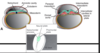
What happens during week one of the prenatal period?

A

Preimpantation stage: zygote, morula, blastocyst

13
Q

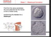
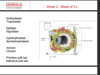
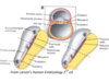
What happens during week 2 of the prenatal period?

A

inner cell mass forms bilaminar embryo

14
Q

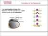
What happens during week 3 during the prenatal period?

A

Bilaminar embryo becomes trilaminar embryo

15
Q

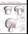
What happens during week 3-8 during the prenatal period?

A

Embryonic period- organogenesis

16
Q

What happens during week 9 to term during the prenatal period?

A

Fetal period- grown and differentation

17
Q

During what time in development is it considered the “all or nothing” period?

A

The first 2 weeks

18
Q

What stage of development is most susceptible to teratogenic agents?

A

The embryonic stage, also termed the “critical period”

19
Q

What stage is most likely to produce minor structural defects or functional abnormalities?

A

The fetal period

20
Q

Gestational age (GA)

A

The age of the embryo/fetus from the presumed first day of the last menstrual period- clinical notion

21
Q

Fertilization age

A

the age of the embryo/fetus from the fertilization day- not used as much in clinic

22
Q

Whats the relationship between fertilization age and gestational age?

A

GA is approx. 2 weeks longer than fertilization age because the oocye is not fertilized until about 2 weeks after last menstrual period (around ovulation time)

23
Q

Naegele’s rule

A

First day of the LMP subtract 3 months then add one year and one week , so LMP- 3 months + 1 year + 1 week

24
Q

What percentage of women deliver on their due date? What percentage deliver within 13 days of their due date?

A

5% due date, 60-70% within 13 days

25
Q

Abortion

A

Expulsion/removal of the embryo/fetus from uterus prior to age of viability (~

26
Q

Miscarriage; what are most caused by?

A

spontaneous expulsion from uterus of embryo/fetus relatively early (usually <20 weeks); most caused by chromosomal abnormalities

27
Q

Premature birth; most common cause?

A

Infants born between stage of viability and 37 weeks –>majority caused by maternal complications of the pregnancy

28
Q

Term birth

A

Infants born after at least 37 weeks of gestation

29
Q

Describe the process of gametogenesis from primordial germ cells up until gonocytes

A

Primordial germ cells are identified during the 4th week in the yolk sac. Week 4-6 they migrate to the future genital ridge are. PGCS continue to multiply by mitosis during their migration. PGCS stimulate the surrounding epithelium to create the somatic support cells, which causes swelling, and this becomes the primitive gonad. PGCs are now called gonocytes.

30
Q

What are the somatic support cells for gonocytes in males and females?

A

ovarian follice (females), sertoli cells (males)

31
Q

Gamete maturation is called what in males? in females?

A

Males, spermatogenesis

Females, oogenesis

32
Q
A
33
Q

What are the two basic processes of gametogenesis that are the same in males and females? What is different between them?

A

several mitotic division, two meiotic divisions; the timing of the maturation of germ cells is different in the male and female

34
Q

Gonocytes have how many chromosomes?

A

23 pairs, total of 46 (diploid). One chromosome of each pair is obtained from maternal gamete and the other from the paternal gamete.

35
Q

Mature sperm and oocytes have how many chromomoses? Why? When does this happen?

A

23 (haploid), during gametogeneis the chromosome # is reduced by half, this happens during meiosis

36
Q
A
37
Q
A
38
Q
A
39
Q

Oogonia divide by what? By the 5th month of pregnancy the number of germ cells in the ovary reaches what?

A

Mitosis, they reach their max (7mil) most degenerate

40
Q

What happens to the surviving oogonias in oogenesis? What are they enclosed in?

A

They enter prophase of meiosis 1; enclosed in follicle

41
Q

By puberty, about how many oogonias remain in females?

A

about 400,000

42
Q

What cell stage are the surviving oocytes arrested in?

A

Prophase of meoisis 1

43
Q

Describe what happens to the primary oocyte during oogenesis

How many follicles resume maturation each month?

A

As a follicle matures, the primary oocyte increses in size and shortly before ovulation, completes the first meoitic division to give rise to a secondary oocyte and the first polar body.

5-12 resume maturation each month, one becomes the dormant one, the other degerates

44
Q

How is cytoplasm distributed amongst the secondary oocyte during oogenesis?

A

The secondary oocyte receives almost all the cytoplasm and the first polar body receives very little. (polar body is small, nonfunctional cell)

45
Q

What happens to the secondary oocyte at ovulation? What stage does it progress to?

A

It enters second meiotic division; progresses only to metaphase, when division is arrested

46
Q

Secondary meiotic division is completed only if what?

A

fertilization occurs

47
Q

Spermatogonias inside embyronic male gonads remain what from 6th week until puberty?

A

Dormant

48
Q

Spermatogenesis takes place ____from puberty until death (___-___ million sperm/day)

A

continuosly; 200-300 million

49
Q

What happens to sertoli cells at puberty, and why?

A

Under the influence of testosterone, Sertoli cells differentiate into a system of seminferous tubles. The dormant PGCs resume development, divide several times by mitosis, and then differntiate into spermatonia

50
Q
A
51
Q
A
52
Q

The secondary oocyte is surrounded by zona pellucida, which is what?

A

a thin layer of transparent gelatinous layer of protein and polysaccharides

53
Q

Corona radiata

A

several layers of ovarian follice

54
Q

What is capacitation, and where does it occur?

A

Functional changes that sperm undergo. This occurs in the female reproductive tract that enable them to fertilize a secondary oocyte

55
Q

What does the acrosome reaction accomplish?

A
56
Q

List the steps in fertilization

A

A. Sperm penetration of corona radiata and zona pelludica

B. Fusion of plasma membranes of sperm and oocyte

C. Formation of male pronucleus…the tail and mitochondria of the spem degrade, so that all mitochondria in the embryo (and all mitochondrial DNA) come together

D. Completeion of second meiotic division of oocyte and formation of female pronucleus

E. Fusion of pronucelei (syngamy) to form a diploid zygote

57
Q

Cortical reaction

A

Oocyte membrane becomes impenetrable to other sperm, happens after fusion of plasma membrane between sperm and oocyte

58
Q

Zona reaction

A

Zona pellucida alters its structure to prevent sperm binding and penetration

59
Q

syngamy

A

Fusion of pronuceli to form a diploid zygote

60
Q

What is the result of cleavage of the zygote?

A

Following fertilization, the zygote undergoes a series of mitotic cell divisions (cleavage) as it migrates toward the uterus. The process of cleavage divides the zygotes cytoplasm into increasingly smaller cells known as blastomeres (blastula).

After the 3rd cleavage, the blastomeres tightly align via tight junctions via a process known as compaction.

About 3 days after fertilization (12-32 cell stage) the embryo is termed a morula, consisting of a solid ball of cells with an inner and outer mass. The inner mass will become the embryo, the outer mass wil become the placenta. The morula enters the uterus by day 4

61
Q

What does the morula form?

A

Fluid from the uterus penetrates the zona pellucia into the morula to form a hollow cavity, transforming the morula into a blastocyst

62
Q
A
63
Q

What is the difference between the trophoblast and embryoblast? What does each come from?

A

The trophoblast is the thin outer cell mass. It will make human corionic gonadotropin (hCG) which is the basis for pregnancy testing.

The embryoblast is the inner cell mass, which is a group of centrally located blastomeres.

64
Q

What is hatching of the embryo and why is it important?

A

“Hatching” is when the blastocyst sheds its zona pellucida, and this is important because it allows the blastocyst to increase its size

65
Q

What produces HcG during week 2 of pregnancy? Why is hCG imporant at this time in the pregnancy? What takes over at week 8?

A

synciotrophoblast, which enters maternal blood via lacunae. hCG stimulates progesteron, which is needed to maintain pregnancy until week 8. After week 8, the placenta takes over progesterone production

66
Q
A
67
Q

What are lacunae, where do they develop, and what do they contain?

A
68
Q
A
69
Q

Explain the steps of implantation

A

Normally, the blastocyst implants along the anterior or posterior wall of the body of the uterus.

In the area over the embryoblast, the trophobolast differentiates into 2 layers, the cytotrophoblast (inner layer of mononucleated cells) and the synctiotrophoblast (outer layer of multinucleated zone that invades the uterine endometrium to implant blastocyst).

70
Q

Cytotrophoblast

A

inner layer of monoucleated cells with mitotic capacity

71
Q

synctiotrophoblast

A

outermultinucleated zone that invades the uterine endometrium to implant the blastocyst

72
Q

What are lacunae, where do they develop, and what do they contain?

A

The synctiotrophoblast invades the maternal endometrium, and as it comes in contact with blood vessels, it creates lacunae, or spaces filled with maternal blood. These form lacunar networks, and these networks function to exchange nutrients and waste with the fetus.

73
Q

When is implantation completed? What marks this completion?

A

By the end of week 2. Marked temporarily by fibrin plug.

74
Q

Explain in vitro vertilization

A

Clomiphene citrate or gonadotropin is administered to stimulate multiple ovulations.

Oocytes are collected by needle aspiration from the ovary

Sperm collected, exposed to ionic solution to induce capcitation

Sperm and oocytes cultured together

cleavage is allowed to proceed in vitro to 8 cell stage

Typically at least 3 embryos are transferred to uterus

75
Q

Whats the clinical correlate of abnormal blastocysts?

A

Most do not produce any sign of pregnancy because trophoblast is defective and could not produde sufficient hCG necessary to maintain corpus luteum.

76
Q

Where and how do amnion and amnionic activity develop?

A

The embryoblast differentiates into 2 cellular layers: dorsal epiblast and ventral hypoblast.

Within the epiblast, clefts develop to form the amniotic cavity and amnion

77
Q

Where and how do the chorion and chorionic cavity develop?

A

Hypoblast cells migrate and line the inner surface of the cytotrophoblast, forming the exocoelomic membrane (Heuser’s membrane), which surrounds a space known as the primitive yolk sac.

The extraembryonic mesoderm (from the primary yolk sac) proliferates to fill the spaces between the cytotrophoblast and either the amnion or the primitive yolk sac.

Cavities form within the extraembryonic mesoderm to form the chorionic cavity

78
Q

What layers form the chorionic membrane (chorionic plate)

A

the extraembryonic mesoderm

the cytotrophoblast

the synctiotrophoblast

79
Q

What are the primary chorionic villi and how do they develop?

A

cytotrophoblasts proliferate to form extensions (primary villi) taht grow into synctiotrophoblast. These primary villi grow out into the lacunae, carrying with them a covering of synctiotrophoblast

80
Q

Describe the development of the primary and secondary yolk sacs

A

The hypoblast cells migrate out along the surface primitive yolk sac (exocoelomic coelom) to form a smaller sac (definitive or secondary yolk sac/umbilical vesicle)

81
Q

What does the connecting stalk form?

A

the umbilical cord

82
Q

Why are most abnormal blastocysts not detected?

A
83
Q

What are hydatidiform moles? How are they formed?

A
84
Q

What is placenta previa?

A

The blastocyst may implant close to the internal os (opening) of the cervix. In this case, the placenta may cover the internal os, resulting in painless vaginal bleeding in the later stages of pregnancy.

85
Q

What is an ectopic pregnancy? Where do 95% occur? What are signs of this?

A
86
Q
A
87
Q

Give a summary of steps of week 2 in embryonic development

A

Embryoblast become bilaminar disc (epiblast and hypoblast)

Amnionic cavity and primary yolk sac (primary umbilical vesicle) form

Extraembryonic mesoderm develops

chorionic cavity develops as spaces within extraembryonic mesoderm appear

definitive yolk sac (secondary umbilical vesicle) forms as the primary yolk sac regresses

88
Q

What 3 things does the process of gastrulation accomplish?

When does it start, and when does it end?

A

It transforms the bilaminar embryonic disk (epiblast and hypoblast) into a trilaminar disk. It establishes 3 germ layers of the embryo (ectoderm, intra-embryonic mesoderm and endoderm) that form all of the tissues and organs of the adult
Also establishes the body axes

Cells are moved to new places allowing for new interactions that allow for processes like neurulation and organogenesis

Begins in week 3 and continues for almost 2 weeks, ending just before the closure of the caual neural tube

89
Q

What is the primitive steak? what 3 things does it consist of?

A

Primitive groove: longitudinal midline groove on the embyonic disk which elongates to occupy half the length of the embry

Primitive node: Elevated area at the cephalic “head” end of the primitive groove

Primitive pit: Depression in the middle of the primitive node

90
Q

Cells from which layer of the bilaminar embryonic disk migrate through the primitive streak and pit? What do they do once they arrive?

A

Cells of the epiblast migrate toward the primitive streak, upon arrival at the primitive groove, they detach from the epiblast and slip beneath it via the process of invagination

91
Q

What happens to the epiblast cells once they have migrated through the primitive groove?

A

Some displace the hypoblast, creating endoderm (which will form the lining of the future gut tube and gut derivatives)

Others come to lie between epiblast and endoderm to form intra embryonic mesoderm

After the endoderm and mesoderm layers are formed, cells remaining in the epiblast become ectoderm

92
Q

What do cells form that move through the primitive pit?

A

At the same time that cells migrate laterally through the primitive groove, they also move cranially through the primitive pit to form:

the pre or pro-chordal plate (located caudal to the buccopharyngeal membrane and cranial to the primitive pit; serves as an important organizer of the head region

The notochord ( solid cord of cells that serves as the basis for the axial skeleton

93
Q

Explain what happens when the intra-embryonic mesoderm spreads out from the primitive streak.

What happens to the length of the primtive streak?

What happens to the primtive streak when gastrulation is complete?

A

As the intra-embryonic mesoderm spreads out from the primitive streak, the whole embryo increases in size and the primitive streak becomes relatively smaller.

The primitive streak regresses caudally, becoming gradually shorter.

When gastrulation is complete, the primtiive streak disappears

94
Q

Which stage is sensitive for teratogens?

A

Gastrulation

95
Q

Conjoined twins result from what?

A

Partial splitting of the primitive node and streak

96
Q

What is sirenomelia?

A

Insufficient mesoderm formed in the caudal region of the embryo, which froms lower limbs and urogenital system and lumbosacral vertebrae

Results in mermaid syndrome, in which lower limbs are fused together

Kidney and bladder development is affected so that many babies that are born with this condition will generally die within a few days of birth

97
Q

Explain how the notochord forms

A

Cells destined to form the notochord migrate through the primitive pit cranially until they reach the pre or pro-chordal plate, where they form the hollow notochordal process

The notochordal process then detaches from the endoderm as a solid cylinder known as the notochord

98
Q

The notochord is important because (3 things)

A

It induces overlying ectoderm to differentiate into neuro ectoderm to form neural plate

induces formation of vertebral bodies

Forms nucleus pulposis of adult interverterbal disks

99
Q

What is sacrococcygeal teratoma?

A

Remnants of the primitive streak in the sacrococcygeal area

Commonly contains tissues derived from all 3 germ layers

Most common tumor in newborns

100
Q

What is chordoma?

A

Tumor arises from remnants of notochord

intracranially or sacrally; more common in adult men in their 50’s

101
Q

What is neurolation?

A

Formation of neural tube. As the notochord develops, it induces the overlying ectoderm to thicken and from the neural plate.

102
Q

What happens to the neural plate early in the 3rd week?

A

The neural plate invaginates to form the median neural groove, flanked by neural folds on either side

103
Q

What happens to the neural folds later in the 3rd week?

A

The neural folds fuse to form the neural tube (CNS precursor), which separates from the surface ectoderm as the folds fuse

104
Q

As the primitive streak regresses, what happens to mesodermal cells?

A

mesodermal cells that migrated laterally through the streak condense into rod or sheet-like sturctes on either side of the notochord

105
Q

What is the paraxial mesoderm?

A

Thick plate of mesoderm located on each side of midline, organized into segments known as somites beginning on day 20

106
Q

What is the intermediate mesoderm, what is it important in?

A

Longitudinal dorsal ridge of mesoderm between paraxial and lateral mesoderm

forms urogenital ridge, which is involved in formation of kidneys and gonads (ovaries and testes)

107
Q

What are the red, blue, and yellow strucutres?

A

ectoderm in blue, mesoderm in red, endoderm in yellow

108
Q

What is the lateral mesoderm?

A

Part highlighted in yellow,

continuous with extraembryonic mesoderm

Large spaces develop within this layer to form intra-embryonic coelom–which will form pleural (around lung), pericardial (around heart) and peritoneal (abdominal) cavities in adult

109
Q

What are the 2 layers of lateral mesoderm?

A

Outer layer is somatic mesoderm, and it lines the outer surface of body cavities

inner layer is called splanchnic (means associated with internal organ) mesoderm, which form the visceral serous coverings of the organs

110
Q

What organ does the splanchnic mesoderm, of the lateral mesoderm, go on to form?

A

heart and great vessels

111
Q

What does the septum transversum of the lateral mesoderm go on to form?

A

It forms the intial partition between the thoracic and abdominal cavities and will form the central tendon of the diaphragm

112
Q

What major event happens during week 4 of embryonic development?

A

Embryonic folding

113
Q

What are the two types of folding that occur during week 4?

A

cranio-caudal (head to tail)

transverse (lateral)

114
Q
A

blue sac is amnionic sac

115
Q

Describe what happens during the transverse folding of the embryo during week 4

A

Ectoderm covers the entire surface of the 3D embryo except for the future umbilical region where the yolk sac and connecting stalk emerge

Fusion of the lateral edges of the yolk sac endoderm finishes creating the gut tube

116
Q

What is gastroschisis? What does it occur from?

A

Occurs if lateral or transverse folds dont completely fuse.

Its an abdominal wall defect, characterized by an opening in the abdominal wall of the fetus. Intestines and other abdominal organs herniate (protrude) through and openining in the abdominal wall and spill out into the amniotic fluid around the fetus

117
Q

How are the foregut and hindgut created during the differentiation of the endoderm? What do each go on to form?

A

When folding occurs, the cranial and caudal portions of the endoderm are converted into blind ended tubes: the foregut and hindgut.

The foregut goes on the create esophagus and stomach, and several digestive accessory digestive organs.

The hindgut will form things like the distal colon and the rectum

118
Q

When does the allantois form, from what layer, and what will it later become as an adult?

A

During week 3, it arises as a ventral endodermal outgrowth from part of the hindgut that forms the urinary bladder

extends into the connecting stalk

119
Q

With what does the midgut communicate?

A

The midgut lies between the fore and hindgut and initally is open to the yolk sac.

Connection between the midgut and yolk sac narrow to form the yolk stalk (vitelline duct)

120
Q

Cells from which layer of the bilaminar embryonic disk migrate through the primitve streak and pit?

What 3 layers do these cells form?

A

Epiblast cells move through the primitive streak and pit.

The epiblast cells form the endoderm first (by displacing the hypoblast beneath), then as they continue to migrate through, they form the mesoderm, and lastly, the remaining epiblast becomes the ectoderm.

121
Q

What layers form the buccopharyngeal and cloacal membranes?

A

Endoderm and ectoderm (in direct contact)

122
Q

How is the notochord formed? What does the notochord induce? What are the adult remnants of the notochord?

A

The notochord forms when the epiblast cells move crainially through the primitive pit, toward the pre or prochordal plate at the endodermal level. It then pinches off from the endoderm and splits the mesoderm into two layers

The notochord induces the nueral tube to form above it, from the ectoderm.

The nucleus pulposa

123
Q

Briefly describe the steps in the process of neurulation.

A

The notochord induces the ectoderm above it to thicken, creating the neural plate. The neural plate then migrates toward the notochord. The neural plate pinches off from the ectoderm, creating neural tube above the notochord, and between the two mesodermal layers

124
Q

What are the 3 types of intraembryonic mesoderm and how are they related to one another anatomically?

A

Paraxial (somites) located on each side of the midline

Intermediate mesoderm

Lateral mesoderm

Paraxials are on each side of the midline, and are called somites

Intermediate mesoderm is below the paraxial (somites), and its smaller

Lateral is split by the intraembryonic coelom into 2 layers: somatic and splanchnic. Somatic forms parietal serous lining of cavities, and splanchnic forms the visceral serous coverings of the organs

125
Q

What do somites form? How are somites related to the notochord?

A

Sclerotome: forms catilage and bone of vertebral column

Myotome: forms skeletal muscles

Dermatome: forms dermis of skin

Notochord splits mesoderm, which gives rise to paraxial mesoderm (somites)

126
Q

What type of mesoderm forms the kidneys?

A

The intermediate mesoderm

127
Q

From what type of mesoderm does the visceral pleura develop?

A

Sphlanchnic mesoderm (of the lateral mesoderm)

128
Q

What is septum transversum? Where is it located? What does it form in the adult?

A

Is a thickened bar of mesoderm that arises early in week 4 rostral to the cardiogenic area; it forms the intial partition between the thoracic and abdominal cavities, and will form the central tendon of the diagphragm

129
Q

What does embryonic folding accomplish?

A

This folding changes the shape of the embryo from a 2 dimensional disk to a three dimensional cylinder that has an antero-lateral body wall and inner gut tube

130
Q

How does the head fold reposition the developing heart, diaphragm and mouth?

A

When folding in week 4, septum transversum is brought caudal (to the right) to the cardiogenic area, so now cardiacogenic area and buccopharyngeal are rostral to septum tansversum. The septum transversum separates the thorax from the abdomin, and becomes the central diagrphram.

131
Q

What germ layer forms the allantois? What does it form in the adult?

A

The germ layer is the endoderm, and it forms the median umbilical ligament

132
Q

With what does the midgut communicate?

A

The midgut communicates with the yok sac, which narrows to form the yok stalk (also known vitelline duct)

133
Q

Hydatidiform moles

A

Hydatidiform moles develop when the trophoblast develops and forms placental membranes with little or no embryonic tissue.

These moles secrete high levels of hCG and may produce benigh or maliginant tumors.