Exam 1 Flashcards
(156 cards)
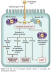
Anatomic pathology
Based on gross, microscopic, chemical, immunologic and molecular examination of organs, tissues and cells.
clinical Pathology
Based on the laboratory analysis of tissue and fluid (blood, urine, body cavities).
Four aspects of disease
–Etiology (cause)
–Pathogenesis (mechanism)
–Morphological changes (structural)
–Clinical-pathological effects (clinical manifestations)
Disease Etiology
Genetic
- Inherited mutations
- Disease-associated gene variants
Acquired
- Infectious
- Nutritional
- Chemical
- Physical
DISEASE
A cluster of signs, symptoms and laboratory findings linked by a common patho-physiologic sequence.
underlying pathology
Most epidemiology is about disease.
ILLNESS
The subjective state of the individual who feels aware of not being well.
The ill individual may or may not be suffering from disease.
social/culteral conceptions of condition
SICKNESS
The social role assumed by an individual suffering from an illness.
what pt birngs to doc
natural history of disease
progression of a disease process in an individual, over time, in the absence of treatment
course a disease takes in individual people from its pathological onset (“inception”) until its eventual resolution through complete recovery or death

CELLULAR HOMEOSTASIS
The steady state in which cells normally exist
The equilibrium of the cells within their own environment (Adequate function preserved)
An increase, decrease, or change in stress on an organ can result in growth adaptations
When disturbed, there is a predisposal for the onset of pathology (Function may be lost)
cellular adaptations: REVERSIBLE
increased demand, trophic stim (horm, GF)
- Hyperplasia: Increase in number of cells
- Hypertrophy: increase in size of cell and then organ
Decreased nutrients, stimulation
- Atrophy: decrease in size, but no loss of function
- Hypoplasia: decrease in the number of cells
chronic irritation:
- Metaplasia: One adult cell type is replaced by another
Dysplasia:
- Abnormal growth with
- loss of cellular orientation/shape/size
- NOT truly adaptive
CELLULAR ADAPTATIONS: irreversible
anaplasia:
- loss in structural differentation & fx
- “primitive”
- may see “giant cells”
neoplasia:
- uncontrolled/excessive/irreversible prolif of cells
- abnorm fx –> death
desmoplasia:
- fibrous tissue formation in response to neoplasm
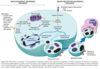
Neoplastic progression (4)
- Hyperplasia/dysplasia
- Carcinoma/preinvasion
- neoplastic cells have not yet invaded basement membrane
- Large nuclei with high amount of chromatin
- Invasive carcinoma
* Basement membrane is invaded via collegenases and hydrolases (metaloproteases) - Metastisis-
* “seed and soil”: carcinomic embolus spreads and invades another organ
HYPERTROPHY
increase cell size –> increased organ size
- Greater synthesis of structural components
- In cells with limited capacity to multiply
causes:
- increased fx demand
- GF stim
- horm stim
types:
- physio: increased demand/stimuli
- increased work load –> increased M fiber size
- patho: chronic hemodynamic overload
- hypertension/valve deficiency –> injury/death (MI)
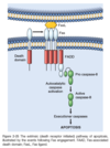
MECHANISMS OF HYPERTROPHY
cause:
- mechanical: increased work/stretch
- GF (trophic): TGF-β, IGF-1, FGF
- vasoactive agents: Noradrenaline, dopamine, adrenaline
signal txduction pathways –> increase protein synth
Ts fac: GATA4, NFAT, MEF2
triggers:
- induction emb/fetal genes: cardiac alpha-actin, ANF
- more E saving contraction: α isoform of myosin heavy chain is replaced by β isoform
- synth contractile proteins
- production of GF
result:
- increase mechanical performance
- decrease work load
“Point of no return” vs lesion
Limits of Hypertrophy
- vasc
- biosynth machineary
- cell injury on persistence of stress
HYPERPLASIA
Cellular proliferation stimulated by growth factors
- important for wound healing
physiological
- horm
- compensatory: residual tissue grouth after removal/partial loss of organ
patho
- excess horm stim/GF
- chronic irritation
- stim antibodies
- viral infection
ENDOMETRIAL HYPERPLASIA
STIMULUS FROM THE HYPOPHYSE HORMONES AND ESTROGEN FROM THE OVARIES
“BPH”
STIMULATED BY ANDROGENS
reversible when no mutations and initial stimulus is removed
assosciated with aging

breast development
physio hyperplasia
prolif gladular epithelium: puberty/preg

compensatory hyperplasia
INDIVIDUALS WHO DONATE A A PART OF THE LIVER HAVE IT RESTORED TO ITS ORIGINAL SIZE
patho hyperplasia: chronic irritation
Bronchial mucous gland hyperplasia in smokers & asthmatics
Thickening of skin following constant scratching
Regenerative nodules in cirrhotic liver due to alcohol
Graves disease
thyroid enlargement due to thyroid stimulating auto-antibodies
mimics TSH
goiter: visibly enlarged thyroid