Exam 1 Spring Flashcards

(380 cards)

1
Q

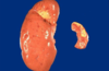
what is anatomic pathology?

A
  1. gross
  2. microscopic
  3. chem
  4. immuno
  5. molecular
2
Q

what is clinical pathology

A

lab analysis of tissue and fluid

3
Q

four aspects of disease

A

etiology (cause)

pathogenesis (mechanism)

morph changes (structural)

clinical-path (calinical manifestations)

4
Q

disease etiology: genetic

A

mutations

disease associated gene varients

5
Q

disease etiology acquired:

A

infectious

nutritional

chemical

physical

6
Q

definition of disease

A

cluster of signs, symps, lab findings linked by common pathophysio seq

7
Q

most of epidemiology is about…

A

disease

8
Q

definition of illness

A

subjective state of individual

ill may or may not be suffering from disease

9
Q

sickness

A

social role assumed by indiviual suffering from disease

10
Q

what the patient brings to the doctor

a person’s subjective experience is called…

A

illness

11
Q

natural hx of disease….

A

progression over tine in absense of tx –> recovery or death

12
Q

nautral course of disease chart

A
13
Q

An increase, decrease, or change in stress on an organ can result in ….

A

growth adaptations

if disturbed –> pathology

14
Q

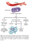
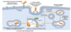
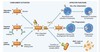
cellular adaptations to stress

A

reversible: number, size, phenotype, metab activity, fx

physio - normal stim

patho - modulate to escape injury

15
Q

when there is increased demand or trophic stimutation on a cell, it resultsi n…

A

hypertrophy and/or hyperplasia

16
Q

a decrease in nutrients and stimulation can result in…

A

hypoplasia

atrophy

17
Q

chronic irritation can result in…

A

metaplasia

18
Q

hypertrophy definition

A

increase cell size –> increased organ size

greater synth structural cmpts

in cells with limited cap to multiply

19
Q

causes of hypertrophy

A

increased fx demand

GF/horm stim

20
Q

physiological hypertrophic example

A

increased work load –> increase M fiber size

21
Q

pathological hypertrophic example

A

chronic hemodynamic overload –> htn, valve deficiency –> injury/death (MI)

22
Q

this is an example of…

A

physiological hypertrophy: increased work load –> increased M fiber size

23
Q

this is an example of?

A

physio hypertrophy: estrogen stim –> uterus growth

24
Q

chronic hemodynamic overload results in…

A

card enlargement (L vent hypertrophy as a result of htn - increase in afterload)

25
mechanisms of hypertrophy
mechanical: increase work load/fx demand, stretch * GF: (trophic) * TGF-beta, IGF-1, FGF * vasoacitve agents * NE, dopa, adrenaline
26
Ts factors for hypertrophy
GATA4 NFAT MEF2
27
during M hypertrophy, what happens to the contractile fibers?
switch from adult (alpha myosin heavy chain) --\> fetal (beta myosin heavy chain) produce more E saving contraction
28
limits of hypertrophy
when enlargement of M cna no longer compensate for increased burden --\> degen changes in myocard fibers --\> frag, loss of myofibr contactile elem **adaptation to stress can progress to fx sig cell injury if stress no relieved**
29
hyperplasia definition
increase # cells in cells cap of replication * imp with wound healing **often with hypertrophy in response to same stimuli**
30
causes of hyperplasia
cellular proliferation stim by GF physio: * horm * compensatory: residual tissue grows after removal/partial loss pathologic: * excess horm stim on GFs (chronic irriation, stim antibodies, virus)
31
examples of physiologic hyperplasia
horm: prolif gland epith of female breast during pub and preg compensatory: liver regen (as early as 12 hours later) * stim: polypeptide GFs from uninjred hepatocytes and nonparenchymal cells
32
pathological examples of hyperplasia
horm excess: endometrium * common cause of abnorm mentrual bleeding * stim by pit horms and ovarian estrogen and inhib by progestrone * usu due to inbalance between estrogen and progestrone GF excess: BPH * angrogens - canbe reversible if no mutations and initial stim removed
33
this is an example of
horm hyperplasia - breast @ pub and preg
34
this is an example of
35
this is an example of
patho hyperplasia: BPH - GF excess (assoc with aging)
36
this is an example of
patho hyperplasia - chronic irritation: thickening of skin after constant scratching
37
this is an example of
patho hyperplasia: regen nodules in cirrhotic liver due to etoh
38
smoking and asthmatic effects on the bronchus is an example of
patho hyperplasia - chronic irritation: bronchial mucous gland hyperplasia
39
Graves disease
thyroid enlgment (goiter) due to TSH auto-antibodies patho hyperplasia
40
hpv - common wart: virus patho hyperplasia
41
how can you tell if a cell with hypertrophy or hyperplasia?
labile cells (dividing) * stem --\> hyperplasia permanent cells (nondividing) * skel/card M --\> hypertrophy stable cells * hepatocyte, smooth M cells --\> both upon stim
42
wound healing is an example of
physio hyperplasia
43
hyperplasia can lead to...
ca if uncontrolled
44
atrophy definition
shrinkage size of cell by loss of cell substance fx is diminished by not lost adaptive respnose
45
causes of atrophy (6)
1. decreased work load: immob limb after injury 2. loss innervation 3. dim blood supply 4. inadeq nut 5. loss endocrine stim 6. aging
46
physiological atrophy example
involution: * mammary gland after lactation * ovary menopause * uterus after parturition
47
this is an example of
path atrophy: decreased workload --\> prolonged immob of limb
48
this is an example of
path atrophy: loss of innervation ---\> wasting
49
this is a progression that is an example of
path atrophy: loss of endocrine stim -- hypopituitary, menopause
50
this is an example of
path atrophy: dim blood supply: heart and brain in atherosclerosis
51
this is an example of
path atrophy: inadequate nut - marasmus
52
this is an example of
path atrophy: intraluminal P - thick secr in CF = atrophy of panc
53
this is an example of
path atrophy: age associated - brain
54
mechanism of atrophy
decreased protein synth * reduced metab activity increased proten degrad * nut defic & disuse --\> activation of ubiquitin-proteasome pathway (ligases) * respon for accerated proteolysis in catabolic conditions: cachexia assoc with ca **stem cells intact**
55
in mahy situations, atrophy is also accompanied by...
increased autophagy --\> increase in number of autophagic vacuoles starved cells eats own cmpts in attempt to survive
56
agenesis
compelte lack of formation of organ
57
aplasia
reduced cell mass but remnant of organ exists
58
hypoplasia
incomplete growth of organ failure of organ to reach normal size
59
definition of metaplasia
one adult cell type replaced by another --\> better able to withstand adverse enviroment reversible if irritant removed predisposition to malignancy
60
causes of metaplasia
often secondary to irritation and/or environmental exposure reprog of stem cells
61
this is an example of
epith metaplasia: chronic smokers - columnar to squamous of bronchial mucosal lining
62
this is an example of
epith metaplasia: barrett's esophagus - sqamous --\> columnar distal end of esophagus
63
this is an example of
mesenchymal metaplasia: new bone formation in injured M/tend/cart
64
this is an example of
mesenchymal metaplasia: myeloid - extramedually hematopoiesis
65
mechanisms of metaplasia
reprog stem cells
66
double edged sword of epithelial metaplasia
smoking: meta to strat-sq keeps cells alive but loses protective mechanisms (mucus secr and ciliary clearance) if persistant --\> ca * squa meta of respiratory often coexists with lung ca of malignant squa cells
67
what vitamin is essential for normal epith differentiation? what is the effect if it is deficient?
vit A may induce squa meta in respiratory epith
68
dysplasia definition
disorded growth/maturation (loss of size, shape, orientation) usually referred to as the prolif of pre-ca cells * accomp by hyperplasia and metaplasia (atyp hyperplasia) **reversible in theory**
69
causes of dysplasia
chronic irritation/inflammation accum of genetic mut: cells turn irresponsible to reg sigs
70
how to differentiation dysplasia from neoplasia?
no invasion of BL
71
cervical intraepithelial neoplasia is an example of
dysplasia --\> precursor to cerv ca
72
risk factors of dysplasia (6)
infection: HPV 16, 18 chemicals: smoking UV light chronic irritation: 3rd degree burn some hyperplasia (endomet) some metaplasia (barrett's)
73
morphology of dysplasia
nuclear: enlarged and hyperchromasic (more basophillic) growth: increase mitosis, disordered prolife poor matur abnorm architecure/arragement
74
diagram comparing types of reversible cell injury
75
mechanisms of accumulation (4)
1. inadeq removal of normal substance 2. accumulation of abnorm endogenous substance 3. failure to degrad metabolite 4. accumulation aborma exogeous substance
76
types of intracellular accumulations (4)
lipid protein glycogen pigments
77
steatosis
fatty change: accum triglyc in parenchymal cells pathogen: abnorm fat metab (reversible) common in: kid, liver, M (skel/card)
78
this is an example of
79
morphology of fatty accumulation
organ enlarges yellow discoloration
80
etiology and steatosis
1. obesity: NADH 2. ETOH 3. protein malnutrition: Kwashiorkor - decreased apolipoprotein to coat VLDL 4. drugs 5. anoxia 6. Reye's syndrome
81
etiology of alchohol of steatosis
accumulation NADH --\> accum DHAP --\> G3P --\> TAG acetyl CoA --\> FA decreased beta-ox of FA
82
common presentations of cholesteral and CE (4)
atherosclerosis: macrophages and smooth M cells in intimal layer bv xanthomas: macrophages in subepith connective tiss/tend cholesterolosis: macrophages in lamina propria of gallbladder niemann-pick: type C: mutation in enz involved in cholesteral trafficking
83
protein accumulation is commonly due to
excess synth of absorption by cells: intermediate filaments
84
morphology of protein accumulation
eoinophillic (hyaline appearance)
85
examples of protein accumulation diseases
1. nephrotic synd: PCT 2. russell bodies - plasma cells 3. alcoholic (mallory) hyaline - liver 4. nuerofibrillary tangles - Alzheier's neurons 5. amyloid - amyloidosis
86
mephrotic syndrome
heavy protein leakage across glom filter - increased protein reabsorption pino vesic fuse with lysosomes --\> pink/hyaline cytoplasmic droplets reversible if proteinuria stops and drops metab
87
russell bodies
Ig in plasma cells - accum in RER rounded, eosin bodies
88
amyloidosis
stained with congo red - apple green birefringence when polarized
89
mallory bodies
alcoholic hyalin - liver * aggregated intermed-fil that resist degrad eosin conden
90
neurofibrillary tangles
Alzhemier's microtub-assoc proteins and neurofil
91
glycogen accumulation
normally stored in cytoplasm deposits = clear vacuoles (best with carmine and PAS stain)
92
glyceogen storage disorder
DM: renal tub epith, islets langerhands, myocardial cells common in liver and skel M
93
anthracosis (coal dust) phago by dust cells coal workers pneumoconiosis
94
this is an example of
tattoo
95
this is an example of
lipofuscin in card myocytes
96
morphology of lipofuscin
perinuc, intralysosomal electron dense granules --\> brown atrophy
97
lipofuscin
yellowish brown wear and tear prigment more with aging and atrophy
98
mechanism of lipofuscin
lipid - protein complex formed by free radical induced peroxidation of lipids in subcell membrane **indicator of free radical injury** common: heart, liver
99
this is an example of
lipofuscin in hepatocytes
100
melanin
brown-black pig prod by melanocytes accum in keratinocytes and macrophages --\> hyperpig
101
melanin accum can be due to...
nevus (birthmark) malignant melanoma increased ACTH * addison's * pituitary adenoma * adrenogenital syndrome
102
hemosiderin
Fe cmpt of heme (golden-brown) * brkdown prod of ferritin (serum) * stored in lyso: **not found in serum**
103
etiology of hemosiderin accum
iron overload stasis dermatitis
104
diseases involving hemosiderin
DVT sideroblastic anemia anemia of chronic disease
105
difference between hemosiderosis and hemochromatosis
siderosis: iron accum in macrophages - **no assoc with dysfx** chromatosis: iron accum in parenchymal cells - may cause dysfx * bronze diabetes * cirrhosis * CHF
106
this is an example of
hemosiderin in liver
107
morphology of hemosiderin accum
golden brown granules - aggregates of ferritin micelles ## Footnote **prussion blue stain**
108
this is an example of
Bilirubin accumulation can affect basal ganglia in kernicterus
109
pathological calcification
abnorm deposits of calcium phosphate dystrophic * dead/dying tissues - absense of Ca2+ metab * serum Ca2+ = norm metastatic * normal tissues - abnorm in Ca2+ metab * serum Ca2+ = high
110
pathogenic steps of dystrophic calcification
init by mito of dying cells memb bound vesicles from dying cells --\> extracell 1. Ca2+ concentrated due to affinity for memb ppls 2. phosphate accum due to phophatase
111
dystrophic calcification is an indicator of
tissue injury --\> may cause organ dysfx
112
examples of dystrophic calcification (5)
1. atherosclerotic plaques 2. dmg heart valves 3. chor panc 4. **psammoma bodies**: meningioma & pap ca of thyroid 5. periventricular calcification in cytomegalovirus infection
113
this is an example of
Psammoma bodies pap ca of thyroid
114
this is an example of
Calcified tricuspid valve
115
diseases associated with metastatic calcification
hypercalcemia * hyperparathyroid * destruction of bone * vit-d intox * sarcoidosis * renal fauilre --\> secondary hyperparathyroidism hyperphosphatemia
116
where are Ca2+ deposits found in metastatic calcification?
bv, kid, lungs, gastric mucosa extensive --\> nephrocalcinosis, respiratory insuff
117
morphology of calcification
gross - fine, white granules/clumps microscopic - intra and extracell basophillic deposits
118
this is an example of
•Gross – Fine white granules or clumps Ca2+ accum
119
this is an example of
•Microscopic – intra or extracellular basophilic deposits Ca2+ accumulation
120
reversible cell injury
reduced ox-phos and cell swelling alt in org-lls
121
necrosis
dmg cell membranes loss of ion homeostasis * lyso enz dig cell --\> leak through PM --\> inflam
122
what pathways of injuries result in neccrosis?
ischemia toxins infections trauma
123
apoptosis
cell suicide - DNA/proteins dmg beyond repair * nuc dissoln --\> frag --\> rapid removal cell debris no cell content leakage --\> no inflam
124
necrosis v apoptosis: cell size
n = enlarged (swell) a = reduced (shrink)
125
necrosis and apop: nucleus changes
n: pyknosis --\> karyorrhexis --\> karyolysis a = frag into nucleosome-size frag (pyknosis can also occur)
126
morph alt in cell injury chart: duration of injury and effect
127
morph alt in reversible injury
gen swellilng PM bleb ribosome detachment clumb nuc chromatin
128
morph alt in cell injury is associated with... (5)
1. decreased ATP 2. loss cell memb integrity 3. defects in protein synth 4. cytoskel dmg 5. dna dmg
129
necrosis is usually due to
mito dmg depletion ATP rupture lysos and PM
130
reversible injury characteristics
cell swell * cannot maint ion/fluid homeostasis: failure of E-dep ion pumps in PM fatty change * lipid vacoules in cytop * in cells dep on fat metab: liver, heart
131
ultrastructural changes of reversible cell injury
1. PM alt: bleb, blunt, loss of microvilla 2. mito changes: swell, small amorphous densities 3. dilations of ER: ribo detach 4. nuc alt: disaggregation of granular and fibrillar elem
132
necrosis
denat intracell proteins enz dig of cell
133
earliest histological evidence of myocard necrosis does not become apparent until...
4-12 hours later
134
morph features of necrosis
increased eosinophillia myelin figures (large whorled ppl masses that replace dead cells) nuc: pyknosis --\> aryohexis --\> karyolysis
135
this is an example of
necrosis: kidneys a: normal b: reversible ischemic injury * surf blebs * **increased eosinophillia** (loss of RNA and denaturaed cytop prot) c: necrosis
136
this is showing a progression to
necrosis notice blebbing and loss of microvilli in **b**
137
coag necrosis
architecture preserved (in the beginning) - firm texture bv obstruction --\> ischemia * local area = **infact**
138
kidney infarct notice how tubues are still look about the same (architexture preserved) but seem to be "filled" (coag) localized = infarct
139
liquefactive necrosis
digestion of dead cells --\> liquid mass (creamy yellow **pus**) * focal bac/fungal infections via stim of accum leukocytes and freeing of enz from them * dead leukocytes make the creamy yellow pus usually involving CNS
140
liq necrosis in brain
141
gangrenous necrosis
usually limb (lower) lost blood supply --\> coag necrosis involving multiple tissue plane --\> addn of bacteria (liq necrosis) --\> wet gangrene
142
caseous necrosis
cheese-like: friable white appearance * structureless collection of frag/lysed cells and amorphous grandular debris enclosed in inflam border usually due to granulomas and TB
143
caseous necrosis: TB of lung
144
fat necrosis
focal areas of fat destruction: usually due to acute panc * release of activated panc lipase into panc and peritoneal cav --\> liq memb of fat cells --\> FA combine ti Ca2+ --\> chalky-white areas (fat saponification)
145
fat necrosis notice white charky deposits: calium soap formation (saponification)
146
fibrinoid necrosis
immune rxns involving bv: antigen-antibody complexes deposit in A walls * complexes + leaked fibrin --\> bright pink, amorphous "fibrinoid" in H&E stains
147
fibrinoid necrosis: A
148
A 33-year-old woman has increasing lethargy and decreased urine output for the past week. Laboratory studies show serum creatinine level of 4.3 mg/dL and urea nitrogen level of 40 mg/dL. A renal biopsy is performed and the specimen is examined using electron microscopy. Which of the following morphologic changes most likely suggests a diagnosis of acute tubular necrosis? A.Mitochondrial swelling B.Plasma membrane blebs C.Chromatin clumping D.Nuclear fragmentation E.Ribosomal disaggregation
Nuclear fragmentation morph changes: * increased eosinophilia * myelin figures * nuc frag
149
A 63-year-old man has a 2-year history of worsening congestive heart failure. An echocardiogram shows mitral stenosis with left atrial dilation. A thrombus is present in the left atrium. One month later, he experiences left flank pain and notes hematuria. Laboratory testing shows elevated serum AST. Which of the following patterns of tissue injury is most likely to be present? A.Liquefactive necrosis B.Caseous necrosis C.Coagulative necrosis D.Fat necrosis E.Gangrenous necrosis
Coagulative necrosis
150
cell responses to injry stimuli depend on
nature of injury duration severity
151
consequences of cell injury dep on injured cell's
type state adaptability
152
what is the fundamental cause of necrotic cell death?
depletion of ATP
153
major causes of ATP depletion
reduced supply O2 and nutrients mito dmg toxin
154
flow chart of bv ischemia
155
mito can be dmged by
increases in cyto Ca2+ ROS O2 deprivation
156
consequences of mito dmg
mito permeability ROS leak proteins to cytosol --\> apoptosis
157
effects of ATP @ 5-10% normal lvls (5 steps and result)
1. reduced act of PM E-dep Na/K ATPase --\> net gain of solute --\> cell swelling 2. change in cell metab --\> decrease in cell ATP, increase AMP --\> increase rate anaerobic glycolysis --\> rapid depletion glycogen stores --\> accumulation lactic acid --\> decreased intracell pH --\> decr activity of many cell enz 3. failure Ca2+ pump 4. ribo detach from RER --\> recued protein synth 5. UPR (unfolded protein response) --\> irreversible dmg to mito and lyso memb --\> necrosis
158
conseq of calcium influx into cell (3)
1. accum in mito: opens up permability txsition pore --\> no ATP gen 2. incrs in cytosolic Ca2+ activated enz * pplases * proteases * endonucleases * ATPases 3. induction apoptosis: direct activation of caspases & incr mito perm
159
most intracellularCa2+ is sequestered...
in mito and ER
160
O2` (radial): production, inactivation, patho effects
prod: ox-phos, phago oxidase in leukocytes inactiv: SOD --\> H2o2 + O2 path effects: * stim prod degrad enz * may directly dmg lipids, proteins, DNA * **acts close to site of production**
161
H2o2: production, inactivation, path effects
prod: SOD, oxidases in peroxisomes inactiv: catalase (perox) & glut-peroxidase (cytosol/mito) --\> h20 + o2 path effects: * conversion to `OH and OCl- (respiratory burst) * **​can act distant from site of production**
162
'OH (radial): prod, inactiv, path effects
prod: * H20 hydrolysis * H2o2 ---(Fenton rxn)---\> * from O2` (radical) inactiv: glut-peroxidase --\> H2o path effects: * **most reactive radical!!** * principle ROS for dmging lipids, proteins, DNA
163
ONOO-: prod, inactiv, path effects
prod: O2` + NO inactiv: peroxiredoxins (cyto/mito) --\> HNO2 path effects: dmg lipids, proteins, DNA
164
free radical generation (6)
1. redox during metabolism 2. ionizing radiation 3. inflammation: NADPH oxidase, intracell oxidases 4. metab of exogenous chem/drugs 5. transition metals: can donate/accept free e- during rxns * Fenton: H2O2 + Fe2+ → Fe3+ + ˙OH + OH− 6. NO
165
free radical removal
1. antiox 2. free iron and copper * normally, these metal rxns are min by binding to storage and txp proteins --\> prev participation in ROS gen rxns 3. ROS enz: catalase, SOD, glut-perox
166
what is a consistent feature of most forms of cell injury? what is the exception?
early loss of selective memb permab --\> overt memb dmg exception = apoptosis
167
mech of membrane dmg
1. ROS - lipid peroxidation 2. decreased ppl synth - defective mito fx/hypoxia 3. increase ppl brkdown - due to activ calcium-dep pplases by increased intracel Ca2+ --\> unesterified free FA, acyl carnitine, lyso-PC 4. cytoskel abnorm - due to activ of proteases by increased intra cel Ca2+
168
consequences of memb dmg
mito: * decrease ATP gen * release proteins --\> apoptosis plasma: * loss osmotic balance: influx fluids/ions, leakage metabolites lysosome: * enz leakage
169
dmg to DNA and proteins that lead to apoptosis
DNA: * exposure to DNA dmg drugs * radiation * ROS proteins: * bad folding: mutations, free rad
170
tissue-specific cellular injury detection ## Footnote cardiac M: liver: hepatocytes:
cardiac M: isoform creatine kinase, troponin liver: isoform alkaline phosphatase hepatocyte: transaminases
171
what is the most common type of cell injury in clinical medicine?
ischemia
172
what usually leads to ischemia? what if it persists?
from hypoxia induced by reduced bloow flow (mech A obstruc) persistant --\> irreversible injury and necrosis
173
compare and contrast hypoxia and ischemia
hypoxia: E prod by anaerotic glycolysis can con't ischemia: compromises delivery of substates of glycolysis * aero E prod compromised **AND** anaero E gen stops after glycolytic stores are drained or inhibit by accumulation of metabolites usually washed out by flowing blood * **why ischemia causes more RAPID AND SEVERE cell and tissue injury than hypoxia**
174
irreversible injury is associated morphologically with... (3)
1. severe swell of mito (large, amophous densities dev in matrix) 2. extensive dmg to PM --\> myelin figures 3. swell of lyso
175
how can reperfusion injury ischemic cells?
restoring blood flow can promote recovery if REVERSIBLY injury **but** can also paradoxically exacerbate injury --\> cell death
176
why is ischemia-reperfusion injury clinically important?
contributes to tiss dmg during MI and cerebral infarctions following therapies to restore blood flow
177
how odes reperfusion injury occur?
new dmging process set in motion --\> cccause cell death that might have recovered otherwise 1. oxidative stress: compromised antiox def mech --\> accumulation free rad 2. intracell calcium overload 3. inflammation: hypoxic cells recruit neutrophils 4. complement sys activation: IgM antibodies deposit in ischemic tissues --\> provide attachment for complement proteins --\> injury & inflammation
178
what organ is afreqent target of drug toxicity?
liver
179
chemiclas can induce cell injury of 2 general mechanisms:
direct * combining with cirtical molecular cmpts * Mercuric chloride * cyanide * antineoplastic chemterapeutic agents * antibiotics conversion to toxic metabolites * usually mod by cyt P450 in smooth ER * cause memb dmg & cell injury via free radicals & lipid peroxidation * CCl4, acetaminophen
180
A 55-year-old man complains of a sudden onset of chest pain 3 hours ago. Laboratory studies show an increased cardiac troponin level. He is given tissue plasminogen activator (t-PA). His troponin level following the therapy is increased further. Which of the following best explains the increase in the troponin level? A)Increased generation of reactive oxygen species B)Glycogen depletion C)Loss of Na+ and water D)Increased oxidase activity E)Calcium influx
Increased generation of reactive oxygen species
181
apoptosis
tightly regulated suicide program --\> apoptotic bodies" (cytop and nucleus parts) --\> tasty morsels for marcophages **no inflamm rxn** (how it differs from necrosis)
182
apoptosis in physio situation
1. embryogensis 2. involution of horm-dep tissues upon horm withdrawl * endometrium, lactating breast, prostate after castration 3. cell loss in prolif cell pops * maintain homeostasis 4. elim self-reactive lymphocytes 5. death of host cells that has served their purpose * neutrophil, lymphocyte
183
apoptosis in patho conditions
1. dna dmg 2. accumulation misfolded proteins: * "ER stress" 3. infections 4. atrophy in parenchymal organs after duct obstruction * panc, parotid, kidney
184
morph and biochem changes in apoptosis (4)
cell shrinkage chromatin condensation cytop blebs/apop bodies phago via macrophages
185
this is showing
apoptosis notice nuclei with peripheral **cresents of compacted chromatin** other nnuc with uniformly dense or fragmented chromatin
186
this is showing
apoptosis: **bright eosinophillic cytoplasm** & **condensed nucleus**
187
this is showing
L: blebbing and formation of apoptotic bodies M: nuc frag R: caspase-3 activation
188
apoptosis results from activation of what enz?
caspases: cysteine proteases that cleave proteins after aspartic residues exist as inactive proenz: cleavage = activation = marker for cells undergoing apoptosis 2 pathways: intrinsic (mito) nad extrinsic (death-receptor-init)
189
intrinsic path of apop
mito: maj mech - increased permeability of mito outer memb --\> release death-inducing (pro-apoptotic) molecules tightly controlled by **BCL2** protein fam * antag anti-apop proteins --\> activate pro-apop prot BAX/BAK --\> form channels in mito --\> leak cyc c --\> caspase activation --\> apoptosis
190
BCL2 family of proteins
* Anti-apoptotic: BCL2, BCL-XL, MCL1 * Pro-apoptotic: BAX and BAK * Sensors: BAD, BIM, BID, Puma, Noxa
191
extrinsic path of apoptosis
init by engagement of PM death receptors: TNF receptor fam with death domain --\> deliver apop sig
192
extrinsic apop path can be inhib by
FLIP (protein) --\> binds pro-caspase-8 --\> does not allow cleavage use to protect themselves from Fas-med apoptosis
193
excution phase of apoptosis
mito: caspase-9, death-receptor: caspase-8 & -10 after initiator caspase cleaved --\> executioner caspases * activate DNase * degrade matrix * frag nuc
194
removal of dead cells by apoptosis
apop bodies easily phago-ed * phosphatidylserine: "flips" to outside of PM * soluble fac * coated with thrombospondin: glycoprotein * natural antibodies: C1q - recog by phagos
195
examples of apoptosis
GF depreviation: * decreased synth BCL2/BCL-XL --\> activate BIM and other pro-apop fact DNA dmg: * involves tumor-suppressor gene TP53 --\> p53 accumulation (protein) --\> BAX/BAK protein misfolding * unfolded protein response overwhelmed --\> "ER stress" --\> activate caspases --\> apop
196
clin-patho correlations: apop induced by TNF receptor family....
FasL on T cells bind FAS on same/neighboring T lympho --\> autoimmune dis
197
clin-patho correlations: cytotoxic T lymphocyte-med apop
CTLS --\> perforin --\> entry of granzymes --\> cleave proteins with asp residues --\> activate caspases
198
disorders associated with dysreg apoptosis
defective --\> increased cell survival * autoimmune increased --\> excessive cell death * neurodegen * ischemic injury * virus
199
necroptosis
hybrid of necrosis (morph) and apoptosis (mech) * invovles RIP1 & 3 nec: loss ATP --\> cell/organelle swell --\> ROS --\> lyso enz --\> PM rupture apop: tirgg by prog signal transduction * **does NOT invovle cacspase activation**
200
necroptosis happens in (6)
1. formation of bone growth plate 2. steatohepatitis 3. acute panc 4. reperfusion injury 5. Parkinsons 6. cytomegalovirus: backup mechanism in host def aga viruses that encode caspase inhibitors
201
pyroptosis
release fever inducing cytokine IL-1 (sim to apoptosis) microbe enters --\> recog via innate immune receptors --\> "inflammasome" (complex) --\> caspase-1 (IL-1-beta converting enz) --\> cleave and activate IL-1 --\> inflam response
202
types of autophagy
chaperone-med microautophage: inward invag of lysosome memb for delivery macroautophagy: major form --\> seques and txp protions of memb-bound autophage vacuole (autophagosome)
203
steps of autophagy
1. form phagophore: isolation memb derived from ER 2. elong of vesicle 3. maturation autophagosome 4. fusion with lysosome 5. degrad of contents
204
dysreg of autophagy in... (3)
ca neurodegen disorders inflam bowel diseases
205
this is showing
fatty liver: accumulation of lipids
206
flow chart of cellular aging
207
this is showing
kidney: protein accumulation * heavy protein leakage across glom filer --\> increased pinocyto reabsorption of proteins --\> pink hyaline dropets in tub * reversible!
208
this is showing
lipofuscin - card myocytes EM shows perinuc, intralyso location of accumulation
209
this is showing
Dystrophic calcification - aortic valve
210
inflammation definition
protective response of vasc tissue --\> brings def cells/molecules --\> elim init cause and necrotic cells/tissues --\> init repair
211
causes of inflammation (5)
1. infections 2. tissue necrosis 3. foreign bodies 4. endgenous substances: gout, atherosclerosis, obesity-relation metab syndrome 5. imune rxns: autoimmune dis, allergies
212
typical inflam rxn: **"five R's"**
recog recruit - leukocytes and plamsa proteins removal regulation resolution/repair
213
inflammation is initated by...
sentinal/resident cells of **innate** immune sys PRRs (pattern recog recep) recog PAMPs/DAMPs (patho/dmg-assoc mol patt) --\> inflamm!
214
examples of PRRs
TLR: recog products of bac (endotox, bac DNA) and virus (dbl stranded RNA) inflammasome: cytop complex recog prod of dead cells (extracel ATP) --\> IL-1 (inflam)
215
major participants in inflam rxn are...
bv: * dilate & dev incr permeability leukocytes: * recruited --\> ingest/destroy
216
inflamm is ordinarily term when...
offending agent elim * mediators broken down & dissipated * leukocyte = short life spans in tissues * anti-inflam mech activated: lipoxins, TGFβ, IL-10
217
mediators' roles in inflammation
initate and amplify inflamm response detm pattern ,severity, clinical, and patho manifestations
218
compare and contract acute and chronic inflammation ## Footnote onset cellular infiltrate tissue injury/fibrosis local/systemic signs
219
inflammation is usually a _____ tissue response... but....
local (may have limited systemic effects like fever (bac), pharyngitis (virus)) SIRS: systemic inflam response synd - ex = sepsis (desseminated bac infection)
220
apop cells frag nuclei with condense chromatin shrunked cell bodies with pieces "falling off"
221
this is showing
hemosiderin in liver cells * a = H&E: golden-brown, fine * b = **prussian blue**
222
harmful consequences of inflammation
TB: protection injures "bystander" tissues autoimmune/allergy: misdirected atherosclerosis, DM2, Alz, ca: chronic inflam --\> chronic disease not ordinarily thought of as inflam
223
5 cardinal manifestations of inflammation
redness (rubor) - dilation small bv & incr blood flow heat (calor) - dilation small bv & incr blood flow swell (tumor) - accum fl/inflamm cells in EXTRAVASC space pain (dolor) - stretch by swelling --\> pain-inducing inflamm mediators loss of fx - swell/pain inhib mvmt
224
vascular changes in acute inflamm
caliber and flow * **transient vasoconstrict** (few sec) --\> vasodil * edema --\> increased concentration RBC --\> incr visc --\> slow blood flow --\> margination of leukocytes (displacement to periphery) --\> sets up for emigration increased permability * exudate: inflam edema
225
exudate versus transudate
exudate: inflammation - * vasodil & statis, increased interendothelian spaces * fl = **HIGH** protein, cell debris and sp-grav transudate: non-inflam - * increased hydrostatic P and decreased colloid osmotic P * fl = **LOW** protein, cell debris, sp-grav
226
227
pus/purulent exudate
exudate with lots of leukocytes (mostly neutrophils), dead cell debris (often microbves)
228
mechanisms of increased vasc permeability
contraction endothelial cells --\> increased interendo spaces endothelian injury/necrosis/detachment transcytosis - incr txp fluids/proteins leakage from immature new bv (angiogenesis)
229
response of lymph sys to inflammation
increased lymph flow --\> drain lueko, debris, microbes --\> possible secondary inflamm --\> lymphangitis and lymphadenitis
230
leukocyte recruitment in inflammation overview
1. margination and rolling 2. stable adhesion 3. transmigration 4. migration to chemotactic stim
231
margination
statis (slow blood due to increased RBCs = incr visc) --\> leukocyte displace to periphery
232
rolling
endothelium activated by cytokines express selectin --\> bind complemntary ligants on leukocytes --\> transiently binding and detachment
233
stable adhesion of leukocytes to endothelium
med by leuko integrins interacting with endothelium ligants (ICAM-1 & VCAM-1) * affinity increased by **cytokines**
234
transmigration
"diapedesis" driven by chemokines prod by extravasc tiss PECAM-1 (CD31) * adhesion mol expr on leuko and endothe --\> homotypic (like-like) binding --\> squeeze through --\> leuko secr collagenases --\> pass through BM
235
chemotaxis of leukocytes during inflammation
chem concentration gradient * bac products * chemokines * complement (C5b) * LT B4 pseudopods --\> anchor to ECM --\> cytoskel pulling to higher density of chemokines
236
this is showing
chemotaxis of leukocytes: pseudopods
237
type of emigrating leukocytes varies with..
age of inflam response * acute inflamm: neutrophils (6-24hrs) --\> macrophages (24-48hrs) * neutro apop @ 24 hrs * macrophages surv for months type of stim * virus: lymphocyte = first * hypersens rxns: eosinophils = main
238
this is showing a progression of...
neutrophils --\> macrophages/monocytes notice multilobed cells on L and larger cells on R
239
leukocyte activation by inflam stimuli enhances.... (4)
1. phago 2. intracell destruction: microbes, dead cells 3. liberate substances that destroy extracel microbes, dead cells 4. prod mediators --\> amp/lim inflam response
240
phagocytosis steps
1. recog & attachment of particle 2. engulf via phago vacuole 3. kill/degrade
241
H₂0₂-MPO-Halide system
phago --\> ox burst: NADPH oxidase --\> super oxide and H2o2 MPO (lyso myeloperoxidase): H2o2 + Cl- ---\> HOCl (bacteriocidal - active ingred in bleach) addnally: phago produce N-derived free rad: NO --\> peroxynitrite
242
o2-independent microbe killing
lyso granules: * lysozyme: bac coat oligoscc * major basic protein: eosin-granule - cytotoxic for parasites * defensins: poke holes in microbe memb
243
secretion of microbicidal substances
leukocytes: * elastase: digest dead tissues neutrophils * NETs (neutrophil extracel traps) :framework of nuclear chromatin with antimicrob peptides and enz * possible souruce of nuclear antigens in autoimmune dis like lupus
244
this is showing
NETs
245
True or False: mechanisms in which leukocytes dmg normal tissues = same as mechanisms involved in antimicrobial defense?
TRUE ## Footnote once leukocytes activated, effector mechaisms do not distinguish b/w offender nad host
246
T lymophocytes produce _____ that also contribute to _______ by....
T lymphocytes that produce **IL-17 (Th17 cells), cells of adaptive immunity,** also contribute to **acute inflammation** by **helping recruit neutrophils and monocytes**
247
principal mediators of inflammation
cell derived * vasoacitve amines * cytokines * arachidonic acid metabolites: PG, LT * platelet-activating factor plasma protein derived * complement * kinins
248
**most** important mediators of acute imflammation are... (4)
1. vasoactive amines 2. arach-acid prod: PG, LT 3. cytokines (& chemokines) 4. products of complement activation
249
histamine
richest source = mast cells * also in basophils and platelets dilate arterioles and increase permability of venules * bind to H1 receptors
250
what kind of drugs are used to tx allergies?
anti**histamine**: H1 receptor antagonist
251
mast cells release granules due to what types of stimuli
physical injury IgE in allergies complement (C3a, C5a)
252
serotonin
vasoactive amine mediator in platelet granules --\> platelet aggregation
253
inflammation and AA (arach-acid)
AA = 20 C PUFA (usually in memb ppls) * **metabolies affect virtually every step of inflammation!** inflam --\> PPL As frees AA from memb ppls * cyclooxygenases: PG, TX * lipoxygenases: HETES, LT, lipoxins
254
big chart of pathway of AA after inflammation activation
255
platelet activating factor
ppl-derived mediator aggrepate platelets & inflamm effects
256
cytokines and chemokines
proteins --\> mediate and regulate immune and inflam rxns chemokines: chemoattractant cytokines that act on **specific** leukocytes
257
cytokines important of acute inflammation
TNF: stim expr endothelian adhesion mol, secr other cytokines, systemic IL-1: sim to TNF but greater role in fever IL-6: systemic (acute phase response) chemokines: recruit leuko --\> inflammation, migration IL-17: recruit neutrophils and monocytes
258
cytokines important in chronic inflammation
IL-12: increased prod IFN-gamma IFN-gamma: activ macrophages IL-17: recruit neutrophils and monocytes
259
circulating proteins of what interrelated systems comtribute to inflamm rxn?
complement kinin coag fibrinolytic
260
complement sys
plasma proteins that circ as inactive precursers C1-C9: seq activated in amp cacade * **critical = C3** * classical: C1 attach to IgM/IgG-antigen complex * alt: microbial surf molecules * lectin: plasma mannose-binding lectin binds carbs on microbes --\> activate C1
261
all 3 complement activation pathways lead to...
formation of C3 convertase: C3 --\> C3a & C3b * C3a released * C3b conval binds complement activated cell/molecule * more binding forms C5 convertase * C5b binds late cmpts --\> forms MAC (multiple C9 molecules)
262
major biologic activities of complement
inflammation * C3a, C5a: anaphylatoxins - stim release of histamine from mast cells * C5a = chemoattractant opsonization * C3b fixed to microbe wall = opsonin --\> prom phago cell lysis * MAC: pore-forming memb attack complex
263
kinins
kininogen ---(kallikrein)---\> bradykinin * dilate bv * incr vasc permea * contract smooth M * pain
264
what initiates the four plasma protein sys that may contribute to inflam response?
activated Hageman factor (Factor XIIa)
265
this is showing
normal lung vascular congestion and statis leukocyte infiltrate
266
this is showing...
acute inflammation
267
Serous inflammation epidermis is separate from dermis by blister (empty space)
268
this is showing
Serous inflammation notice empty bubble b/w epidermis and dermis
269
morphology of this kind of inflammation?
Serous inflammation watery, protein-poor fluid = effusion * plasma * secretion of mesothelial cavity lining cells
270
this is
Fibrinous inflammation
271
this type of inflammation occurs when
fibrinous exudate: dev when vascular leaks are large or local procoag stimulus fibrin = eosin meshwork and threads
272
this is showing
Purulent inflammation abscess: local - typically central LARGE necrotic region rimmed by neutrophils * over time, can be walled off and replaced with CT
273
this is showing
Lung abscesses: purulent inflamm
274
this is showing:
Purulent inflammation: Pneumonia
275
this is showing
ulcer: occur when tissue/inflam **on/near a surface** notice HOLE * cell necrosis and sloughing of necrotic and inflam tissue
276
outcomes of acute inflammation
1. complete 2. CT replacement: scar/fibrosis * substantial tissue destruction or abundant fibrin exudation 3. chronic inflamm
277
causes of chronic inflammation
1. pesistant infection 2. hypersens diseases 3. prolonged exposure to tox agents
278
persistant infection examples: chronic inflammation
unresolved acute infections: necrotizing pna --\> chronic lung abscess diff to eradicate microbes: TB * can have granulomatous rxn
279
morph features of chronic inflammation
1. infiltration with mononuc cells: macro, lympho, plasma 2. tissue destruction 3. attempts @ healing: replace dmged tissue --\> fibrosis, angiogenesis
280
major features distinguising acute and chronic inflammation
neutrophil infiltrate v mononuc infiltrate edema v tissue destruction minimal repair v fibrosis and angiogenesis
281
what are the dominant cell in most chronic inflam rxns?
macrophages * APCs in T-cell activation * secr inflam mediators: init and propagate rxn * TNF, IL-1, chemokines, eicosanoids * init repair: secr GFs, fibrosis/scar formation **will cuase tiss destruction when inapprop/excessively activated**
282
resident macrophages
mononuclear phagocyte sys: * Kupffer: liver * spleen * sinus histiocytes: lymph nodes * microglia: CNS * dusk cells: lungs
283
compare and contract life spans: circ monocyte v tissue macrophages
about a day v months to years
284
differentiation of monocytes during inflammation?
bone marrow monocytes --\> blood --\> (under infl of adhesion mol and cytokines) tissues --\> differentiate into macrophages --\> replace neutrophils by 48 hours --\> become predom inflamm cell type
285
macrophages are derived from what during early development?
hematopoietic stem cells in bone marrow progenitor in emb yolk sac and fetal liver
286
tissue macrophages are * * * than monocytes
larger longer life spans greater fx capacity
287
this is showing
monocytes v activated macrophage
288
major pathways of macrophage activation
classic: M1 * phago & destroy microbes * potentiate inflam rxns alternative: M2 * tissue repair * resolution inflamm
289
290
lymphocytes in chronic inflammation
activation T & B lympho --\> amp and propag chronic inflam rxn (& granulomatous inflamm) * T: secr cytokines * B/plasma
291
T cells and the roles of thier cytokines in inflammation
3 subsets of CD4+ T cells: * TH1: IFN-γ * activate classic macrop pathway * TH2: secrete IL-4, IL-5, and IL-13 * recruit/activate eosinophils * activate alt macrop pathway * TH17: secrete IL-17 * recruit neutrophils and monocytes
292
relationship b/w lympho and macrop in chronic inflammation
activate each other in bidirection way --\> amplify and prolong
293
what do you see? what is this indicative of?
chronic inflammation: mononuc cells: macrop, lympho, plasma
294
these types are cells are abundant in immune rxns mediated by \_\_\_\_\_\_
eosinophils: IgE, parasites, allergies super pink dots = major basic protein
295
tertiary lymph organs - hashimoto's thyroiditis
296
Tertiary Lymphoid Organs
clusters of lymphocytes, APCs and plasma cells in chronic inflamm rxns --\> form lymphoid tissue
297
what is this comparing?
inflammation of the lung: acute (left) * alveolar spaces filled with neutrophils chronic (right) * lymphoid aggregate of monocytes: \* * destruction of lung tissue: normal = lost (no nice alveolar spaces) * lining of alveolar by plump (cuibodal-like) cells: arrowheads * fibrosis/scaring: arrows
298
Chronic pancreatitis showing inflammation and progressive fibrosis
299
granulomatous inflamm
collections of activated macrophages (usually T lympho) sometimes assocaited with centrol necrosis * attempt to "wall off" diff to eradicate agent strongly activate T cells activate macrophages --\> epithelioid and nultinuc GIANT CELLS
300
this is showing
Typical TB granuloma notice area of central necrosis (nothingness) surrouneded by TALL DARK CELLS
301
the 2 types of granulomas
foreign body: foreign bodies in ABSENSE of T cell-med immune rxn immune: different to eradicate agents in PERSISTANT T cell-med immune response
302
pathogenesis of immune granulomas
**strong** TH1-cell activation --\> LOTS of cytokines like IF-γ activated macrophages may dev --\> epitheloid, multinuc giant cells
303
types of diseases that cause granulomas (6)
TB: mycobacterium tuberculosis leprosy: mycobacterium leprae syphillis: treponema pallidum cat-stratch disease: gram negative bacillus sarcoidosis: unknown etiology crohns: autoimmune
304
supplement techniques used to ID specific etiologic agent of granulomatous process why is this needed?
usually mophology distinctive enough, but there are many atyp presentations 1. special stains: acid-fast for tubercle bacilli 2. culture methods: TB, fungal 3. molecular techniques: PCR for TB 4. serologic studies: syphillis
305
Sarcoidosis - granuloma in lymph node
306
Granuloma
307
TB - grnauloma with central necrosis
308
acid-fast stain: tubercle bacilli pulm TB
309
Foreign Body Granuloma - Suture Granuloma
310
Foreign Body Granuloma – Talc Granuloma in Intravenous Drug User
311
Foreign Body Granuloma – Talc Granuloma in Intravenous Drug User
312
TB: granuloma central area of caseous necrosis, GIANT cells, peripheral accum of lymophocytes
313
Eotaxin
specific chemokine for eosinophils
314
Eosinophil-Rich Inflammation in Urinary Bladder with Schistosomiasis (eggs)
315
good and bad of major basic protein
good: toxic to parasites bad: may lyse host epithelial cells --\> allergic tissue dmg
316
Eosinophil-Rich Inflammation in Bronchus of Asthmatic Patient notice the lots of PINK granuoles (major basic protein)
317
mast cells and atopic persons (prone to allergic rxns)
mast cells "armed" with IgE for environ antigen --\> triggers to release histamine and other inflam changes --\> acute hypersens
318
mast cell
319
neutrophil role in chronic inflammation
usually are hallmark of acute inflammation may be part of chronic from persistance of microbes/necrotic cells "acute on chronic inflamm" ex: chronic osteonyelitis
320
Acute on chronic inflammation – ulcerative colitis * lots of monocytes AND neutrophils (in lumen)
321
acute phase response
inflamm (even localized) associated with sys rxns invluve: TNF, IL-1, and IL-6
322
The acute-phase response: Typical features
**FIELD RAMS** fever increased HR/BP elev plasma lvls of acute-phase proteins leukocytosis decreased sweating rigors/chills (shivering and perception of being cold) anorexia malaise somnolence (sleepy/drowsy)
323
Fever
thought to be protective bac prod (exogenous pyrogens) --\> cytokine release (ENDOgenous pyrogens) --\> PGE2 synth in hypothalamus --\> neurotransmitters --\>reset temp set pt to higher lvl
324
acute-phase proteins
mostly synth in liver * CRP, fibrinogen, SAA CRP & SAA: opsonins for complement elev CRP --\> marker for increased risk of MI, CVA due to atherosclerosis fibrinogen binds RBCs --\> stacks (rouleaux) --\> sediment rapidly * basis for ESR (erythrocyte sedimentation rate) - test for sys inflam
325
leukocytosis
"left shift" - incr due to accelerated release for cells from bone marrow postmitotic reserve pool, including some immature forms prolong infection --\> release CSFs --\> incr bone marrow prod leukocytes * bac: neutrophilia * virus: lymophcytosis * asthma, hay fever, parasite: eosinophilia * infections (typhoid fever): leukopenia
326
repair processes
regen: store to original condition by prolif via stem cells CT deposit: scarring/fibrosis
327
if birosis dev in tissue space occupied by inflamm exudate, it is called
organization
328
ability to regen is detn by....
1. prolif signals from GFs and ECM 2. intrinsic prolif cap of cells 3. integrity of supporting CT framework
329
labile cells
con't lost and replace as long as pool of stem cells preserved ex: hematopoitic bone marrow cells, majority of surf epithlia (skin, GIT, GUT)
330
stable cells
quiescent (in G0 phase) * cap of dividing in response to injury/loss of tissue mass ex: kid, liver, panc, endothelial cells, fibroblast, smooth M cells * with exception of **liver**, stable tissues have lim cap to regen after injury
331
permanent cells
term differentiate and nonprolif postnatal ex: majority of neurons and card M
332
GFs and tumors
GF stim act of genes for cell growth/division many pathway genes are proto-oncogenes: gain-of-fx mutations can convert them into ongogenes capable of driving tumor formation
333
GFs are typically produced..
by cells near site of dmg: * most important = macrop * also in epithelian and stromal cells some bind to ECM --\> stim cell prolif via integrins
334
stem cell characteristics
self-renewal asymm division
335
2 fund varieties of stem cells
emb (ES) * inner cell mass of blastocyst --\> all cells of body tissue * stem cell niches --\> maint populatio and replace dmged cells
336
examples of stem cell niches
337
mesenchymal stem cells
--\> chondro, osteo, adipo, myo prod stromal scaffolding after tiss regen
338
obstacles to success of regn medicine
diff to introduce fxally integrative replacement cells @ dmg site HLA (histocompatibility) prove imm reject by host (immunogenicity of most stem cells)
339
iPS
induced pluripotent stem cells --\> somatic cells reprog in vitro --\> "stem-ness" of ES cells ex: insulin secreting beta-cells for DM
340
genome editing
nuclease Cas9 + CRISPRs (guide RNAs) = selective alter/correct DNA seq technique: gen iPS cells --\> gene edit --\> correct mutation --\> txplant
341
regen in surface epithelia with labile cell populations
rapidly replaced **IF** BM intact
342
regen in parenchymal organs with stable cell pops
usually limited * kidney removed --\> remaining kidney =hypertrophy/plasia of prox duct cells **liver is unique!**
343
what causes this to happen?
prolif of remaining hepatocytes & repopulation from progenitor cells (canals of Hering)
344
repair by CT deposits
"patches" : fibrosis/scarring * not cap of prolif * dmg to structural framework
345
macrophages and repair
clear offending agents and dead tissues provide GFs and cytokines --\> cell prolif & CT synthesis **M2 most involved in repair**
346
steps in scar formation
1. angiogensis * "leaky" b/c VEGF also incr vascular permea - may acct for edema in healing wounds after acute inflamm response resolved 2. migration/prolif of fibroblasts 3. collagen and ECM synth * granulation tissue 4. remodeling of CT
347
granulation tissue
pink, soft, granular gross appearnce: seen beneath scab * prolif of fibroblasts * thin, delicate caps (angiogenesis) * loss ECM mixed with inflam cells (mainly macrophages) minimal collagen (trichrome blue) @ this point
348
steps in scar formation: repair begins within _______ and granulation tissue is apparent by \_\_\_\_\_\_
24 hours 3-5 days
349
Granulation tissue
350
Scar - post myocardial infarction ## Footnote notice the abundance of CT
351
trichrome: a: granulation: bv, edema, loose ECM, very little collagen (blue) b: mature scar: LOTS of dense collagen (blue), scattered vasc channels
352
angiogensis
new bv dev from existing vessels important for: * healing * collateral circ * increase tumor size
353
angiogenesis steps
1. vasodilate 2. separate pericytes (angiopoietin) & breakdown BM (MMPs) --\> vessel sprouting 3. vessel sprouting with leading "tip" (VEGF, Notch signals) 4. remodeling: elongation of vascular stalk 5. recruit periendothelial cells: pericytes, smooth M 6. formation new vessel 7. suppression of further angiogenesis
354
TGF-β
most important cytokine for synth & deposit of CT & ECM proteins scar formation fibrosis after chronic inflammation: kid, liver, lung
355
what is critical to dev of strength in a healing wound site?
collagen
356
scar maturation
granulation --\> scar * progressive vasc regression * myofibroblasts (**actin**) = scar contraction
357
remodeling of CT
balance b/w synth and degrad of ECM cmpts * TGF-β = (+) ECM deposit * MMP = degrade * inhib by TIMPS (tiss inhib of metalloproteinases)
358
systemic facotrs that influence tissue repair
DM: one of most important sys causes of abnorm wound healing nutrition: vit C deficiency inhib collagen synth --\> slows healing GCs (steroids): anti-inflam: prescribed for corneal infections to reduce opacity * can result in weakness of scars due to inhib of TGF-β and dminished fibrosis poor perfusion
359
local factors that influence tissue repair
infection: prolongs infalamm and increases local tissue injury mech factors: pull apart wounds foreign bodies size/location/type of wound
360
arterial ulcer: poor circulation impairs wound healing
361
reparing the liver is an example of: regen or fibrotic repair
both!
362
healing by first intention
injury involves **only epithelial layer** ex: sx 3 processes: inflam, prolif, maturation of scar
363
steps of healing by first intention
1. wound --\> coag --\> clot * stops bleed & scaffolding for repair * VEGF 2. neutrophils clear debris @ 24 hours & basal cells of epidermis incr in mitotic activity and deposit BM, meeting at midline @ 24-48 hrs 3. granulation @ day 3 (macrophages replace neutrophils) - angiogensis and ECM deposition, collagen 4. peak neovasculaization peak @ day 5 5. "blanching" during 2nd week: con't collage accumulation and fibroblast prolif **WITH** decreased vascularity 6. by end of 1st month, scar largely devoid of inflam cells **but** dermal appendages destroyed = permanently lost 7. strength of scar increases with time
364
healing by second intention
when dmg is more extensive, repair = regen adn scarring * parenchymal organs: large wounds, abscess, ulcer, infarct
365
what kind of healing is this demostrating?
2nd - diabetic ulcer
366
how does 2ndary healing different from primary healing?
fibrin clot: larger - more exudate and necrotic debris inflam: more intense granulation tissue: larger amount formed matrix: collagen III ---\> I **wound contraction**
367
wound strength: ## Footnote sutured: 1 week: 3 months:
sutured: 70% 1 week: 10& (after suture removal) 3 months: 70-80%
368
fibrosis examples in parenchymal organs
liver cirrhosis systemic sclerosis idiopathic pulm fibrosis ESRD constrictive pericarditis
369
liver cirrhosis
370
if the R image is normal (what is this organ), what is the L image?
lung: L = pulm fibrosis
371
Constrictive pericarditis
372
deficient tissue repair results in...
wound dehiscence ulceration
373
excessive tissue repair results in
hypertrophic scars exhuberant granulation tissue keloids desmoids/fibromatosis
374
Wound dehiscence - deficient scar formation
375
Incisional hernia - deficient scar formation
376
Excessive granulation tissue
377
hypertrophic scar: excessive scar tissue * **within confines of original wound**
378
Keloid - excessive scar tissue formation * **beyound boundaries of original wound**
379
Note the thick connective tissue deposition in the dermis keloid - excessive collagen deposits in scar formation
380
contracture: excessive scar tissue contracting