Exam 1 Flashcards

(64 cards)

1
Q

Why doesn’t complete destruction of the geniculostriate pathway in humans leave the person completely blind?

A

There is another optic pathway, the Tectopulvinar pathway.

2
Q

antagonists

A

decrease neurotransmission effectiveness

3
Q

V2 (thin, thick, pale strips)

A
  • Thin: color
  • Thick: form
  • Pale: motion

N’C

F’ick

PaM

4
Q

Name & describe 3 types agnosia

A

Agnosia: inability to combine individual visual impressions into complete patterns

  • Visual form: inability to recognize line drawings of objects
  • Prosopagnosia: facial-recognition deficit
  • Object agnosia:
    • Apperceptive: Failure in object recognition but basic visual functions (acuity, color, motion) preserved
    • Associative: Inability to recognize an object despite its apparent perception
      • can copy a drawing accurately but can’t ID it
      • Results from lesions higher in the processing hierarchy, such as the anterior temporal lobe
5
Q

blobs

A

color perception

6
Q

Agonist

A

drugs that increase effectiveness of neurotransmissions
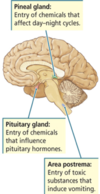
7
Q

area postrema

A

medullary structure in the brain that controls vomiting

8
Q

Astereognosis

A

Astereognosis (or tactile agnosia if only one hand is affected) is the inability to identify an object by active touch of the hands without other sensory input, such as visual or sensory information.

9
Q

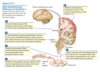
Draw and briefly describe Von Economo’s cytoarchitectonic regions of the parietal lobes and their associated functions.

A
  • PE: Somatosensory
  • PF: receives heavy input from the primary somatosensory cortex (through PE) and a small visual input through area PG.
  • PG: “parieto-temporo-occipital crossroads;” controls spatially guided behavior with respect to visual and tactile information.
10
Q

Apraxia

A

inability to perform particular purposive actions, as a result of brain damage.

11
Q

simultanagnosia

A

inability to perceive the visual field as a whole

12
Q

Simultaneous Extinction

A
  • Somatoperceptual disorder in which two stimuli would be reported if applied singly, but only one would be reported if both were applied together​​
    • Associated with damage to areas of PE and PF
13
Q

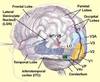
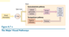
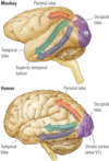
Describe the structural and functional differences between the ventral, dorsal, and STS visual streams. Draw the streams.

A
  • Ventral: includes inferior temporal pathway and STS pathway, concerned w/ object perception (including color & faces) and perceiving certain types of movements
  • Dorsal (parietal pathway): participates in the visual guidance of movement
  • Superior temporal sulcus: multimodal
14
Q

oculomotor apraxia

A

difficulty in fixating the eyes

15
Q

optic ataxia

A

inability to move the hand to a specific object by using vision

16
Q

How would you describe the functions of the left and right posterior zones of the parietal lobe in humans? Give an example from everyday life.

A
  • Integrate info from vision w/ somatosensory info for movement & spatial function
    • mental imagery; Esp. object rotation & navigation through space
  • Damage to R parietal lobe:
    • contralateral neglect:
      • cause difficulty in making things (constructional apraxia),
      • denial of deficits (anosognosia),
      • and drawing ability.
  • Patients w/posterior parietal damage:
    • L-R Confusion & cannot do mental rotations
17
Q

Anosognosia

A

Unawareness of denial of illness

18
Q

Name the cranial nerves and draw a “face” picture of them that helps describe their function

A
  1. Olfactory: Smell
  2. Optic: Visual fields and ability to see
  3. Oculomotor: Eye movements; eyelid opening
  4. Trochlear: Eye movements
  5. Trigeminal: Facial sensation
  6. Abducens: Eye movements
  7. Facial: Eyelid closing; facial expression; taste sensation
  8. Auditory/vestibular: Hearing; sense of balance
  9. Glossopharyngeal: Taste sensation; swallowing
  10. Vagus: Swallowing; taste sensation
  11. Accessory: Control of neck and shoulder muscles
  12. Hypoglossal: Tongue movement

https://blog.cognifit.com/12-pairs-of-cranial-nerves/

19
Q

Interblobs

A
  • form and motion perception
20
Q

nocioception

A
  • Perception of pain, temperature, and itch.
  • anterior spinothalamic tract
21
Q

Hapsis

A
  • Perceptual ability to discriminate objects on the basis of touch
  • posterior spinothalamic tract
22
Q

Proprioception

A
  • Perception of the position and movement of the body, limbs, and head
  • Anterior Spinothalamic Tract
23
Q

What is a homunculus and how is the 2 point test relevant to the homunculus

A
  • Homunculus: “Little Human”
    • Represents relative sensitivity of body parts, so sizes are disproportionate
    • Larger area = higher sensitivity
  • The ability to recognize the presence of two pencil points close together, a measure called two-point sensitivity, is highest on the parts of the body having the most touch receptors.
24
Q

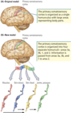
Differentiate the role of premotor, primary, and prefrontal cortex in motor movement

A
  • Posterior Cortex: Specifies movement goals and sends information to the prefrontal cortex
  • Prefrontal Cortex: Generates plans for movement
  • Premotor Cortex: Movement sequences
  • Primary Motor Cortex: Executes movements
25
The first stage in recovery from contralateral neglect is termed \_\_\_\_\_\_. **a**. extinction **b**. reintegration **c**. morphosynthesis **d**. allesthesia
**Allesthesia:** person’s beginning to respond to stimuli on the neglected side as if the stimuli were on the unlesioned side. The person responds and orients to visual, tactile, or auditory stimuli on the left side of the body as if they were on the right.
26
Describe Charles Bonnet Syndrome
* common condition among people who've **lost their sight** * **Abnormal activity in** **fusiform** gyrus causes people to hallucinate faces * When you lose sight, **brain receives less info** from eyes; brain can **fill in gaps** w/ new fantasy pictures, patterns or old pictures
27
Your patient has had a stroke involving the left parietal cortex, particularly anterior areas 1, 2, 3a and adjacent cortex (parts of 43 and 5). What is your expectation regarding possible cognitive defects and how would you test for this?
* Areas 3, 2, 1 (*postcentral gyrus*): Impairment in all **somatic sensations in contralateral** side of body. * Area 43: **Gustatory** * 5: Contralateral astereognosis and sensory neglect (damage in nondominant hemisphere).
28
Describe the feedback circuit that allows the cerebellum to correct movements.
**Intention, Action, Feedback:** By comparing the message for the intended movement with the movement that was actually performed, the cerebellum sends an error message to the cortex to improve the accuracy of a subsequent movement.
29
What is the Gerstmann syndrome? Why, do you suppose, these symptoms appear together following parietal damage? (see page 391)
* Gerstmann Syndrome * **F**inger agnosia of fingers on either hand * **A**graphia: inability to write * **R**ight-Left Confusion * **A**calculia: inability to do simple math problems – Disturbed language function – Results from a left parietal lobe lesion (Roughly the angular gyrus (PG)) https://www.youtube.com/watch?v=GLJdcmSIoNQ
30
Explain how activation of the GABA receptor can cause sedation. Why is it dangerous to mix benzodiazepines and barbiturates?
Limits the excitability of neuronal activity in all areas of the brain, resulting in sedation. Both drugs work to suppress the CNS in different ways. When taken together, they may suppress them enough to stop breathing, blood flow...
31
In order to be identified as a neurotransmitter, a substance must meet several tests. Which of the following is not a test? **A.** It must be present in the terminals. **B.** Its structure must be known. **C.** It must be released when neuron fires an action potential. **D.** There must be an inactivating mechanism in the cleft.
**B.** Its structure must be known
32
What is the functional significance of brain regions that lack a blood-brain barrier? Give an example.
**area** **postrema** of the lower brainstem allows toxic substances in the blood to **trigger a vomiting response**. **pineal gland:** open to the hormones that **modulate the** **day–night** **cycles** this gland controls.
33
What are the three main symptoms originally described by Balint in 1909 that led to the concept of Balint's syndrome? Damage to what area of the brain is associated with Balint Syndrome?
* 3 main symptoms: * inability to perceive the visual field as a whole (**simultanagnosia**) * difficulty in fixating the eyes (**oculomotor apraxia**), * inability to move the hand to a specific object by using vision (**optic ataxia**) * Damage to bilateral parietooccipital lobes
34
1. What is meant by the term blindsight? 2. What brain systems are impacted? 3. Give an example from one or more of the case studies given.
1. blindsight aka **agnosopsia:** 1. blindness due to damage to the primary visual cortex but the actual visual system (the eyes) are undamaged; person can sometimes respond to visual stimuli that they can 'see' with their eyes but their primary visual cortex cannot translate to the brain (i.e., *perceived location without being able to perceive content*) 2. V1 (maybe V2) 3. D.K.s right calcarine fissure was removed to surgically excise an angioma; was then able to "perceive" stimuli and could discriminate a 10degree difference in orientation
35
What are the seven major events in synaptic transmission?
1. **Neurotransmitter synthesis** can take place in the cell, axon, or terminal 2. **Stored** in storage granules or in vesicles until it is 3. **Released** from the terminals presynaptic membrane to 4. **Act on a receptor** embedded in postsynaptic membrane. Excess neurotransmitter in the synapse is either 5. **Deactivated** or 6. Taken back into presynaptic terminal (**reuptake**) for reuse. 7. The synapse also has a mechanism for **degrading** excess neurotransmitter and removing unneeded-by-products.
36
Identify, in correct order, the major connections of the geniculostriate system from eye to cortex.
* Geniculostriate Pathway * Optic nerves leave the eye and cross at the optic chiasm * The right half of each eye’s visual field is transmitted to the left hemisphere; the left half of each eye’s visual field is transmitted to the right hemisphere See: [http://pip.ucalgary.ca/psyc-369/mod2-the-visual-system/unit2.3-visual-pathways/Geniculostriate.html](http://pip.ucalgary.ca/psyc-369/mod2-the-visual-system/unit2.3-visual-pathways/Geniculostriate.html)
37
* What is a scotoma? * What do such phenomena tell us about the organization of the visual system?
* Small blind spots in visual field; lesion on occipital lobe * Our brains fill in missing bits of information?
38
How would damage to the dorsal portion of my right occipital lobe impact my visual field?
can result in loss of the left visual field of both eyes See also: [interactive brain](https://tbi.cemmlibrary.org/Interactive-Brain/Interactive-Brain)
39
Briefly list the main functional role of each of the following: V1, V2, V3, V4, V5 (a sentence or two for each area is sufficient, if it is the right sentence or two).
* **V1:** (striate cortex) is the first processing level in the hierarchy, receiving the largest input from the lateral geniculate nucleus of the thalamus and projecting to all other occipital regions * **V2**: second processing level, also projects to all other occipital regions * **V3**: (aka MT; middle temporal) concerns dynamic form—the shape of objects in motion. * **V4** damage: loss of color cognition (only see shades of gray) * Cannot see, *imagine*, or recall color * **V5** damage: erases the ability to perceive objects in motion; can only see objects at rest
40
Compare the behavioral capacities of high and low decerebrate animals.
* **High**: * Moves toward auditory and visual stimuli but shows no evidence of vision (can see but can't process) * Can effectively perform: * voluntary movements * automatic movements * **Low**: Difficulty maintaining consciousness, inactive if left alone, can't regulate temperature, sensory stimulation elicits movement and typical affective behavior
41
What is the evidence that the dopamine system is involved in drug addiction?
Support: * Animals are easily trained to press a lever for stimulation of the dopamine system * Abused drugs release dopamine or prolong its availability * Drugs that are dopamine antagonists are not abused
42
What are four ways neurotransmitters can be removed from the synaptic cleft?
* **Diffusion**: diffuses away from synaptic cleft * **Degradation**: enzymes in cleft break down neurotransmitter * **Reuptake**: transmitter taken back into presynaptic axon terminal for reuse * **Glial cells** take up some neurotransmitters
43
Anosodiaphoria
indifference to illness
44
Calcarine Sulcus
* Contains much of primary visual cortex (V1) * Separates upper and lower visual fields
45
The visual system consists of many subsystems. For each subsystem listed on the left, choose the most appropriate alternative. Visual cortex A) orientation of head B) voluntary eye movements C) pupil size D) pattern perception Frontal eye fields A) orientation of head B) voluntary eye movements C) pupil size D) pattern perception Superior colliculus A) orientation of head B) voluntary eye movements C) pupil size D) pattern perception
The visual system consists of many subsystems. For each subsystem listed on the left, choose the most appropriate alternative. Visual cortex A) orientation of head B) voluntary eye movements C) pupil size **D) pattern perception** Frontal eye fields A) orientation of head **B) voluntary eye movements** C) pupil size D) pattern perception Superior colliculus **A) orientation of head** B) voluntary eye movements C) pupil size D) pattern perception
46
* 3 symptoms of Parkinson’s * Name region most associated with the disease
* Results in **muscular rigidity** * Difficulty **initiating** and **performing** movements (hypokinetic symptoms) Region most associated: * Basal Ganglia
47
What causes Huntington’s disease and what are 3 symptoms?
Causes: * Destroys cells in the **caudate putamen** (cellular degeneration in the basal ganglia) Three symptoms: 1. involuntary, and 2. exaggerated movements (hyperkinetic symptoms) 3. Leads to deficits on tests of implicit memory
48
Differentiate the behaviors associated with left and right temporal-parietal junction (TPJ):
* **right TPJ**: processing of info in terms of the ability of an individual to pay **attention** * **left TPJ**: role in reasoning of other’s beliefs, intentions, and desires
49
Diagnostic criteria & symptoms of Posterior Cortical Atrophy
* insidious onset and gradual progression * visual deficits (in the absence of ocular disease) * relatively preserved episodic memory, verbal fluency, and personal insight * presence of symptoms including: * visual agnosias * simultanagnosia * optic ataxia * ocular apraxia * dyspraxia, and * environmental disorientation; and absence of stroke or tumour
50
4 diagnostic features of Lewy Body Dementia
* Parkinsonian Symptoms * Visual Hallucinations (“halLEWYcinations”) * Cognitive Decline * Waxing/Waning Symptoms wPVC
51
Motor system: What is it?
usually reserved for those parts of the nervous system, charted in Figure 9.1B. * Basal ganglia (force) * Motor cortex (commands) * Cerebellum (accuracy * Brainstem and spinal cords (movements) * Motor neurons
52
Draw a graph showing the relationship between wanting and liking over time?
**Wanting** = craving for drug **Liking** = pleasure produced by taking the drug
53
How is the dopamine system differentiated from the endogenous opioid system?
* **Dopamine system**: associated w/ the incentive, preparatory aspect of reward (experienced as thrill, urgency, or craving; i.e., **wanting**) * **Opioid system**: associated w/ more hedonic aspects of reward (feelings of enjoyment following drug use; i.e., **liking**)
54
Fruits or Faces: Explain.
* [Split](https://www.youtube.com/watch?v=ZMLzP1VCANo)[brain behavioral](https://www.youtube.com/watch?v=ZMLzP1VCANo)[experiments](https://www.youtube.com/watch?v=ZMLzP1VCANo) **Right** sees *gestalt* **Left** sees units
55
Damage to Fusiform causes...
* Face blindness
56
Five categories of vision
* **Vision for action** (e.g., grasping) * **Action for vision** (e.g., scanning faces) * **Visual...** * **Recognition** * **Space** (spatial location) * allocentric * egocentric * **Attention**
57
Draw the Visual Processing Hierarchy
58
Cranial Nerves Sentence Mnemonic
**O**hh - **Olfactory** **O**hh - **Optic** **O**hh - **Oculomotor** **T**o - **Trochlear** **T**ouch - **Trigeminal** **A**nd - **Abducens** **F**eel - **Facial** **V**ery - **Vestibulocochlear** **G**ood - **Glossopharyngeal** **V**elvet - **Vagus** **S**uch - **Spinal Accessory** **H**eaven - **Hypoglossal**
59
How/ what? Grasping dorsal / ventral
* **Dorsal Stream** * Visual guidance of movements for grasping * **Ventral Stream** * *IT* Inferior Temporal Cortex * Object Perception * ***STS* Superior Temporal Sulcus** * Visuospatial functions
60
Somatosensory Agnosias
Loss of sense of one's own body; most commonly affects left side of body. Types: * **Anosognosia** (nosos, "disease" & gnōsis, "knowledge"): * **Anosodiaphoria**: indifference to own illness * **Autopagnosia**: loss power to recognize or orient a bodily part
61
Behaviors associated with right/left brain
**Left**: * Language (temporal lobe) * Verbal memory (temporal lobe) * Speech processing (temporal lobe) * Motor functions **Right**: * Theory of Mind * More important role in facial processing (temporal lobe) * Nonverbal memory (temporal lobe) * Spatial functions R & L motor cortices control limbs on contralateral side body
62
Left hemi, angular gyrus (Brodmann area 39) damage. What results?
**Gerstmann Syndrome** * **Finger agnosia** of fingers on either hand​ * **Right-Left Confusion** * **Agraphia** (inability to write) * **Acalculia:** (inability to do simple math problems) * **Disturbed language function** ​​ * Results from a left parietal lobe lesion (roughly the angular gyrus [PG]). **-----** * Damage to right intraparietal sulcus and the right angular gyrus results in **Contralateral neglect.**
63
What does the connectivity between brain regions tell us?
64
Temporal Lobe Functions
* **Process** auditory input * **Visual** object recognition * Long-term storage of sensory input- i.e., **memory**