eyes and ears Flashcards

1
Q

absence of the eye

A

Anophthalmos

2
Q

incomplete separation, or early fusion, of paired globes

A

Synopthalmia

3
Q

– abnormally small eye

A

micropthalmia

– inherited defect in Collie dogs

4
Q

inversion of the eyelids → trichiasis

A

Entropion

5
Q

– eversion of the eyelids

A

ectropion

6
Q

– rubbing of the eyelashes against the eye surface

A

Trichiasis

7
Q

failure of complete fusion of the lips of the embryonic
choroid fissure

A

Coloboma

8
Q

which part of the eye is most affected in coloboma

A
  • the posterior portions of the eye (optic disc, iris, ciliary body) most often affected
  • – inherited in Charolais cattle
9
Q

what are the lesions of coloboma

A
  • cavitation of the choroid and sclera

– cavity lined by a thinned retinal layer

  • visual defects

– only in very severe cases

10
Q

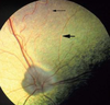
– etiopathogenesis Collie eye anomaly

A
  • improper development of the optic cup
  • abnormal formation of choroid and retina
  • chorioretinal dysplasia or choroidal hypoplasia
11
Q

Collie eye anomaly lesions/defects:

A
  • abnormal retinal vessels
  • areas of chorioretinal dysplasia or hypoplasia
  • ectasia
               – optic disc
                – sclera
  • posterior staphyloma
  • ± severe visual impairment
12
Q

sequela of collie eye anomali

A
  • retinal degeneration and detachment
  • intraocular hemorrhage
13
Q

delayed or incomplete atrophy of the anterior
perilenticular vascular network results in

A

Persistent pupillary membrane

atrophy is frequently incomplete at birth

14
Q

Persistent pupillary membrane lesions

A
  • bloodless strands

– short, threadlike protrusions from the area of
the minor arterial circle (iris collarette)

15
Q

Persistent pupillary membrane clinical significance

A
  • obstructed vision
  • corneal or lens opacity

– due to dysplasia of corneal endothelium or lens
because of contact with the strands

16
Q

Partial or complete absence of an eyelid

A

Eyelid agenesis and coloboma

partial defect (coloboma) involving the upper
eyelid is the most common
» localized corneal dessication followed by
cutaneous metaplasia

17
Q

abnormal or prolonged fusion or adhesion of
the eyelids

A

Ankyloblepharon

18
Q

what is the function of physiological Ankyloblepharon

A

essential to protect the immature cornea from
infectious keratitis, dessication, and corneal
rupture

Congenital ankyloblepharon in dogs and cats persists into the 2nd week of life

19
Q

inward rolling of the eyelid margin (inversion or
infolding) because of inadequate overall length

A

Entropion

20
Q

what is the sequela of entropion

A

irritation of the cornea by the eyelid skin,
cilia, and/or hair

very common anomaly in purebred dogs

21
Q

undue laxity of an excessively long
eyelid resulting in an outward gaping of the
eyelid margin

A

ectropion

22
Q

what is the sequela of ectropion

A

chronic conjunctivitis and keratitis from
exposure to debris

23
Q

presence of an ectopic row of cilia originating
from the ducts of the Meibomian glands

A

Distichiasis

24
Q

what is sequela of Distichiasis

A

corneal ulceration

25
misdirection of the normal cilia
Trichiasis
26
what is the sequela of trichiasis
corneal irritation leading to keratitis and even ulceration
27
abnormally placed cilia within the lamina propria of the conjunctiva
ectopic cilia
28
what is the sequela of ectopic cilia
their emergence through the palpebral conjunctiva can cause severe corneal irritation
29
suppurative adenitis of the **adnexal glands** of Moll or Zeis
external Hordeolum (stye)
30
suppurative adenitis of the **Meibomian gland**
internal Hordeolum (stye)
31
pathogenesis of internal focal hordeolum
persistent inflammation \>\> sebaceous secretions extend into the adjacent soft tissues \>\> _granulomatous response_
32
differential diagnosis for focal hordeolum
adenoma » fungal granuloma » lymphoid hyperplasia
33
sterile granulomatous inflammation of the Meibomian gland
Focal Chalazion
34
differential diagnosis of chalazion
Meibomian gland adenomas
35
Diffuse blepharitis Types
ulcerative, seborrheic , allergic , actinic, parasitic
36
a shetland is presented wih erythema,crusting, vesiculation, and erosion of the dorsum of the nose,inner aspect of the pinna, perioral and periocular region. which diases do you suspect
diffuse Blepharitis
37
* most common tumor of canine eyelid (80% of all eyelid tumors)
Meibomian gland adenoma it is the exact counterpart of sebaceous adenomas
38
2nd most common tumor of the canine eyelid
melanocytoma universally benign
39
in which dog breeds are dermoids found
: St. Bernard, Dalmatian – lateral canthus or limbus
40
benign, raised, wedge-shaped growth of the conjunctiva
Pterygium
41
what causes Pterygium (conjuctival disease)
1. trauma 2. irritation 3. allergens like pollen 4. toxins 5. infections\>bacteria, virus,mycobacterium, parasites
42
failure of in growth of the future iris epithelium
iris hypoplasia more frequent in horses; inherited
43
Choroidal hypoplasia associated with
inadequate pigmentation of the retinal pigment epithelium and choroids – CEA
44
posterior uveitis or choroiditis
inflammation of ciliary body and choroid
45
inflamamtion of iris and ciliary body
anterior uvitis or iridocyclitis
46
what causes conjuctivitis
can be associated with inflammation elsewhere, e.g., upper respiratory disease with concurrent keratoconjunctivitis
47
neoplasms of the conjuctiva are common in which animals
cattle followed by horses, dogs and horses
48
which conjuctival tumer is common in cattle
SCC
49
.SCC Predisposing factors include
* actinic iradiattion and hypomelanosis * associated with IBR virus * genetic predisposition in herefords
50
A horse present with begin mass at the limbal region or the leading edge of the 3rd eyelid that progress to the conjunctiva.which tumer do you suspect?
SCC of the conjuctiva
51
a faint loss of transparency of cornea that can be seen with oblique illumination
nebulla
52
a light gray spot in the cornea that can be visualized in natural light
macula
53
any dense white spot that may indicate moderate to severe corneal disease
Leukoma
54
gray-blue opacity of the cornea and may have a “cobblestone” appearance.
corneal edema
55
what causes superficial keratitis
: due to corneal ulceration (ulcerative keratitis)
56
what causes deep keratitis
: due to infection (mycotic or bacterial) or intraocular inflammation
57
stereotyped sequence of keratitis
1) edema 2) leukocyte immigration from tears and distant limbic venules 3) corneal stromal vascularization 4) fibrosis 5) epithelial metaplasia with pigmentation
58
discribe the epithelial type of keratitis
* usually transient and mild non ulcerative keratitis * superficial punctate keratitis consists of multiple fine epithelial opacities that are foci of epithelial hydropic change * intercellular fluid accumulation (bullous keratopathy) is seen as a sequel to corneal edema
59
describe Stromal keratitis
* it is a non ulcerative keratitis which can either be deep or superficial
60
are are the two types of non ulcerative keratitis
epithelial and stromal keratitis
61
describe the etiopathogenesis of ulcerative keratitis
* loss of epithelium initiates a series of reactions caused by tear imbibition, * local production of cytokines * opportunistic microbial contamination of the wound
62
sequela of ulcerative keratitis
* involve cornea, conjunctiva, and uvea * the ulcer may: 1. heal with vascularization and scarring; granulation tissue fills the ulcerative defect. 2. persist as a stubborn but nonprogressive lesion 3. progress to involve more of the stroma and epithelium * stromal liquefaction that reaches the Descemet’s membrane results in its forward bulging as a descemetocele or keratocele due to increased intraocular pressure; this appears grossly as a transparent center to the ulcer
63
focal adhersion of the iris to the injured cornea due to anterior uvitis is called
anterior synechia
64
WHAT PREDISPOSE THE CORNEA TO ANTERIOR synechia
* the Descemet’s membrane, although resistant to penetration of microbial agents themselvesis permeable to inflammatory mediators and microbial toxins which diffuse into the anterior chamber; * these chemicals, combined with a vasoactive sensory neural reflex from irritated cornea, are responsible for the vasodilation and exudation in anterior uvea e) the anterior uveitis may result in sufficient fibrin exudation so as to predispose to focal adhesions
65
a fibrous tunic (i.e., corneal) defect lined by uvea (i.e., iris) or incorporation of the iris into the corneal stroma
anterior staphyloma it is a a permanent anterior synechia
66
what causes anterior staphyloma
in the case of corneal perforation, the iris flows forward to plug the defect and subsequently becomes incorporated into the corneal scarring, this defect is a permanent anterior synechia
67
potrution of the descemet'se membrane through an ulcerated membrane
: descemetocele
68
anterior synechia (iris adhering to the cornea) lined by epithelial cells
corneal fistula
69
discuss the summary flow chart for ulcerative keratitis
70
diffuse, blue-gray clouding of the cornea associated with inflammatory changes in the deep corneal layers
stromal (interstitial) keratitis caused by iridocyclitis is characterized by deep vascularization, i.e., ingrowth of vessels (usually short and straight) from the ciliary body.
71
pannus keratitis
* it is a type of superficial stromal keratitis * seen mostly in GSD * chronic, nonulcerative proliferative inflammation * there is no ulceration but pigmentation is often marked
72
pathogenesis of pannus keratitis( superficial stromal keratitis)
* immune-mediated; cell mediated injury to altered corneal antigens following prolonged exposure to UV light; * severity is exacerbated by dust and high altitudes
73
what are the lesions of pannus keratitis( superficial stromal keratitis)
* vascularized opacity in the corneal stroma (bilateral); * marked pigmentation which eventually infiltrates the entire corne * a converting the superficial stroma to an opaque membrane resembling granulation tissue; * histologically, there is an intense mononuclear inflammatory infiltrate
74
what are the gross lesions of eosinophilic keratitis
* . predominantly in cats, occasionally in horses * . grossly, granular white proliferative lesion extending inwardly along what seems to be the corneal surface from the medial or lateral limbus * microscopically, infiltration of eosinophils, plasma cells, mast cells, and macrophages 4. etiopathogenesis is unknown
75
what causes deep stromal keratitis
a) results from extension from anterior uveitis or from endothelial damage by uveitis, trauma, or glaucoma b) common in horses which have suffered one or more bouts of equine recurrent ophthalmitis
76
what causes Keratoconjunctivitis sicca, KCS
2) decreased lacrimal secretion which normally has protective lubricant and bacteriostatic functions in the conjunctival sac common in toy breeds
77
what is a sequela of keratoconjuctivity sicca
* chronic keratoconjunctivitis with congestion, * epithelial keratinization, * dry lusterless cornea and a thick, white, sticky, ropy mucoid exudates * . Chronic corneal changes may include ulceration, melanosis and lipoidal dystrophy.
78
summary: Non-ulcerative keratitis (followed by iridocyclitis)
79
what are the lesons of iridocyclitis
* marked hyperemia/congestion of iridal and ciliary vessels –altered formation of aqueous * reflected by a drop in intraocular pressure –exudation * cellular (mononuclear or neutrophils) •±exudation of fibrin
80
what is aqueous flare
lesion of iridocyclitis (antirior uvitis) characterised by accumulation of exudate (fibrin and inflammatory cells) in the aqueous
81
yellow-white or pink flocculant sediment in the anterior chamber
hypopyon a iridocyclitis lesion
82
• blood or neutrophils in the anterior chamber
hyphema it is aIridocyclitis (anterior uveitis)
83
stippled appearance of the cornea with adherance of clumps of inflammatory cells to the posterior surface of the cornea
keratitis punctata a lesion of anterior uvitis
84
“mutton-fat” keratic precipitates characteristics
Iridocyclitis (anterior uveitis) lesions: * white, yellowish greasy precipitates of inflammatory cells (granulomatous response) * adherent to the corneal endothelium
85
sequela of Iridocyclitis (anterior uveitis)
* glaucoma (increased intraocular pressure) * lens luxation
86
Iridocyclitis (anterior uveitis) pathogenesis and types
* fibrinous exudation over the iris 1. anterior synechia (adhesion of iris to cornea) 2. posterior synechia (adhesion of iris to lens) * partial synechia → small visual defects * total synechiae→ blockage of aqueous flow and glaucoma
87
iris bombé
sequela to Iridocyclitis (anterior uveitis) 360 ° (complete) posterior synechiae • bowing of the iris anteriorly * apposition of the iris to the lens preventing aquous from flowing from the posteria to the anteria chamber.pressure rises resulting in bowing of the iris
88
occlusio pupillae
a complete fibrovascular membrane across the pupil fibrovascular membrane on the iris face → pupillary block → neovascular glaucoma it is a sequela to iridocyclitis
89
eversion of the pigmented posterior epithelium of the iris at the pupillary margin
Ectropion uveae
90
discuss changes that can conceal changese of iridocyclitis deep in the eye
the blue-gray corneal opacity due to edema, interstitial keratitis and fibrosis – examples: * in MCF of cattle – iridocyclitis with hypopyon may be obscured by the onset of interstitial keratitis * dogs recovering from ICH – iridocyclitis due to type III hypersensitivity
91
significance effect of uveitis on cornea
edema and hyperemia due to endothelial damage | (as in ICH, FIP)
92
effects of uveitis on cornea look at the picture
* stromal fibrosis * vascularization
93
discuss the effect of uvitis shown below
corneal ullceration
94
bullous keratopathy
edema due to percolation of stromal fluid into the corneal epithelium
95
hypotonic, shrunken, disorganized eye
phthisis bulbi
96
lesions of Choroiditis (posterior uveitis)
* marked exudation * choroid * vitreous * retinal detachment • local or general * retinal degeneration • esp. horses (no retinal arteries) * atrophy/proliferation of choroidal pigment cells
97
which parts of the eye are affected by the equine recurent uvitis
* damage to the choroid, retina and optic nerve leading to blindness
98
etiopathogenesis of equine recurent uvitis
* immunologic reaction against intraocular leptospiral antigens – a delayed reaction to a systemic infection with Leptospira interrogans serovar pomona * intraocular dead or dying microfilaria of Onchocerca cervicalis act as antigens * deficiencies of vitamins A, B2 (riboflavin) and C have been incriminated but not proven
99
lesions of equine recurent uvitis
anterior neutrophilic uveitis which rapidly becomes predominantly lymphocytic
100
diagnoses of equine recurent uvitis
presence of an eosinophilic hyaline membrane within the apical cytoplasm of the nonpigmented ciliary epithelium and resembles amyloid
101
sequelae of equine recurrent uveitis
* interstitial keratitis * cataract * lens luxation * synechiae * retinal separation
102
what causes glaucoma
* sustained increased in intraocular pressure due to – decreased drainage of aqueous humor – increased production of aqueous
103
glaucoma is predisposed by
* ocular disease * uveitis + synechiae
104
what cuases Congenital glaucoma
abnormal development of anterior chamber
105
what causes primary glacoma
malformation of filtration angle inherited, bilateral disease breed predisposition: cocker spaniels usually no ocular disease
106
what are the lexamples of primary glaucoma
* _goniodysgenesis_ – detectable maldevelopment of the trabecular meshwork » incomplete remodeling of the solid mass of anterior chamber mesenchyme * _open-angle glaucoma_ – no primary histologic lesions » no visible abnormality in the structure of the trabecular meshwork or other portions of the aqueous outflow pathways
107
what causes secondary glaucoma
due to anything capable of obstructing the flow of aqueous through the pupil (pupillary block) or – its exit through the trabecular meshwork (trabecular occlusion)
108
discuss eye lesions than can cause secondary glaucoma
* inflammatory » synechiae, iritis, iridocyclitis, corneal ulcer * degenerative * neoplastic (uveal melanoma, lymphosarcoma) * occlusive » pannus, inflammatory debris, tumor cells » lens (luxation, intumescent cataract)
109
buphthalmos
Glaucoma lesions/sequelae: * enlarged globe, * thinning of the sclera
110
lesions of glaucoma on the cornea
chemosis • pannus • dessication • ulceration
111
lesions of glaucoma on the lens
* cataract * subluxation/luxation
112
what are the effects of glaucoma on the uvea
* uveal tract reaction - atrophy and scarring
113
lesions of glaucoma on the retina
* retinal degeneration • ganglion cell layer, inner nuclear layer
114
effects of glaucoma on the optic nerve
degeneration of branches of optic nerve and the optic disc (cupping of the optic disc) – blindness
115
faulty differentiation of the retina (photoreceptor cells) with proliferation of one or more of its constituents
Retinal dysplasia
116
what are the lesions of retinal dysplasia
lesions are characterized by rosettes composed of a variable number of neuronal retinal cells, often with accompanying retinal folds
117
retinal dysplasia is associated with........
* a single ocular defect or * multiple ocular anomalies – microphthalmia – posterior coloboma
118
what causes secondary retinal dysplasia
* in utero viral infections BVD in calves, bluetongue in lambs * vitamin A deficiency * x-ray irradiation * certain drugs * intrauterine trauma
119
Retinal dysplasia lesions
* rosettes with primitive, undifferentiated neuronal cells * + retinal folds
120
retinal dysplasia lesions on anterior segment of the eye
* corneal scar * hypoplastic iris * cataractous lens * dysplastic retina
121
what causes renitis
* systemic infections which attack nervous tissue • canine distemper * neuronal degeneration with gliosis • MCF, FIP * ocular toxoplasmosis – choroidal infections * toxoplasmosis * tuberculosis
122
loss of photoreceptors and other retinal neurons with formation of spaces within the retina
retinal degeneration there is decreased thickness of the neurosensory retina (retinal atrophy)
123
causes of retinal degeneration
* 2° to retinitis or chorioretinitis – senility * focal retinal degeneration * nutritional deficiency vitamins A, E, taurine * toxicity - pressure atrophy due to glaucoma – excessive exposure to light – hypertension – irradiation – infectious agents – hereditary (most important in dogs)
124
what are the characteristics of generalised Progressive retinal atrophy (PRA)
* early onset * photoreceptor dysplasia
125
what are the lesions of central Progressive retinal atrophy (PRA)
later onset • retinal pigment epithelium dystrophy → photoreceptor degeneration
126
discuss etiopathogenesis of retinal degeneration
* photoreceptor dysplasia * it is nherited in Irish setters, miniature and toy poodles, cockerspaniels, etc. * photoreceptors never reach morphologic or physiologic maturity * with time, photoreceptor loss extends to 2° atrophy of nuclear and plexiform layers of the retina
127
what are the lesions of generalised progressive retinal atrophy (PRA)
* rods affected first degeneration of the processes and cell bodies in the outer nuclear layer this results in poor dim- light vision or night blindness * involvement of the cones→loss of day vision * degeneration of RPE, disappearance of retinal vessels leading to total blindness * progressive loss of the inner nuclear layer * degeneration of the pigment epithelium * narrowing and disappearance of the retinal vessels leading to total blindness
128
what is the effect of generalised PRA on the rods
* degeneration of the processes and cell bodies in the outer nuclear layer * resulting in poor dim-light vision or night blindness
129
what are the effects of generalised PRA on the cones
loss of day vision
130
discuss the lesions seen below
* _Retinal degeneration_ * **hyper-reflectivity** of the tapetal retina dorsal to the disc * **narrowing** of the retinal vessels * **pallor** to the optic disc
131
discuss the cause of central progressive retinal atrophy
* inherited in collies, labradors and English springers
132
what is the primary lesion of central progressive retnal atrophy
primary pigment epithelial dystrophy
133
DISCUSS THE PATHOGENESIS FOR CENTRAL PRA
* normally RPE engulfs and degrades shed photoreceptor outer segments by intracellular phagocytosis resulting in a gradual buildup of intracellular lipopigments throughout life **– abnormally** • defective intracellular phagocytosis of shed photoreceptor outer segments → excessive membrane peroxidation → lipopigments thus accumulate to excess in the RPE