female reproductive physiology Flashcards

1
Q

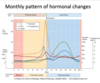
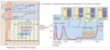
diagram the effect in a female with the release of GnRH

A
  1. estrogen and progesterone can exert BOTH positive and negative feedback control on: hypothalamus and pituitary
    1. depends on the hormone production by the growing oocyte during the menstual cycle
2
Q

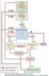
what relases LH/FSH and what cells do they effect and how?

A
  1. estrogen and progesterone can exert BOTH positive and negative feedback control on: hypothalamus and pituitary
    1. depends on the hormone production by the growing oocyte during the menstual cycle
3
Q

LH affects what two cells, describe the relationship.

A
  1. estrogen and progesterone can exert BOTH positive and negative feedback control on: hypothalamus and pituitary
    1. depends on the hormone production by the growing oocyte during the menstual cycle
  2. LH is not the major
4
Q

what are the two hormones that exhibit positive and negative feedback. Explain

A
  1. estrogen and progesterone can exert BOTH positive and negative feedback control on: hypothalamus and pituitary
    1. depends on the hormone production by the growing oocyte during the menstual cycle
5
Q

describe the release of gonadotropin in the anterior pituitary

A

GnRH-> Gprotein on the anteior pituitary->Gq->PLC

  • PLC
    • DAG
      • PKC
        • additive affect
          • follicular estrogen has an additive affect
          • Activin from the ovary has an additive affect
        • inhibitory affect
          • luteal phase estrogen and progesterone have an inhibitory affect
          • inhibin from the ovary has an inhibitory affect
      • gene transcription of LH and FSH
      • LH and FSH are synthesized, dimerized and packaged
    • IP3
      • leads to a release of Ca++ from the sarcoplasmic reticulum
  • Ca++ leads to the exocytosis of the gonadotropin vesicles
6
Q

what is the affect of the following with respect to the HPA in a female

A

GnRH-> Gprotein on the anteior pituitary->Gq->PLC

  • PLC
    • DAG
      • PKC
        • additive affect
          • follicular estrogen has an additive affect
          • Activin from the ovary has an additive affect
        • inhibitory affect
          • luteal phase estrogen and progesterone have an inhibitory affect
          • inhibin from the ovary has an inhibitory affect
      • gene transcription of LH and FSH
      • LH and FSH are synthesized, dimerized and packaged
    • IP3
      • leads to a release of Ca++ from the sarcoplasmic reticulum
  • Ca++ leads to the exocytosis of the gonadotropin vesicles
7
Q

a patient presents with a tumor generating an incredible amount of progesterone and estrogen. will this be inhibitory or stimulatory of LH and FSH

A

GnRH-> Gprotein on the anteior pituitary->Gq->PLC

  • PLC
    • DAG
      • PKC
        • additive affect
          • follicular estrogen has an additive affect
          • Activin from the ovary has an additive affect
        • inhibitory affect
          • luteal phase estrogen and progesterone have an inhibitory affect
          • inhibin from the ovary has an inhibitory affect
      • gene transcription of LH and FSH
      • LH and FSH are synthesized, dimerized and packaged
    • IP3
      • leads to a release of Ca++ from the sarcoplasmic reticulum
  • Ca++ leads to the exocytosis of the gonadotropin vesicles
8
Q

A patient demonstrates a mutation in the IP3 of the cell signaling which of the following is true?

  1. the patient will have high levels of serum LH and FSH
  2. The patietn will have high levels intracellular LH and FSH in the anterior pituitary
  3. the patient will have high levels of estrogen
A

The patient will have high levels of intracellular LH and FSH in the anterior pituitary. IP3 is needed to send the vesicles filled with LH and FSH out via exocytosis

GnRH-> Gprotein on the anteior pituitary->Gq->PLC

  • PLC
    • DAG
      • PKC
        • additive affect
          • follicular estrogen has an additive affect
          • Activin from the ovary has an additive affect
        • inhibitory affect
          • luteal phase estrogen and progesterone have an inhibitory affect
          • inhibin from the ovary has an inhibitory affect
      • gene transcription of LH and FSH
      • LH and FSH are synthesized, dimerized and packaged
    • IP3
      • leads to a release of Ca++ from the sarcoplasmic reticulum
  • Ca++ leads to the exocytosis of the gonadotropin vesicles
9
Q

where is estrogen synthesized in a female?

A

Bulk is generated by the granulosa cells

women have two forms of estrogen

  1. weak - estrone E1
  2. strong - Estradiol E2
10
Q

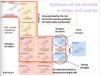
what 18 carbon compound is essential for the female reproductive tract?

A

women have two forms of estrogen

  1. weak - estrone E1
  2. strong - Estradiol E2
11
Q

describe the synthesis of weak and strong female hormone. Describe the carbon number for each

A

women have two forms of estrogen

  1. weak - estrone E1
  2. strong - Estradiol E2
12
Q

an increase in cAMP occurs from what cells in response to what hormones?

A

formation of estrogen

  1. granulosa cells
    1. provide a protective/nursing function and express FSH receptors
      1. induces expression of LH receptors on granulosa cells in late follicular phase.
      2. Allows granulosa cells to maintain high aromatase function as FSH falls and allows cells to respond to LH surge.
        1. FSH decreases in response to inhibin
    2. LACK HYDROXYLASE and DESMOLASE
      1. means that DHEA and androsteridiol must be generated in the theca and transfered over to the granulosa.
  2. theca cells
    1. considered nurse cells
    2. express LH receptors and produce androgens (progesteron +androstenedione)
    3. LACK THE ENZYME AROMATASE
      1. means the last product generated is testosterone and andrsteridiol for the granulosa cells (not sure what androsterone is for)
  3. biochemistry= constant in theca but receptors and function change in granulosa
    1. LH stimulaes theca cells ->increase cAMP -> increase syntheis of LDL and HDL receptors along with STaR protein
    2. theca cells increase synthesis of androstenedione
    3. androstenedione diffuses freely to granulosa cells
    4. FSH stimulates granulosa cells increasing cAMP leading to an increase in
      1. aromatase function
      2. LH receptors
    5. aromatase converts androstenedione to erstrone
    6. 17B-HSDehydrogenase converts estone to estradiol
    7. FSH incduced LH receptor expression on the granulosa cells in late follicular phase->allows
      1. high aromatase function when FSH falls
      2. Response to LH surge
    8. Estradiol diffuses into blood vessels
13
Q

what enzymes are lacking in the theca and granulosa cells? Explain the dynamic between the two

A

formation of estrogen

  1. granulosa cells
    1. provide a protective/nursing function and express FSH receptors
      1. induces expression of LH receptors on granulosa cells in late follicular phase.
      2. Allows granulosa cells to maintain high aromatase function as FSH falls and allows cells to respond to LH surge.
        1. FSH decreases in response to inhibin
    2. LACK HYDROXYLASE and DESMOLASE
      1. means that DHEA and androsteridiol must be generated in the theca and transfered over to the granulosa.
  2. theca cells
    1. considered nurse cells
    2. express LH receptors and produce androgens (progesteron +androstenedione)
    3. LACK THE ENZYME AROMATASE
      1. means the last product generated is testosterone and andrsteridiol for the granulosa cells (not sure what androsterone is for)
  3. biochemistry= constant in theca but receptors and function change in granulosa
    1. LH stimulaes theca cells ->increase cAMP -> increase syntheis of LDL and HDL receptors along with STaR protein
    2. theca cells increase synthesis of androstenedione
    3. androstenedione diffuses freely to granulosa cells
    4. FSH stimulates granulosa cells increasing cAMP leading to an increase in
      1. aromatase function
      2. LH receptors
    5. aromatase converts androstenedione to erstrone
    6. 17B-HSDehydrogenase converts estone to estradiol
    7. FSH incduced LH receptor expression on the granulosa cells in late follicular phase->allows
      1. high aromatase function when FSH falls
      2. Response to LH surge
    8. Estradiol diffuses into blood vessels
14
Q

LH leads to an increase in which receptors and proteins?

A

formation of estrogen

  1. granulosa cells
    1. provide a protective/nursing function and express FSH receptors
      1. induces expression of LH receptors on granulosa cells in late follicular phase.
      2. Allows granulosa cells to maintain high aromatase function as FSH falls and allows cells to respond to LH surge.
        1. FSH decreases in response to inhibin
    2. LACK HYDROXYLASE and DESMOLASE
      1. means that DHEA and androsteridiol must be generated in the theca and transfered over to the granulosa.
  2. theca cells
    1. considered nurse cells
    2. express LH receptors and produce androgens (progesteron +androstenedione)
    3. LACK THE ENZYME AROMATASE
      1. means the last product generated is testosterone and andrsteridiol for the granulosa cells (not sure what androsterone is for)
  3. biochemistry= constant in theca but receptors and function change in granulosa
    1. LH stimulaes theca cells ->increase cAMP -> increase syntheis of LDL and HDL receptors along with STaR protein
    2. theca cells increase synthesis of androstenedione
    3. androstenedione diffuses freely to granulosa cells
    4. FSH stimulates granulosa cells increasing cAMP leading to an increase in
      1. aromatase function
      2. LH receptors
    5. aromatase converts androstenedione to erstrone
    6. 17B-HSDehydrogenase converts estone to estradiol
    7. FSH incduced LH receptor expression on the granulosa cells in late follicular phase->allows
      1. high aromatase function when FSH falls
      2. Response to LH surge
    8. Estradiol diffuses into blood vessels
15
Q

What occurs in a patient with a mutant aromatase?

A

no formation of estrogen and possibly a masculization. no mensing. brittle bones.

  1. granulosa cells
    1. provide a protective/nursing function and express FSH receptors
      1. induces expression of LH receptors on granulosa cells in late follicular phase.
      2. Allows granulosa cells to maintain high aromatase function as FSH falls and allows cells to respond to LH surge.
        1. FSH decreases in response to inhibin
    2. LACK HYDROXYLASE and DESMOLASE
      1. means that DHEA and androsteridiol must be generated in the theca and transfered over to the granulosa.
  2. theca cells
    1. considered nurse cells
    2. express LH receptors and produce androgens (progesteron +androstenedione)
    3. LACK THE ENZYME AROMATASE
      1. means the last product generated is testosterone and andrsteridiol for the granulosa cells (not sure what androsterone is for)
  3. biochemistry= constant in theca but receptors and function change in granulosa
    1. LH stimulaes theca cells ->increase cAMP -> increase syntheis of LDL and HDL receptors along with STaR protein
    2. theca cells increase synthesis of androstenedione
    3. androstenedione diffuses freely to granulosa cells
    4. FSH stimulates granulosa cells increasing cAMP leading to an increase in
      1. aromatase function
      2. LH receptors
    5. aromatase converts androstenedione to erstrone
    6. 17B-HSDehydrogenase converts estone to estradiol
    7. FSH incduced LH receptor expression on the granulosa cells in late follicular phase->allows
      1. high aromatase function when FSH falls
      2. Response to LH surge
    8. Estradiol diffuses into blood vessels
16
Q

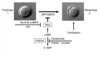
what are five functions of the ovarian follicle?

A

The ovarian follicle is the functional unit of the ovary

  1. ovarian follicle=one germ cell completely surround by a cluster of endocrine cells
  2. function
    1. maintains and nurtures the oocyte
    2. matures the oocyte and releases it at the approtpriate time
    3. prepares the vagina and fallopian tubes to assist in fertilization
    4. prepares the uterus to accept and implant a zygote
    5. maintains hormonal support of the fetus until placenta takes over
17
Q

one germ cell completely surround by a cluster of endocrine cells

A

The ovarian follicle is the functional unit of the ovary

  1. ovarian follicle=one germ cell completely surround by a cluster of endocrine cells
  2. function
    1. maintains and nurtures the oocyte
    2. matures the oocyte and releases it at the approtpriate time
    3. prepares the vagina and fallopian tubes to assist in fertilization
    4. prepares the uterus to accept and implant a zygote
    5. maintains hormonal support of the fetus until placenta takes over
18
Q

the functional unit of the ovary

A

The ovarian follicle is the functional unit of the ovary

  1. ovarian follicle=one germ cell completely surround by a cluster of endocrine cells
  2. function
    1. maintains and nurtures the oocyte
    2. matures the oocyte and releases it at the approtpriate time
    3. prepares the vagina and fallopian tubes to assist in fertilization
    4. prepares the uterus to accept and implant a zygote
    5. maintains hormonal support of the fetus until placenta takes over
19
Q

function of the ovarian follicle include

  1. maintain and nurture
  2. matures ___ and releases it
  3. prepares the ____ and ____ to assist in fertilization
  4. prepares the ___ to thicken
  5. assists fetus until the ____ takes over
A

The ovarian follicle is the functional unit of the ovary

  1. ovarian follicle=one germ cell completely surround by a cluster of endocrine cells
  2. function
    1. maintains and nurtures the oocyte
    2. matures the oocyte and releases it at the approtpriate time
    3. prepares the vagina and fallopian tubes to assist in fertilization
    4. prepares the uterus to accept and implant a zygote
    5. maintains hormonal support of the fetus until placenta takes over
20
Q

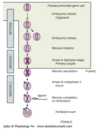
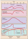
Describe what occurs during the follwing dates

  1. gestaion
    1. week 5-6
    2. month2-6
  2. after puberty
    1. day 28 of cycle
A
  1. gestation
    1. week 5-6
      1. primordial germ cells divide mitotically
        1. by week20-24 = 7million
    2. 2month-6month gestation
      1. oogonia enter prophase of meiosis 1 and become primary oocytes and remain in prophase until sexual maturity
    3. primary oocytes + layer of granulosa cells = primordial follicle
      1. basil lamina is formed on the outside of the granulosa cells
  2. after puberty
    1. formation of secondary follicle from primary follicle is characterized via
      1. addition of thecal layer of cells
    2. post pubertal follicular development is under the control of cyclic FSH release
    3. day 28 of cycle = graafian follicle development
      1. occurs 70-85 days post menarche(first menstrual cycle)
      2. vesicles of the secondary follicle coalesce forming an antrum rich in estrogen
        1. estrogen is due to theca and granulosa cells in the cumulusoophorus
      3. graafian follicle increases in size
      4. theca cells strech and blister in a bulge formation
21
Q

post pubertal follicular development is under the control of cyclic ___ release

A
  1. gestation
    1. week 5-6
      1. primordial germ cells divide mitotically
        1. by week20-24 = 7million
    2. week 2month-6month gestation
      1. oogonia enter prophase of meiosis 1 and become primary oocytes and remain in prophase until sexual maturity
    3. primary oocytes + layer of granulosa cells = primordial follicle
      1. basil lamina is formed on the outside of the granulosa cells
  2. after puberty
    1. formation of secondary follicle from primary follicle is characterized via
      1. addition of thecal layer of cells
    2. post pubertal follicular development is under the control of cyclic FSH release
    3. day 28 of cycle = graafian follicle development
      1. occurs 70-85 days post menarche(first menstrual cycle)
      2. vesicles of the secondary follicle coalesce forming an antrum rich in estrogen
        1. estrogen is due to theca and granulosa cells in the cumulusoophorus
      3. graafian follicle increases in size
      4. theca cells strech and blister in a bulge formation
22
Q

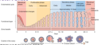
describe the flow of maturation in oogenesis

  1. primordial germ
  2. oogonia
  3. primary oocytes
  4. primordial follicle
  5. primary follicle
  6. secondary follicle
  7. graafian follicle
A
  1. gestation
    1. week 5-6
      1. primordial germ cells divide mitotically
        1. by week20-24 = 7million
    2. week 2month-6month gestation
      1. oogonia enter prophase of meiosis 1 and become primary oocytes and remain in prophase until sexual maturity
    3. primary oocytes + layer of granulosa cells = primordial follicle
      1. basil lamina is formed on the outside of the granulosa cells
  2. after puberty
    1. formation of secondary follicle from primary follicle is characterized via
      1. addition of thecal layer of cells
    2. post pubertal follicular development is under the control of cyclic FSH release
    3. day 28 of cycle = graafian follicle development
      1. occurs 70-85 days post menarche(first menstrual cycle)
      2. vesicles of the secondary follicle coalesce forming an antrum rich in estrogen
        1. estrogen is due to theca and granulosa cells in the cumulusoophorus
      3. graafian follicle increases in size
      4. theca cells strech and blister in a bulge formation
23
Q

what occurs 3-4 months after menarche?

A
  1. gestation
    1. week 5-6
      1. primordial germ cells divide mitotically
        1. by week20-24 = 7million
    2. week 2month-6month gestation
      1. oogonia enter prophase of meiosis 1 and become primary oocytes and remain in prophase until sexual maturity
    3. primary oocytes + layer of granulosa cells = primordial follicle
      1. basil lamina is formed on the outside of the granulosa cells
  2. after puberty
    1. formation of secondary follicle from primary follicle is characterized via
      1. addition of thecal layer of cells
    2. post pubertal follicular development is under the control of cyclic FSH release
    3. day 28 of cycle = graafian follicle development
      1. occurs 70-85 days post menarche(first menstrual cycle)
      2. vesicles of the secondary follicle coalesce forming an antrum rich in estrogen
        1. estrogen is due to theca and granulosa cells in the cumulusoophorus
      3. graafian follicle increases in size
      4. theca cells strech and blister in a bulge formation
24
Q

describe the phases that the oocyte is arrested in and why

A
  1. oogonium
    1. meiosis begins
    2. Integral proteins are low in concentration so the oocyte “arrests” in prophase 1 for up to 50 years
  2. primary oocyte
    1. as oocyte grows it synthesizes proteins required to complete meiosis, but high cAMP levels actively maintain arrested state
    2. just prior to ovulation
      1. oocyte completes meiorsis 1 and extrudes 1st polar body
      2. arresting in metaphase 2
  3. secondary oocyte
    1. completes meiosis at fertilization and extrudes second polar body
25
Q

What occurs in the oocyte with a decrease/increase in cAMP levels?

A
  1. oogonium
    1. meiosis begins
    2. Integral proteins are low in concentration so the oocyte “arrests” in prophase 1 for up to 50 years
  2. primary oocyte
    1. as oocyte grows it synthesizes proteins required to complete meiosis, but high cAMP levels actively maintain arrested state
    2. just prior to ovulation
      1. oocyte completes meiorsis 1 and extrudes 1st polar body
      2. arresting in metaphase 2
  3. secondary oocyte
    1. completes meiosis at fertilization and extrudes second polar body
      1. cAMP decreases allowing meiosis to finish
26
Q

How many follicles does a woman start with and how many are left at the point of menarche?

A

ovarian reserve in women

  1. 7million primary oocytes by 5th month of gestation
    1. >6million follicles experience atresia
  2. ovarian reserve
    1. less than 300k primordial follicles at menarche
    2. atresia > 270kfollicles
  3. growth
    1. less 30k primordial follicles
  4. ovarian ovulation
    1. less than 500 dominant follicles
27
Q

describe the ovarian reserve in women

A

ovarian reserve in women

  1. 7million primary oocytes by 5th month of gestation
    1. >6million follicles experience atresia
  2. ovarian reserve
    1. less than 300k primordial follicles at menarche
    2. atresia > 270kfollicles
  3. growth
    1. less 30k primordial follicles
  4. ovarian ovulation
    1. less than 500 dominant follicles
28
Q

What if the frequency of GnRH release for the stimulation of follicular development?

A

GnRH released in pulsatile manner

  1. high frequency
    1. 1 every 60-90 min promotes LH production
  2. low frequency
    1. every 120 min promotes FSH production
29
Q

Stimulation of GnRH in what frequency leads to an increase of 17a-OHProgesterone? What cell is affected?

A

GnRH released in pulsatile manner

  1. high frequency
    1. 1 every 60-90 min promotes LH production
  2. low frequency
    1. every 120 min promotes FSH production
30
Q

Describe the two organs involved with hypothalamic -pituitary-axis cycle

A
  1. two organs
    1. ovary
      1. follicular and luteal phases separated by oculation
    2. uterus -endometrial cycle
      1. menstrual
      2. proliferatie and secretory phases
  2. monthly pattern results from the interaction of ovarian steroids and peptides and their ability to exert both posistive and negative feedback on the hypothalamus and pituitary
  3. three phases of the munstrual cycle
    1. overall duration of “normal” cycle =28 days
      1. can range from21-35days
    2. phases
      1. follucular phase
        1. begins with onset of menstrual bleeding
        2. ~15 days
        3. most variable part of cycle (9-23day)
      2. ovulatory phase
        1. ~1-3days
        2. culminate in ovulation
      3. luteal phase
        1. lasts 13-14 days and is a fixed amount of time, this will not adjust in length
        2. ends with menstrual bleeding
31
Q

list and describe the two organs and their phase involvement in menstrual cycle

A
  1. two organs
    1. ovary
      1. follicular and luteal phases separated by oculation
    2. uterus -endometrial cycle
      1. menstrual
      2. proliferatie and secretory phases
  2. monthly pattern results from the interaction of ovarian steroids and peptides and their ability to exert both posistive and negative feedback on the hypothalamus and pituitary
  3. three phases of the munstrual cycle
    1. overall duration of “normal” cycle =28 days
      1. can range from21-35days
    2. phases
      1. follucular phase
        1. begins with onset of menstrual bleeding
        2. ~15 days
        3. most variable part of cycle (9-23day)
      2. ovulatory phase
        1. ~1-3days
        2. culminate in ovulation
      3. luteal phase
        1. lasts 13-14 days and is a fixed amount of time, this will not adjust in length
        2. ends with menstrual bleeding
32
Q
  1. begins with onset of menstrual bleeding
  2. ~15 days
  3. most variable part of cycle (9-23day)
A
  1. two organs
    1. ovary
      1. follicular and luteal phases separated by oculation
    2. uterus -endometrial cycle
      1. menstrual
      2. proliferatie and secretory phases
  2. monthly pattern results from the interaction of ovarian steroids and peptides and their ability to exert both posistive and negative feedback on the hypothalamus and pituitary
  3. three phases of the munstrual cycle
    1. overall duration of “normal” cycle =28 days
      1. can range from21-35days
    2. phases
      1. follucular phase
        1. begins with onset of menstrual bleeding
        2. ~15 days
        3. most variable part of cycle (9-23day)
      2. ovulatory phase
        1. ~1-3days
        2. culminate in ovulation
      3. luteal phase
        1. lasts 13-14 days and is a fixed amount of time, this will not adjust in length
        2. ends with menstrual bleeding
33
Q
  1. ~1-3days
  2. culminate in ovulation
A
  1. two organs
    1. ovary
      1. follicular and luteal phases separated by oculation
    2. uterus -endometrial cycle
      1. menstrual
      2. proliferatie and secretory phases
  2. monthly pattern results from the interaction of ovarian steroids and peptides and their ability to exert both posistive and negative feedback on the hypothalamus and pituitary
  3. three phases of the munstrual cycle
    1. overall duration of “normal” cycle =28 days
      1. can range from21-35days
    2. phases
      1. follucular phase
        1. begins with onset of menstrual bleeding
        2. ~15 days
        3. most variable part of cycle (9-23day)
      2. ovulatory phase
        1. ~1-3days
        2. culminate in ovulation
      3. luteal phase
        1. lasts 13-14 days and is a fixed amount of time, this will not adjust in length
        2. ends with menstrual bleeding
34
Q

lasts 13-14 days and is a fixed amount of time, this will not adjust in length

ends with menstrual bleeding

A
  1. two organs
    1. ovary
      1. follicular and luteal phases separated by oculation
    2. uterus -endometrial cycle
      1. menstrual
      2. proliferatie and secretory phases
  2. monthly pattern results from the interaction of ovarian steroids and peptides and their ability to exert both posistive and negative feedback on the hypothalamus and pituitary
  3. three phases of the munstrual cycle
    1. overall duration of “normal” cycle =28 days
      1. can range from21-35days
    2. phases
      1. follucular phase
        1. begins with onset of menstrual bleeding
        2. ~15 days
        3. most variable part of cycle (9-23day)
      2. ovulatory phase
        1. ~1-3days
        2. culminate in ovulation
      3. luteal phase
        1. lasts 13-14 days and is a fixed amount of time, this will not adjust in length
        2. ends with menstrual bleeding
35
Q

describe the phases of mentrual cycle

A
  1. two organs
    1. ovary
      1. follicular and luteal phases separated by oculation
    2. uterus -endometrial cycle
      1. menstrual
      2. proliferatie and secretory phases
  2. monthly pattern results from the interaction of ovarian steroids and peptides and their ability to exert both posistive and negative feedback on the hypothalamus and pituitary
  3. three phases of the munstrual cycle
    1. overall duration of “normal” cycle =28 days
      1. can range from21-35days
    2. phases
      1. follucular phase
        1. begins with onset of menstrual bleeding
        2. ~15 days
        3. most variable part of cycle (9-23day)
      2. ovulatory phase
        1. ~1-3days
        2. culminate in ovulation
      3. luteal phase
        1. lasts 13-14 days and is a fixed amount of time, this will not adjust in length
        2. ends with menstrual bleeding
36
Q

describe some important events in the follicular stage(follicular can be group into early, intermediate and late)

A

ovarian cycle

  1. follicular phase
    1. 1-13 early follicular phase
      1. high Progesterone in luteal phase is thought to slow GnRH pulses resulting in selective stimulation of FSH production
      2. No fertilization =regression of corpus luteum
      3. inhibin, E and P are low
      4. menses occurs here
      5. low hormone levels = gonadotroph does not experience -feedback = increase in FSH secretion
      6. characterized by
        1. recruitment and growth of 15-20 antral follicles
        2. growth of a dominanat follicle until ovulation
      7. rise in FSH recruits a crop of large antral follicles that begin rapid FSH dependent growth
        1. they produce low levels of E and inhibinB
        2. Take not in the photo around day 1 estradiol increase a little bit
    2. mid-follicular phase
      1. rising E and inhibin B = (-) feedback on FSH secretion
      2. prolonged low levels of P and E increase the frequency of GnRH pulses
        1. selective increae in LH synthesis and secretion
      3. LH/FSH ratio increases during follicular phase
        1. note the change from day 2-12, that LH is increasing and FSH is decreasing = inhibin decreases the FSH
      4. decrease FSH leads to atresia of developing follicle
        1. largest follicle with the most FSH receptors will survive
    3. late-follicular phase
      1. FSH also induces expression of LH receptors on granulosa cells of dominant follicle making it more sensittive to the LH surge
        1. which is why the follicle with the most FSH receptors continues to maturation
      2. dominant follicle produces increasing amount of
        1. estradiol 17-B
        2. inhibin
      3. once estradiol 17-B reaches ~20pg/mL for 48 hrs estrogen begins to have a positive feedback on gonadotroph
      4. High E and little P has + effect on LH and FSH
  2. ovulation
    1. germinal vesicle breakldown occurs after LH surge = 1.5 days-> cumulus oocyte is released from wall of follicle
    2. two structures
      1. corpus luteum-
        1. two actions increase LDL and HDL and StAR
          1. basil lamina degrade of granulosa cells
            1. causing release of VEGF
          2. increase in GF
        2. restructization of the theca and granulosa cells ->estrogen and progesterone generation
      2. oocyte
        1. traveling towards to the uterine tubule
    3. ovarian cycle: luteal phase
      1. early luteal phase: initial decrease in E
        1. terminates + feedback on LH
        2. corpus luteu matures and increases levels in P and E = - feedback on both LH and FSH
      2. late luteal phase:
        1. Corpus luteum starts to regress = decrease in P and E
      3. no fertilization
        1. regression of corpus luteum
        2. inhibin, P and E are low
        3. gonadotroph now experiences no - feedback = increase in FSH
    4. menses - see the uterine cycle
      1. coincides with the follicular phase
37
Q

what phase is marked by atresia of developing follicles? Describe this process

A

ovarian cycle

  1. follicular phase
    1. 1-13 early follicular phase
      1. high Progesterone in luteal phase is thought to slow GnRH pulses resulting in selective stimulation of FSH production
      2. No fertilization =regression of corpus luteum
      3. inhibin, E and P are low
      4. low hormone levels = gonadotroph does not experience -feedback = increase in FSH secretion
      5. characterized by
        1. recruitment and growth of 15-20 antral follicles
        2. growth of a dominanat follicle until ovulation
      6. rise in FSH recruits a crop of large antral follicles that begin rapid FSH dependent growth
        1. they produce low levels of E and inhibinB
        2. Take not in the photo around day 1 estradiol increase a little bit
    2. mid-follicular phase
      1. rising E and inhibin B= - feedback on FSH secretion
      2. prolonged low levels of P and E increase the frequency of GnRH pulses
        1. selective increae in LH synthesis and secretion
      3. LH/FSH ratio increases during follicular phase
        1. note the change from day 2-12, that LH is increasing and FSH is decreasing = inhibin decreases the FSH
      4. decrease FSH leads to atresia of developing follicle
        1. largest follicle with the most FSH receptors will survive
    3. late-follicular phase
      1. FSH also induces expression of LH receptors on granulosa cells of dominant follicle making it more sensittive to the LH surge
        1. which is why the follicle with the most FSH receptors continues to maturation
      2. dominant follicle produces increasing amount of
        1. estradiol 17-B
        2. inhibin
      3. once estradiol 17-B reaches ~20pg/mL for 48 hrs estrogen begins to have a positive feedback on gonadotroph
      4. High E and little P has + effect on LH and FSH
  2. ovulation
    1. germinal vesicle breakldown occurs after LH surge = 1.5 days-> cumulus oocyte is released from wall of follicle
    2. two structures
      1. corpus luteum-
        1. two actions increase LDL and HDL and StAR
          1. basil lamina degrade of granulosa cells
            1. causing release of VEGF
          2. increase in GF
        2. restructization of the theca and granulosa cells ->estrogen and progesterone generation
      2. oocyte
        1. traveling towards to the uterine tubule
    3. ovarian cycle: luteal phase
      1. early luteal phase: initial decrease in E
        1. terminates + feedback on LH
        2. corpus luteu matures and increases levels in P and E = - feedback on both LH and FSH
      2. late luteal phase:
        1. Corpus luteum starts to regress = decrease in P and E
      3. no fertilization
        1. regression of corpus luteum
        2. inhibin, P and E are low
        3. gonadotroph now experiences no - feedback = increase in FSH
    4. menses - see the uterine cycle
      1. coincides with the follicular phase
38
Q

diagram and explain the follicular and luteal phases

A

ovarian cycle

  1. follicular phase
    1. 1-13 early follicular phase
      1. high Progesterone in luteal phase is thought to slow GnRH pulses resulting in selective stimulation of FSH production
      2. No fertilization =regression of corpus luteum
      3. inhibin, E and P are low
      4. low hormone levels = gonadotroph does not experience -feedback = increase in FSH secretion
      5. characterized by
        1. recruitment and growth of 15-20 antral follicles
        2. growth of a dominanat follicle until ovulation
      6. rise in FSH recruits a crop of large antral follicles that begin rapid FSH dependent growth
        1. they produce low levels of E and inhibinB
        2. Take not in the photo around day 1 estradiol increase a little bit
    2. mid-follicular phase
      1. rising E and inhibin B= - feedback on FSH secretion
      2. prolonged low levels of P and E increase the frequency of GnRH pulses
        1. selective increae in LH synthesis and secretion
      3. LH/FSH ratio increases during follicular phase
        1. note the change from day 2-12, that LH is increasing and FSH is decreasing = inhibin decreases the FSH
      4. decrease FSH leads to atresia of developing follicle
        1. largest follicle with the most FSH receptors will survive
    3. late-follicular phase
      1. FSH also induces expression of LH receptors on granulosa cells of dominant follicle making it more sensittive to the LH surge
        1. which is why the follicle with the most FSH receptors continues to maturation
      2. dominant follicle produces increasing amount of
        1. estradiol 17-B
        2. inhibin
      3. once estradiol 17-B reaches ~20pg/mL for 48 hrs estrogen begins to have a positive feedback on gonadotroph
      4. High E and little P has + effect on LH and FSH
  2. ovulation
    1. germinal vesicle breakldown occurs after LH surge = 1.5 days-> cumulus oocyte is released from wall of follicle
    2. two structures
      1. corpus luteum-
        1. two actions increase LDL and HDL and StAR
          1. basil lamina degrade of granulosa cells
            1. causing release of VEGF
          2. increase in GF
        2. restructization of the theca and granulosa cells ->estrogen and progesterone generation
      2. oocyte
        1. traveling towards to the uterine tubule
    3. ovarian cycle: luteal phase
      1. early luteal phase: initial decrease in E
        1. terminates + feedback on LH
        2. corpus luteu matures and increases levels in P and E = - feedback on both LH and FSH
      2. late luteal phase:
        1. Corpus luteum starts to regress = decrease in P and E
      3. no fertilization
        1. regression of corpus luteum
        2. inhibin, P and E are low
        3. gonadotroph now experiences no - feedback = increase in FSH
    4. menses - see the uterine cycle
      1. coincides with the follicular phase
39
Q

describe the important events in the luteal cycle

A

ovarian cycle

  1. follicular phase
    1. 1-13 early follicular phase
      1. high Progesterone in luteal phase is thought to slow GnRH pulses resulting in selective stimulation of FSH production
      2. No fertilization =regression of corpus luteum
      3. inhibin, E and P are low
      4. low hormone levels = gonadotroph does not experience -feedback = increase in FSH secretion
      5. characterized by
        1. recruitment and growth of 15-20 antral follicles
        2. growth of a dominanat follicle until ovulation
      6. rise in FSH recruits a crop of large antral follicles that begin rapid FSH dependent growth
        1. they produce low levels of E and inhibinB
        2. Take not in the photo around day 1 estradiol increase a little bit
    2. mid-follicular phase
      1. rising E and inhibin B= - feedback on FSH secretion
      2. prolonged low levels of P and E increase the frequency of GnRH pulses
        1. selective increae in LH synthesis and secretion
      3. LH/FSH ratio increases during follicular phase
        1. note the change from day 2-12, that LH is increasing and FSH is decreasing = inhibin decreases the FSH
      4. decrease FSH leads to atresia of developing follicle
        1. largest follicle with the most FSH receptors will survive
    3. late-follicular phase
      1. FSH also induces expression of LH receptors on granulosa cells of dominant follicle making it more sensittive to the LH surge
        1. which is why the follicle with the most FSH receptors continues to maturation
      2. dominant follicle produces increasing amount of
        1. estradiol 17-B
        2. inhibin
      3. once estradiol 17-B reaches ~20pg/mL for 48 hrs estrogen begins to have a positive feedback on gonadotroph
      4. High E and little P has + effect on LH and FSH
  2. ovulation
    1. germinal vesicle breakldown occurs after LH surge = 1.5 days-> cumulus oocyte is released from wall of follicle
    2. two structures
      1. corpus luteum-
        1. two actions increase LDL and HDL and StAR
          1. basil lamina degrade of granulosa cells
            1. causing release of VEGF
          2. increase in GF
        2. restructization of the theca and granulosa cells ->estrogen and progesterone generation
      2. oocyte
        1. traveling towards to the uterine tubule
  3. ovarian cycle: luteal phase
    1. early luteal phase: initial decrease in E
      1. terminates + feedback on LH
      2. corpus luteu matures and increases levels in P and E = - feedback on both LH and FSH
    2. late luteal phase:
      1. Corpus luteum starts to regress = decrease in P and E
    3. no fertilization
      1. regression of corpus luteum
      2. inhibin, P and E are low
      3. gonadotroph now experiences no - feedback = increase in FSH
  4. menses - see the uterine cycle
    1. coincides with the follicular phase
40
Q

what occurs during the 26th-28th day of the luteal phase? describe before and after this stage.

A

ovarian cycle

  1. follicular phase
    1. 1-13 early follicular phase
      1. high Progesterone in luteal phase is thought to slow GnRH pulses resulting in selective stimulation of FSH production
      2. No fertilization =regression of corpus luteum
      3. inhibin, E and P are low
      4. low hormone levels = gonadotroph does not experience -feedback = increase in FSH secretion
      5. characterized by
        1. recruitment and growth of 15-20 antral follicles
        2. growth of a dominanat follicle until ovulation
      6. rise in FSH recruits a crop of large antral follicles that begin rapid FSH dependent growth
        1. they produce low levels of E and inhibinB
        2. Take not in the photo around day 1 estradiol increase a little bit
    2. mid-follicular phase
      1. rising E and inhibin B= - feedback on FSH secretion
      2. prolonged low levels of P and E increase the frequency of GnRH pulses
        1. selective increae in LH synthesis and secretion
      3. LH/FSH ratio increases during follicular phase
        1. note the change from day 2-12, that LH is increasing and FSH is decreasing = inhibin decreases the FSH
      4. decrease FSH leads to atresia of developing follicle
        1. largest follicle with the most FSH receptors will survive
    3. late-follicular phase
      1. FSH also induces expression of LH receptors on granulosa cells of dominant follicle making it more sensittive to the LH surge
        1. which is why the follicle with the most FSH receptors continues to maturation
      2. dominant follicle produces increasing amount of
        1. estradiol 17-B
        2. inhibin
      3. once estradiol 17-B reaches ~20pg/mL for 48 hrs estrogen begins to have a positive feedback on gonadotroph
      4. High E and little P has + effect on LH and FSH
  2. ovulation
    1. germinal vesicle breakldown occurs after LH surge = 1.5 days-> cumulus oocyte is released from wall of follicle
    2. two structures
      1. corpus luteum-
        1. two actions increase LDL and HDL and StAR
          1. basil lamina degrade of granulosa cells
            1. causing release of VEGF
          2. increase in GF
        2. restructization of the theca and granulosa cells ->estrogen and progesterone generation
      2. oocyte
        1. traveling towards to the uterine tubule
    3. ovarian cycle: luteal phase
      1. early luteal phase: initial decrease in E
        1. terminates + feedback on LH
        2. corpus luteu matures and increases levels in P and E = - feedback on both LH and FSH
      2. late luteal phase:
        1. Corpus luteum starts to regress = decrease in P and E
      3. no fertilization
        1. regression of corpus luteum
        2. inhibin, P and E are low
        3. gonadotroph now experiences no - feedback = increase in FSH
    4. menses - see the uterine cycle
      1. coincides with the follicular phase
41
Q

describe the selection of an oocyte for ovulation. think of it as 1= at ovulation

2= post ovulation

A

oogenesis: selection for ovulation

  1. MOST RAPID STAGE lasting 7-10 days
  2. the Graafian follicle that is selected for ovulation is determined by the number of estrogen and FSH receptors
  3. ovulation occurs under influence of LH
    1. at ovulation
      1. 1st meitotic division (2n->1n) is complete
        1. generating a secondary oocyte
      2. secondary oocyte begins the 2nd meitotic division but is suspended at metaphase 2
      3. germinal vesicle breakdown occurs ~30 hrs after LH surge=dissolution of nuclear membrane and intterphase of nuclear structure
    2. after ovulation
      1. secondary oocyte travels down ampulla of oviduct
      2. sperm penetration of the zona pellucida completes the 2nd meitotic division forming and ootid and the 2nd polar body which degenerattes
  4. remainig follicle in the ovary->corpu luteum
    1. at the rupture is the corpus hemorrhagium
      1. wall of follicle and ovary at the stigma are broken down with release of cumulus-oocyte (ovulation) 1.5 days after LH surge
    2. under the influence of LH
      1. granulosa and theca cells form the corpus luteum (yellow body)
        1. yellow due to increase in cholesterol synthesis and use to generate sex steroids
        2. enzymatic degradation of basal lamina of granulosa cells causes release of VEGF, angiopoetin 2 and fibroblast growth factor
          1. these are increase LDL,HDL and StAR acttivity
    3. transient decrease in aromatase
      1. stops + feedback of estrogen on gonadotroph and allows the granulosa and theca cells to restructure into corpus luteum
      2. note the drop in estrogen for ~ 2 days
    4. produces progesterone and
      1. pregnancy
        1. persists if pregnancy ensues
      2. no pregnancy
        1. corpus luteum -> corpus albicans (white body) towards end of luteum
42
Q

what is the most rapid stage lasting 7-10 days?

A

oogenesis: selection for ovulation

  1. MOST RAPID STAGE lasting 7-10 days
  2. the Graafian follicle that is selected for ovulation is determined by the number of estrogen and FSH receptors
  3. ovulation occurs under influence of LH
    1. at ovulation
      1. 1st meitotic division (2n->1n) is complete
        1. generating a secondary oocyte
      2. secondary oocyte begins the 2nd meitotic division but is suspended at metaphase 2
      3. germinal vesicle breakdown occurs ~30 hrs after LH surge=dissolution of nuclear membrane and intterphase of nuclear structure
        1. after ovulation
      4. secondary oocyte travels down ampulla of oviduct
      5. sperm penetration of the zona pellucida completes the 2nd meitotic division forming and ootid and the 2nd polar body which degenerattes
  4. remainig follicle in the ovary->corpu luteum
    1. at the rupture is the corpus hemorrhagium
      1. wall of follicle and ovary at the stigma are broken down with release of cumulus-oocyte (ovulation) 1.5 days after LH surge
    2. under the influence of LH
      1. granulosa and theca cells form the corpus luteum (yellow body)
        1. yellow due to increase in cholesterol synthesis and use to generate sex steroids
        2. enzymatic degradation of basal lamina of granulosa cells causes release of VEGF, angiopoetin 2 and fibroblast growth factor
          1. these are increase LDL,HDL and StAR acttivity
    3. transient decrease in aromatase
      1. stops + feedback of estrogen on gonadotroph and allows the granulosa and theca cells to restructure into corpus luteum
      2. note the drop in estrogen for ~ 2 days
    4. produces progesterone and
      1. pregnancy
        1. persists if pregnancy ensues
      2. no pregnancy
        1. corpus luteum -> corpus albicans (white body) towards end of luteum
43
Q

describe germinal breakdown with regards to hormone and time

A

oogenesis: selection for ovulation

  1. MOST RAPID STAGE lasting 7-10 days
  2. the Graafian follicle that is selected for ovulation is determined by the number of estrogen and FSH receptors
  3. ovulation occurs under influence of LH
    1. at ovulation
      1. 1st meitotic division (2n->1n) is complete
        1. generating a secondary oocyte
      2. secondary oocyte begins the 2nd meitotic division but is suspended at metaphase 2
      3. germinal vesicle breakdown occurs ~30 hrs after LH surge=dissolution of nuclear membrane and intterphase of nuclear structure
        1. after ovulation
      4. secondary oocyte travels down ampulla of oviduct
      5. sperm penetration of the zona pellucida completes the 2nd meitotic division forming and ootid and the 2nd polar body which degenerattes
  4. remainig follicle in the ovary->corpu luteum
    1. at the rupture is the corpus hemorrhagium
      1. wall of follicle and ovary at the stigma are broken down with release of cumulus-oocyte (ovulation) 1.5 days after LH surge
    2. under the influence of LH
      1. granulosa and theca cells form the corpus luteum (yellow body)
        1. yellow due to increase in cholesterol synthesis and use to generate sex steroids
        2. enzymatic degradation of basal lamina of granulosa cells causes release of VEGF, angiopoetin 2 and fibroblast growth factor
          1. these are increase LDL,HDL and StAR acttivity
    3. transient decrease in aromatase
      1. stops + feedback of estrogen on gonadotroph and allows the granulosa and theca cells to restructure into corpus luteum
      2. note the drop in estrogen for ~ 2 days
    4. produces progesterone and
      1. pregnancy
        1. persists if pregnancy ensues
      2. no pregnancy
        1. corpus luteum -> corpus albicans (white body) towards end of luteum
44
Q

remaining follicle post ovulation leaves a structure in the ovary. Describe the important steps taken to develop and hormones involved

A

oogenesis: selection for ovulation

  1. MOST RAPID STAGE lasting 7-10 days
  2. the Graafian follicle that is selected for ovulation is determined by the number of estrogen and FSH receptors
  3. ovulation occurs under influence of LH
    1. at ovulation
      1. 1st meitotic division (2n->1n) is complete
        1. generating a secondary oocyte
      2. secondary oocyte begins the 2nd meitotic division but is suspended at metaphase 2
      3. germinal vesicle breakdown occurs ~30 hrs after LH surge=dissolution of nuclear membrane and intterphase of nuclear structure
        1. after ovulation
      4. secondary oocyte travels down ampulla of oviduct
      5. sperm penetration of the zona pellucida completes the 2nd meitotic division forming and ootid and the 2nd polar body which degenerattes
  4. remainig follicle in the ovary->corpu luteum
    1. at the rupture is the corpus hemorrhagium
      1. wall of follicle and ovary at the stigma are broken down with release of cumulus-oocyte (ovulation) 1.5 days after LH surge
    2. under the influence of LH
      1. granulosa and theca cells form the corpus luteum (yellow body)
        1. yellow due to increase in cholesterol synthesis and use to generate sex steroids
        2. enzymatic degradation of basal lamina of granulosa cells causes release of VEGF, angiopoetin 2 and fibroblast growth factor
          1. these are increase LDL,HDL and StAR acttivity
    3. transient decrease in aromatase
      1. stops + feedback of estrogen on gonadotroph and allows the granulosa and theca cells to restructure into corpus luteum
      2. note the drop in estrogen for ~ 2 days
    4. produces progesterone and
      1. pregnancy
        1. persists if pregnancy ensues
      2. no pregnancy
        1. corpus luteum -> corpus albicans (white body) towards end of luteum
45
Q

describe the structures in the ovary that occur under pregnancy and no pregnancy

A

oogenesis: selection for ovulation

  1. MOST RAPID STAGE lasting 7-10 days
  2. the Graafian follicle that is selected for ovulation is determined by the number of estrogen and FSH receptors
  3. ovulation occurs under influence of LH
    1. at ovulation
      1. 1st meitotic division (2n->1n) is complete
        1. generating a secondary oocyte
      2. secondary oocyte begins the 2nd meitotic division but is suspended at metaphase 2
      3. germinal vesicle breakdown occurs ~30 hrs after LH surge=dissolution of nuclear membrane and intterphase of nuclear structure
        1. after ovulation
      4. secondary oocyte travels down ampulla of oviduct
      5. sperm penetration of the zona pellucida completes the 2nd meitotic division forming and ootid and the 2nd polar body which degenerattes
  4. remainig follicle in the ovary->corpu luteum
    1. at the rupture is the corpus hemorrhagium
      1. wall of follicle and ovary at the stigma are broken down with release of cumulus-oocyte (ovulation) 1.5 days after LH surge
    2. under the influence of LH
      1. granulosa and theca cells form the corpus luteum (yellow body)
        1. yellow due to increase in cholesterol synthesis and use to generate sex steroids
        2. enzymatic degradation of basal lamina of granulosa cells causes release of VEGF, angiopoetin 2 and fibroblast growth factor
          1. these are increase LDL,HDL and StAR acttivity
    3. transient decrease in aromatase
      1. stops + feedback of estrogen on gonadotroph and allows the granulosa and theca cells to restructure into corpus luteum
      2. note the drop in estrogen for ~ 2 days
    4. produces progesterone and
      1. pregnancy
        1. persists if pregnancy ensues
      2. no pregnancy
        1. corpus luteum -> corpus albicans (white body) towards end of luteum
46
Q

what leads to a decrease in aromatase activity of the granulosa cells and why?

A

oogenesis: selection for ovulation

  1. MOST RAPID STAGE lasting 7-10 days
  2. the Graafian follicle that is selected for ovulation is determined by the number of estrogen and FSH receptors
  3. ovulation occurs under influence of LH
    1. at ovulation
      1. 1st meitotic division (2n->1n) is complete
        1. generating a secondary oocyte
      2. secondary oocyte begins the 2nd meitotic division but is suspended at metaphase 2
      3. germinal vesicle breakdown occurs ~30 hrs after LH surge=dissolution of nuclear membrane and intterphase of nuclear structure
        1. after ovulation
      4. secondary oocyte travels down ampulla of oviduct
      5. sperm penetration of the zona pellucida completes the 2nd meitotic division forming and ootid and the 2nd polar body which degenerattes
  4. remainig follicle in the ovary->corpu luteum
    1. at the rupture is the corpus hemorrhagium
      1. wall of follicle and ovary at the stigma are broken down with release of cumulus-oocyte (ovulation) 1.5 days after LH surge
    2. under the influence of LH
      1. granulosa and theca cells form the corpus luteum (yellow body)
        1. yellow due to increase in cholesterol synthesis and use to generate sex steroids
        2. enzymatic degradation of basal lamina of granulosa cells causes release of VEGF, angiopoetin 2 and fibroblast growth factor
          1. these are increase LDL,HDL and StAR acttivity
    3. transient decrease in aromatase
      1. stops + feedback of estrogen on gonadotroph and allows the granulosa and theca cells to restructure into corpus luteum
      2. note the drop in estrogen for ~ 2 days
    4. produces progesterone and
      1. pregnancy
        1. persists if pregnancy ensues
      2. no pregnancy
        1. corpus luteum -> corpus albicans (white body) towards end of luteum
47
Q

describe the hormones and their contribution for the uterine cycle. (2 phases)

A

uterine cycle

  1. phase
    1. proliferative phase
      1. coincides with follicular phase
      2. rising E in mid-late follicular phase induce all cell types in the stratum basale to grow and divide
      3. endometrial lining starts to grow
      4. cell prolliferation occurs
        1. directly
          1. from estrogen receptor
        2. indirectly production of growth factors like
          1. IGF1
          2. VEGF
      5. arteries become more abundant and uterine glands form
      6. E induces expression of P receptors
        1. this PRIMES the uterus to repond to pregesteron during the luteal phase of the cycle
    2. secretory phase
      1. preperation for implantation
      2. under influence of P and E
      3. coincides with luteal phase
      4. P induces
        1. differentiation of epithelial and stromal cells
        2. uterine glands to fill with glycogen vacuoles - supports the embryo
        3. inactivating isoforms of 17B-HSD
          1. so active estradiol is converted to inactive estrone
            1. important for protecting the endometrium from estrogen induced uterine cancer
      5. P inhibits
        1. proliferative actions of E by down regulating the ER
48
Q

answering the following in regard to the proliferative phase

  1. coincides with the ____
  2. what type of estrogen induces stratum basale cells to grow?
  3. what grow as a response? cell proliferation occurs directly and indirectly
  4. estrogen induces expression of whatt receptors?
A

uterine cycle

  1. phase
    1. proliferative phase
      1. coincides with follicular phase
      2. rising E in mid-late follicular phase induce all cell types in the stratum basale to grow and divide
      3. endometrial lining starts to grow
      4. cell prolliferation occurs
        1. directly
          1. from estrogen receptor
        2. indirectly production of growth factors like
          1. IGF1
          2. VEGF
      5. arteries become more abundant and uterine glands form
      6. E induces expression of P receptors
        1. this PRIMES the uterus to repond to pregesteron during the luteal phase of the cycle
    2. secretory phase
      1. preperation for implantation
      2. under influence of P and E
      3. coincides with luteal phase
      4. P induces
        1. differentiation of epithelial and stromal cells
        2. uterine glands to fill with glycogen vacuoles - supports the embryo
        3. inactivating isoforms of 17B-HSD
          1. so active estradiol is converted to inactive estrone
            1. important for protecting the endometrium from estrogen induced uterine cancer
      5. P inhibits
        1. proliferative actions of E by down regulating the ER
49
Q

answer the following questions with regard to the secretory phase

  1. preperation for ____
  2. under the influence of ___ and ____
  3. Progesterone
    1. induces -3 things
    2. inhibits - 1 thing
A

uterine cycle

  1. phase
    1. proliferative phase
      1. coincides with follicular phase
      2. rising E in mid-late follicular phase induce all cell types in the stratum basale to grow and divide
      3. endometrial lining starts to grow
      4. cell prolliferation occurs
        1. directly
          1. from estrogen receptor
        2. indirectly production of growth factors like
          1. IGF1
          2. VEGF
      5. arteries become more abundant and uterine glands form
      6. E induces expression of P receptors
        1. this PRIMES the uterus to repond to pregesteron during the luteal phase of the cycle
    2. secretory phase
      1. preperation for implantation
      2. under influence of P and E
      3. coincides with luteal phase
      4. P induces
        1. differentiation of epithelial and stromal cells
        2. uterine glands to fill with glycogen vacuoles - supports the embryo
        3. inactivating isoforms of 17B-HSD
          1. so active estradiol is converted to inactive estrone
            1. important for protecting the endometrium from estrogen induced uterine cancer
      5. P inhibits
        1. proliferative actions of E by down regulating the ER
50
Q

explain the following about uterine cycle menstruation

  1. a sudden withdrawl of P is an indication of what?
  2. what does this lead to?
A

uterine cycle

  1. menstruation
    1. in a non-fertile cycle the corpus luteum dies and there is a sudden withdrawal of P
    2. this leads to a loss of the lamina functionalis and sloughin go the uterine lining
      1. lasts 4-5 days
    3. ~25-35 ml of blood lost during this time
  2. implantation window
    1. there is onlt a brief time period of endometrial receptivitty for implantation
    2. as early as day 16 and is late as day 19
      1. 3 day window!!
      2. this is around mid-late secretory phase of the uterine cycle
    3. fertilization normally occurs within one day of ovulation so the effective window is less than 4 days
51
Q

answer the following with regard to uterine cycle implantation window

  1. when does implatation occur?
  2. when doesfertilization occur?
A

uterine cycle

  1. menstruation
    1. in a non-fertile cycle the corpus luteum dies and there is a sudden withdrawal of P
    2. this leads to a loss of the lamina functionalis and sloughin go the uterine lining
      1. lasts 4-5 days
    3. ~25-35 ml of blood lost during this time
  2. implantation window
    1. there is onlt a brief time period of endometrial receptivitty for implantation
    2. as early as day 16 and is late as day 19
      1. 3 day window!!
      2. this is around mid-late secretory phase of the uterine cycle
    3. fertilization normally occurs within one day of ovulation so the effective window is less than 4 days
52
Q

what changes in the oviduct during the menstural cycle?

A

other changes during the menstrual cycle

  1. oviduct
    1. increase the muscular and ciliary activity for egg, sperm and zygote transfer
  2. vagina
    1. E stimulates proliferation of epithelium and increases rthe cells glycogen content
    2. P increases desquamation of the epithelial cells
    3. the glycogen is metabolized by the lactobacilli, and converted to lactic acid thus maintaing an acidic enviornment that is hostil to sperm
      1. this also inhibits infection by non-comensal bacteria and fungi
53
Q

what does E and P have on the vagina during menstrual cycle?

A

other changes during the menstrual cycle

  1. oviduct
    1. increase the muscular and ciliary activity for egg, sperm and zygote transfer
  2. vagina
    1. E stimulates proliferation of epithelium and increases rthe cells glycogen content
    2. P increases desquamation of the epithelial cells
    3. the glycogen is metabolized by the lactobacilli, and converted to lactic acid thus maintaing an acidic enviornment that is hostil to sperm
      1. this also inhibits infection by non-comensal bacteria and fungi
54
Q

what are the hormones that ellicit a metabolic effect during menstruation?

A

other changes during the menstrual cycle

  1. oviduct
    1. increase the muscular and ciliary activity for egg, sperm and zygote transfer
  2. vagina
    1. E stimulates proliferation of epithelium and increases rthe cells glycogen content
    2. P increases desquamation of the epithelial cells
    3. the glycogen is metabolized by the lactobacilli, and converted to lactic acid thus maintaing an acidic enviornment that is hostil to sperm
      1. this also inhibits infection by non-comensal bacteria and fungi
  3. metabolic
    1. P - increase basal body temp 1 degree c
  4. cervix
    1. E
      1. stimulates production of thin, slightly alkaline mucus that creates an ideal enviornment for sperm
    2. P
      1. stimulates production of scant, viscous acidic mucus that does not promote sperm growth
  5. breasts
    1. E
      1. enhances DUCT GROWTH
    2. P
      1. responsible for ALVEOLAR DEVELOPMENT
55
Q

what affects do E and P hvve on the cervix during menstrual cycle?

A

other changes during the menstrual cycle

  1. oviduct
    1. increase the muscular and ciliary activity for egg, sperm and zygote transfer
  2. vagina
    1. E stimulates proliferation of epithelium and increases rthe cells glycogen content
    2. P increases desquamation of the epithelial cells
    3. the glycogen is metabolized by the lactobacilli, and converted to lactic acid thus maintaing an acidic enviornment that is hostil to sperm
      1. this also inhibits infection by non-comensal bacteria and fungi
  3. metabolic
    1. P - increase basal body temp 1 degree c
  4. cervix
    1. E
      1. stimulates production of thin, slightly alkaline mucus that creates an ideal enviornment for sperm
    2. P
      1. stimulates production of scant, viscous acidic mucus that does not promote sperm growth
  5. breasts
    1. E
      1. enhances DUCT GROWTH
    2. P
      1. responsible for ALVEOLAR DEVELOPMENT
56
Q

describe what affects E and P have on the BREASTS

A

other changes during the menstrual cycle

  1. oviduct
    1. increase the muscular and ciliary activity for egg, sperm and zygote transfer
  2. vagina
    1. E stimulates proliferation of epithelium and increases rthe cells glycogen content
    2. P increases desquamation of the epithelial cells
    3. the glycogen is metabolized by the lactobacilli, and converted to lactic acid thus maintaing an acidic enviornment that is hostil to sperm
      1. this also inhibits infection by non-comensal bacteria and fungi
  3. metabolic
    1. P - increase basal body temp 1 degree c
  4. cervix
    1. E
      1. stimulates production of thin, slightly alkaline mucus that creates an ideal enviornment for sperm
    2. P
      1. stimulates production of scant, viscous acidic mucus that does not promote sperm growth
  5. breasts
    1. E
      1. enhances DUCT GROWTH
    2. P
      1. responsible for ALVEOLAR DEVELOPMENT
57
Q

describe the effects of E on bone

A
  1. Biological effects of E and P on non reproductive tissues
  2. bone
    1. epiphyseal plate closure of long bones in both sexes.
    2. estrdiol has a bone anabolic effect and calsitropic effect
      1. E2 stimulates
        1. intestinal Ca absorption
        2. renal Ca reabsorption
      2. E2 promotes bone formation
        1. survival of osteoblasts
        2. apoptosis of oseteoclasts
  3. liver
    1. E improves circulating lipid profiles
      1. E increases
        1. LDL receptor, enhancing clearane
        2. circulating levels of HDL
  4. integument
    1. E and P maintain skin health
      1. E and P
        1. collagen synthesis in the dermis and suppress matric metalloproteases
      2. E
        1. increase
          1. glycosaminoglycan production
        2. promote
          1. wound healing
  5. cardiovascular
    1. E promotes
      1. vasodilation
      2. increased production of NO
    2. premenopausal women have significantly lower cardiovascular disease than men orpostmenapausal women
  6. CNS
    1. E = neuroprotective, inhibits neronal cell death in response to hypoxia
      1. E
        1. positive effect on angiogenesis
        2. known to block MAO, degrade seotonin
          1. resulting in elevation of mood
      2. P
        1. acts on hypothalamus to alter thermoregualtory set-point
          1. elevating body temperature
      3. progestins
        1. increase MAO producing depression and irritability
      4. think about how the menstrual cycle can have on this cascade
  7. kidney
    1. P
      1. competatative inhibitor of aldosterone and increases hormone sensitive lipase
  8. adipose
    1. lipolytic effect
    2. decrease lipoprotein lipase activity and increase hormone sensitive lipase
    3. loss of E results in accumulation of adipose, esp in abdominal area
58
Q

Describe the effects lf E on the liver

A

Biological effects of E and P on non reproductive tissues

  1. bone
    1. epiphyseal plate closure of long bones in both sexes.
    2. estrdiol has a bone anabolic effect and calsitropic effect
      1. E2 stimulates
        1. intestinal Ca absorption
        2. renal Ca reabsorption
      2. E2 promotes bone formation
        1. survival of osteoblasts
        2. apoptosis of oseteoclasts
  2. liver
    1. E improves circulating lipid profiles
      1. E increases
        1. LDL receptor, enhancing clearane
        2. circulating levels of HDL
  3. integument
    1. E and P maintain skin health
      1. E and P
        1. collagen synthesis in the dermis and suppress matric metalloproteases
      2. E
        1. increase
          1. glycosaminoglycan production
        2. promote
          1. wound healing
  4. cardiovascular
    1. E promotes
      1. vasodilation
      2. increased production of NO
    2. premenopausal women have significantly lower cardiovascular disease than men orpostmenapausal women
  5. CNS
    1. E = neuroprotective, inhibits neronal cell death in response to hypoxia
      1. E
        1. positive effect on angiogenesis
        2. known to block MAO, degrade seotonin
          1. resulting in elevation of mood
      2. P
        1. acts on hypothalamus to alter thermoregualtory set-point
          1. elevating body temperature
      3. progestins
        1. increase MAO producing depression and irritability
      4. think about how the menstrual cycle can have on this cascade
  6. kidney
    1. P
      1. competatative inhibitor of aldosterone and increases hormone sensitive lipase
  7. adipose
    1. lipolytic effect
    2. decrease lipoprotein lipase activity and increase hormone sensitive lipase
    3. loss of E results in accumulation of adipose, esp in abdominal area
59
Q

define the effects of E and P on the skin

A

Biological effects of E and P on non reproductive tissues

  1. bone
    1. epiphyseal plate closure of long bones in both sexes.
    2. estrdiol has a bone anabolic effect and calsitropic effect
      1. E2 stimulates
        1. intestinal Ca absorption
        2. renal Ca reabsorption
      2. E2 promotes bone formation
        1. survival of osteoblasts
        2. apoptosis of oseteoclasts
  2. liver
    1. E improves circulating lipid profiles
      1. E increases
        1. LDL receptor, enhancing clearane
        2. circulating levels of HDL
  3. integument
    1. E and P maintain skin health
      1. E and P
        1. collagen synthesis in the dermis and suppress matric metalloproteases
      2. E
        1. increase
          1. glycosaminoglycan production
        2. promote
          1. wound healing
  4. cardiovascular
    1. E promotes
      1. vasodilation
      2. increased production of NO
    2. premenopausal women have significantly lower cardiovascular disease than men orpostmenapausal women
  5. CNS
    1. E = neuroprotective, inhibits neronal cell death in response to hypoxia
      1. E
        1. positive effect on angiogenesis
        2. known to block MAO, degrade seotonin
          1. resulting in elevation of mood
      2. P
        1. acts on hypothalamus to alter thermoregualtory set-point
          1. elevating body temperature
      3. progestins
        1. increase MAO producing depression and irritability
      4. think about how the menstrual cycle can have on this cascade
  6. kidney
    1. P
      1. competatative inhibitor of aldosterone and increases hormone sensitive lipase
  7. adipose
    1. lipolytic effect
    2. decrease lipoprotein lipase activity and increase hormone sensitive lipase
    3. loss of E results in accumulation of adipose, esp in abdominal area
60
Q

define the effects of E on the cardiovascular system. Compare to post menapausal woman and men

A

Biological effects of E and P on non reproductive tissues

  1. bone
    1. epiphyseal plate closure of long bones in both sexes.
    2. estrdiol has a bone anabolic effect and calsitropic effect
      1. E2 stimulates
        1. intestinal Ca absorption
        2. renal Ca reabsorption
      2. E2 promotes bone formation
        1. survival of osteoblasts
        2. apoptosis of oseteoclasts
  2. liver
    1. E improves circulating lipid profiles
      1. E increases
        1. LDL receptor, enhancing clearane
        2. circulating levels of HDL
  3. integument
    1. E and P maintain skin health
      1. E and P
        1. collagen synthesis in the dermis and suppress matric metalloproteases
      2. E
        1. increase
          1. glycosaminoglycan production
        2. promote
          1. wound healing
  4. cardiovascular
    1. E promotes
      1. vasodilation
      2. increased production of NO
    2. premenopausal women have significantly lower cardiovascular disease than men orpostmenapausal women
  5. CNS
    1. E = neuroprotective, inhibits neronal cell death in response to hypoxia
      1. E
        1. positive effect on angiogenesis
        2. known to block MAO, degrade seotonin
          1. resulting in elevation of mood
      2. P
        1. acts on hypothalamus to alter thermoregualtory set-point
          1. elevating body temperature
      3. progestins
        1. increase MAO producing depression and irritability
      4. think about how the menstrual cycle can have on this cascade
  6. kidney
    1. P
      1. competatative inhibitor of aldosterone and increases hormone sensitive lipase
  7. adipose
    1. lipolytic effect
    2. decrease lipoprotein lipase activity and increase hormone sensitive lipase
    3. loss of E results in accumulation of adipose, esp in abdominal area
61
Q

describe the effects of E and P on the CNS system of a female with respect to menstrual cycle

A

Biological effects of E and P on non reproductive tissues

  1. bone
    1. epiphyseal plate closure of long bones in both sexes.
    2. estrdiol has a bone anabolic effect and calsitropic effect
      1. E2 stimulates
        1. intestinal Ca absorption
        2. renal Ca reabsorption
      2. E2 promotes bone formation
        1. survival of osteoblasts
        2. apoptosis of oseteoclasts
  2. liver
    1. E improves circulating lipid profiles
      1. E increases
        1. LDL receptor, enhancing clearane
        2. circulating levels of HDL
  3. integument
    1. E and P maintain skin health
      1. E and P
        1. collagen synthesis in the dermis and suppress matric metalloproteases
      2. E
        1. increase
          1. glycosaminoglycan production
        2. promote
          1. wound healing
  4. cardiovascular
    1. E promotes
      1. vasodilation
      2. increased production of NO
    2. premenopausal women have significantly lower cardiovascular disease than men orpostmenapausal women
  5. CNS
    1. E = neuroprotective, inhibits neronal cell death in response to hypoxia
      1. E
        1. positive effect on angiogenesis
        2. known to block MAO, degrade seotonin
          1. resulting in elevation of mood
      2. P
        1. acts on hypothalamus to alter thermoregualtory set-point
          1. elevating body temperature
      3. progestins
        1. increase MAO producing depression and irritability
      4. think about how the menstrual cycle can have on this cascade
  6. kidney
    1. P
      1. competatative inhibitor of aldosterone and increases hormone sensitive lipase
  7. adipose
    1. lipolytic effect
    2. decrease lipoprotein lipase activity and increase hormone sensitive lipase
    3. loss of E results in accumulation of adipose, esp in abdominal area
62
Q

describe the effect of E on the kidneys

A

Biological effects of E and P on non reproductive tissues

  1. bone
    1. epiphyseal plate closure of long bones in both sexes.
    2. estrdiol has a bone anabolic effect and calsitropic effect
      1. E2 stimulates
        1. intestinal Ca absorption
        2. renal Ca reabsorption
      2. E2 promotes bone formation
        1. survival of osteoblasts
        2. apoptosis of oseteoclasts
  2. liver
    1. E improves circulating lipid profiles
      1. E increases
        1. LDL receptor, enhancing clearane
        2. circulating levels of HDL
  3. integument
    1. E and P maintain skin health
      1. E and P
        1. collagen synthesis in the dermis and suppress matric metalloproteases
      2. E
        1. increase
          1. glycosaminoglycan production
        2. promote
          1. wound healing
  4. cardiovascular
    1. E promotes
      1. vasodilation
      2. increased production of NO
    2. premenopausal women have significantly lower cardiovascular disease than men orpostmenapausal women
  5. CNS
    1. E = neuroprotective, inhibits neronal cell death in response to hypoxia
      1. E
        1. positive effect on angiogenesis
        2. known to block MAO, degrade seotonin
          1. resulting in elevation of mood
      2. P
        1. acts on hypothalamus to alter thermoregualtory set-point
          1. elevating body temperature
      3. progestins
        1. increase MAO producing depression and irritability
      4. think about how the menstrual cycle can have on this cascade
  6. kidney
    1. P
      1. competatative inhibitor of aldosterone and increases hormone sensitive lipase
  7. adipose
    1. lipolytic effect
    2. decrease lipoprotein lipase activity and increase hormone sensitive lipase
    3. loss of E results in accumulation of adipose, esp in abdominal area
63
Q

define the effects of E on adipose of females during menstrual. cycle

A

Biological effects of E and P on non reproductive tissues

  1. bone
    1. epiphyseal plate closure of long bones in both sexes.
    2. estrdiol has a bone anabolic effect and calsitropic effect
      1. E2 stimulates
        1. intestinal Ca absorption
        2. renal Ca reabsorption
      2. E2 promotes bone formation
        1. survival of osteoblasts
        2. apoptosis of oseteoclasts
  2. liver
    1. E improves circulating lipid profiles
      1. E increases
        1. LDL receptor, enhancing clearane
        2. circulating levels of HDL
  3. integument
    1. E and P maintain skin health
      1. E and P
        1. collagen synthesis in the dermis and suppress matric metalloproteases
      2. E
        1. increase
          1. glycosaminoglycan production
        2. promote
          1. wound healing
  4. cardiovascular
    1. E promotes
      1. vasodilation
      2. increased production of NO
    2. premenopausal women have significantly lower cardiovascular disease than men orpostmenapausal women
  5. CNS
    1. E = neuroprotective, inhibits neronal cell death in response to hypoxia
      1. E
        1. positive effect on angiogenesis
        2. known to block MAO, degrade seotonin
          1. resulting in elevation of mood
      2. P
        1. acts on hypothalamus to alter thermoregualtory set-point
          1. elevating body temperature
      3. progestins
        1. increase MAO producing depression and irritability
      4. think about how the menstrual cycle can have on this cascade
  6. kidney
    1. P
      1. competatative inhibitor of aldosterone and increases hormone sensitive lipase
  7. adipose
    1. lipolytic effect
    2. decrease lipoprotein lipase activity and increase hormone sensitive lipase
    3. loss of E results in accumulation of adipose, esp in abdominal area
64
Q

describe the HPA role of estrogen from the dominant follicle to the following statements

  1. amount secreted towards the end of the follicular stage
  2. effect on anterior pituitary
A
  1. role of estrogen from dominant follicle
    1. E secretion by dominant follicle increases rapidly near end of follilcular stage
    2. promotes positive feedback of anterior pituitary and senstizes it to GnRH. Signal HPA that follicular development is complete
    3. this induces the LH surge and ~36 hrs later ovulation takes place
    4. termination of LH surge due ot loss of positive feedback of estradiol and pogesterone increases
65
Q

describe the HPA role of estrogen from the dominant follicle to the following statements

  1. event 36 hrs post LH surge
  2. what occurs with E and P post LH surge?
A
  1. role of estrogen from dominant follicle
    1. E secretion by dominant follicle increases rapidly near end of follilcular stage
    2. promotes positive feedback of anterior pituitary and senstizes it to GnRH. Signal HPA that follicular development is complete
    3. this induces the LH surge and ~36 hrs later ovulation takes place
    4. termination of LH surge due ot loss of positive feedback of estradiol and pogesterone increases
66
Q

what is the method of transport for E and P?

Halflife? How is it metabolized?

A
  1. transport of E and P
    1. transporters
      1. E
        1. 60%-SHBG
        2. 20%-albumin
        3. 20%free
      2. P
        1. CBP
    2. circulating T1/2
      1. 5 min
    3. aromatization of androgens near fat tissues (breasts) increase levels of estrogen
    4. E and P are conjugated in the liver with fulfate or glucuronide and excreted in the urine
  2. mechanism of action of E and P
    1. pass through cell membrane
    2. bind to ER or PR receptors in the cytoplasm
    3. unbound receptors are complexed with chaperone proteins
    4. ligand binding dissociated the receptors from the chaperones and induces dimerizaion and nuclear translocation
    5. hormon-receptor complex binds the response element on DNA ot initiate transcription
67
Q

mechanism of action of E and P

A
  1. transport of E and P
    1. transporters
      1. E
        1. 60%-SHBG
        2. 20%-albumin
        3. 20%free
      2. P
        1. CBP
    2. circulating T1/2
      1. 5 min
    3. aromatization of androgens near fat tissues (breasts) increase levels of estrogen
    4. E and P are conjugated in the liver with fulfate or glucuronide and excreted in the urine
  2. mechanism of action of E and P
    1. pass through cell membrane
    2. bind to ER or PR receptors in the cytoplasm
    3. unbound receptors are complexed with chaperone proteins
    4. ligand binding dissociated the receptors from the chaperones and induces dimerizaion and nuclear translocation
    5. hormon-receptor complex binds the response element on DNA ot initiate transcription
68
Q

what is the possible role of oxytocin during female sex act

A

role of oxytocin in female sex act

  1. pituitary secretes ocytocin at time of orgasm
    1. causes increased uterine contractility
      1. facilitating sperma transport
    2. uterine and tubual activity serve a major role in sperm transport
69
Q

what hormonal markers are the sign on menapause?

A

changesi n hormone status throughout life

  1. puberty
    1. transition from non-cycle-> cyclic
    2. begins about 8-9 and ends at 11-16
    3. primary sex characterisitcs
      1. pulsatile release of GnRH results in pulsatile LH and FSH release leading to an increase in E and androgen by ovaries
      2. menarche
        1. begining of menstrual cycle
    4. secondary sex characteristics
      1. thelarche
        1. due to E
        2. first dign of puberty
      2. adrenarche
        1. pubic hair growth
        2. DHEA and androgens
    5. growth spurt
      1. occurs earlier in females b/c of earlier onset of GnRH release
    6. metabolic actions of E
      1. protein
        1. anabolic
      2. lipids
        1. anti-lipolytic (promotes fat storage
      3. stimulates epihpyseal closure
    7. last step of process is maturation of HP-o-A
      1. decrease in the sensitivity of gonadotrophs to feedback inhibition by E
  2. menopause (climacteric)
    1. signals the termination of reproductive function. End ofmenses and childbearing
    2. there is a massive loss of oocytes ovet the reproductive years so that onle a few primary follicle remain at time of menopause
      1. due to atresia
    3. most lost due to atresia during reporductive life
    4. loss of functional follicles causes menopause
      1. level of ovarian steroirds falls
        1. E and P
      2. gonadotropin levels rise
        1. LH and FSH
70
Q

what are the changes in a female during puberty regarding the hormones and actions involved in primary and secondary sex characteristics

A

changesi n hormone status throughout life

  1. puberty
    1. transition from non-cycle-> cyclic
    2. begins about 8-9 and ends at 11-16
    3. primary sex characterisitcs
      1. pulsatile release of GnRH results in pulsatile LH and FSH release leading to an increase in E and androgen by ovaries
      2. menarche
        1. begining of menstrual cycle
    4. secondary sex characteristics
      1. thelarche
        1. due to E
        2. first dign of puberty
      2. adrenarche
        1. pubic hair growth
        2. DHEA and androgens
    5. growth spurt
      1. occurs earlier in females b/c of earlier onset of GnRH release
    6. metabolic actions of E
      1. protein
        1. anabolic
      2. lipids
        1. anti-lipolytic (promotes fat storage
      3. stimulates epihpyseal closure
    7. last step of process is maturation of HP-o-A
      1. decrease in the sensitivity of gonadotrophs to feedback inhibition by E
  2. menopause (climacteric)
    1. signals the termination of reproductive function. End ofmenses and childbearing
    2. there is a massive loss of oocytes ovet the reproductive years so that onle a few primary follicle remain at time of menopause
      1. due to atresia
    3. most lost due to atresia during reporductive life
    4. loss of functional follicles causes menopause
      1. level of ovarian steroirds falls
        1. E and P
      2. gonadotropin levels rise
        1. LH and FSH
71
Q

describe the metabolic actions of E with regards to

  1. protein
  2. lipids
  3. bones
A

changesi n hormone status throughout life

  1. puberty
    1. transition from non-cycle-> cyclic
    2. begins about 8-9 and ends at 11-16
    3. primary sex characterisitcs
      1. pulsatile release of GnRH results in pulsatile LH and FSH release leading to an increase in E and androgen by ovaries
      2. menarche
        1. begining of menstrual cycle
    4. secondary sex characteristics
      1. thelarche
        1. due to E
        2. first dign of puberty
      2. adrenarche
        1. pubic hair growth
        2. DHEA and androgens
    5. growth spurt
      1. occurs earlier in females b/c of earlier onset of GnRH release
    6. metabolic actions of E
      1. protein
        1. anabolic
      2. lipids
        1. anti-lipolytic (promotes fat storage
      3. stimulates epihpyseal closure
    7. last step of process is maturation of HP-o-A
      1. decrease in the sensitivity of gonadotrophs to feedback inhibition by E
  2. menopause (climacteric)
    1. signals the termination of reproductive function. End ofmenses and childbearing
    2. there is a massive loss of oocytes ovet the reproductive years so that onle a few primary follicle remain at time of menopause
      1. due to atresia
    3. most lost due to atresia during reporductive life
    4. loss of functional follicles causes menopause
      1. level of ovarian steroirds falls
        1. E and P
      2. gonadotropin levels rise
        1. LH and FSH
72
Q

what is the last step of process in maturation of HPA axis of a female

A

changesi n hormone status throughout life

  1. puberty
    1. transition from non-cycle-> cyclic
    2. begins about 8-9 and ends at 11-16
    3. primary sex characterisitcs
      1. pulsatile release of GnRH results in pulsatile LH and FSH release leading to an increase in E and androgen by ovaries
      2. menarche
        1. begining of menstrual cycle
    4. secondary sex characteristics
      1. thelarche
        1. due to E
        2. first dign of puberty
      2. adrenarche
        1. pubic hair growth
        2. DHEA and androgens
    5. growth spurt
      1. occurs earlier in females b/c of earlier onset of GnRH release
    6. metabolic actions of E
      1. protein
        1. anabolic
      2. lipids
        1. anti-lipolytic (promotes fat storage
      3. stimulates epihpyseal closure
    7. last step of process is maturation of HP-o-A
      1. decrease in the sensitivity of gonadotrophs to feedback inhibition by E
  2. menopause (climacteric)
    1. signals the termination of reproductive function. End ofmenses and childbearing
    2. there is a massive loss of oocytes ovet the reproductive years so that onle a few primary follicle remain at time of menopause
      1. due to atresia
    3. most lost due to atresia during reporductive life
    4. loss of functional follicles causes menopause
      1. level of ovarian steroirds falls
        1. E and P
      2. gonadotropin levels rise
        1. LH and FSH
73
Q

describe the day night cycle for the following with regard to LH/gonadotrophin secretions for the following

  1. childhood
  2. puberty
  3. reproductive
  4. menapause
A

changesi n hormone status throughout life

  1. puberty
    1. transition from non-cycle-> cyclic
    2. begins about 8-9 and ends at 11-16
    3. primary sex characterisitcs
      1. pulsatile release of GnRH results in pulsatile LH and FSH release leading to an increase in E and androgen by ovaries
      2. menarche
        1. begining of menstrual cycle
    4. secondary sex characteristics
      1. thelarche
        1. due to E
        2. first dign of puberty
      2. adrenarche
        1. pubic hair growth
        2. DHEA and androgens
    5. growth spurt
      1. occurs earlier in females b/c of earlier onset of GnRH release
    6. metabolic actions of E
      1. protein
        1. anabolic
      2. lipids
        1. anti-lipolytic (promotes fat storage
      3. stimulates epihpyseal closure
    7. last step of process is maturation of HP-o-A
      1. decrease in the sensitivity of gonadotrophs to feedback inhibition by E
  2. menopause (climacteric)
    1. signals the termination of reproductive function. End ofmenses and childbearing
    2. there is a massive loss of oocytes ovet the reproductive years so that onle a few primary follicle remain at time of menopause
      1. due to atresia
    3. most lost due to atresia during reporductive life
    4. loss of functional follicles causes menopause
      1. level of ovarian steroirds falls
        1. E and P
      2. gonadotropin levels rise
        1. LH and FSH