Final Review Flashcards

1
Q

Where are cotton wool spots usually found?

A

Within 3-5DD from the disc, as that is where the NFL is thickest

2
Q

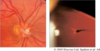
Where are histo streaks usually found?

A

Mid periphery to equator

3
Q
A

Lattice degeneration

4
Q

What is the leading cause of rhegmatogenous RD?

A

Horse-shoe tears

5
Q

Where are hard exudates found?

A

Outer plexiform layer of retina

6
Q
A

Snailtrack degeneration

7
Q

When is peripapillary atrophy benign?

A

When mild and associated with myopia

8
Q

Synchesis can often cause what?

A

PVD

9
Q

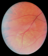
Horse shoe tears can develop along which margin of WSP?

A

Posterior margin

10
Q
A

Boat Heme (D-shaped)

11
Q

What is the key difference between toxo and histo?

A

Active toxo causes inflammatory A/C reaction - KPs, cells, flare

12
Q
A

Histoplasmosis

13
Q
A

Remnant of Hyaloid Artery

14
Q

WSP is a sign of what?

A

Vitreoretinal traction

15
Q

Bone spicules are a part of what disease?

A

Retinitis pigmentosa

16
Q

What are the four types of hemorrhages?

A

Pre-retina (boat shaped) Flame Dot & blot Sub-retinal

17
Q
A

Peripheral “histo streak”

18
Q

Where is PPA most common?

A

On the temporal side

19
Q

Where are vitreoretinal tufts usually seen?

A

Near equator and ora, but may be seen anywhere

20
Q

Name three benign “dark” findings

A

CHRPE Bear Tracks Pigment Crescent

21
Q

Where does the apex of a horse shoe tear point?

A

Toward the posterior pole

22
Q

What are the defining features of central retinal artery occlusion?

A

Cherry red spot Blanched fundus

23
Q

What color are atrophic retinal holes?

A

Red

24
Q

Which are brighter, hard exudates or drusen?

A

Hard exudates

25
Q

Retinal holes found with snailtrack can be associated with RD if they are located where?

A

On the edge

26
Q

What are the vitreous attachments from strongest to weakest?

A

Vitreous base Peripapillary Macula Vessels Mid periphery stronger than posterior pole

27
Q

What supplies blood to the macula?

A

Choroidal blood flow

28
Q

How big are atrophic retinal holes?

A

Pinpoint to 2DD

29
Q
A

HST

30
Q

What characterizes honeycomb degeneration?

A

Network of perivascular pigmentation

31
Q

Why do horse-shoe tears “cause” RD?

A

Allows an influx of fluid

32
Q
A

Hard Exudates

33
Q

Syneresis is due to what?

A

Shrinkage of the vitreous

34
Q
A

Pavingstone degeneration

35
Q

Where is toxo found?

A

Posterior pole

36
Q

What do hard exudates look like?

A

Yellow, distinct spots

37
Q

Where is lattice most frequently found?

A

Superior temporal Bilateral

38
Q

What supplies blood to the retina?

A

Central retinal artery

39
Q

If a cuff around an atrophic retinal hole exists, when is it at risk for RD?

A

If it is greater than 1DD Retina has become opaque

40
Q

HTN retinopathy often shows which signs?

A

Cotton wool spots Flame hemes

41
Q

Vitreoretinal tufts are often benign, but can result in ___ if ___ is present.

A

HST PVD

42
Q

Drusen are thought to be due to what?

A

The eye’s failure to eliminate waste

43
Q
A

Myelinated NFL

44
Q
A

Drusen

45
Q
A

Myelinated NFL

46
Q
A

Nevus

47
Q

What does histo look like?

A

Black “clouds”

48
Q
A

Toxoplasmosis

49
Q

Where is snailtrack found?

A

Between ora and equator

50
Q

In what diseases are cotton wool spots common?

A

HTN DM Vein occlusions Vascular disease (lupus, HIV) Interferon therapy

51
Q

How long does it take for a cotton wool spot to fade?

A

6-8 weeks

52
Q

Where in the retina does honeycomb degeneration usually occur?

A

3 and 9, but spreads 360 degrees

53
Q

What forms hard exudates?

A

Deposition of lipids and lipoproteins

54
Q

What does toxo look like?

A

Black with white inside

55
Q
A

Lattice Degeneration

56
Q
A

Snalitrack

57
Q

When is PPA pathological?

A

When associated with glaucoma and histoplasmosis

58
Q
A

Dot and Blot Heme

59
Q

What is the “histo triad”?

A

Peripapillary atrophy Maculopathy Punch-out lesion in the retina or histo streaks

60
Q

Where are drusen depositied?

A

Between RPE and Bruch’s membrane of choroid

61
Q
A

Branched Retinal Artery Occlusion

62
Q

What are 4 predisposing factors for RD?

A

High myopia Trauma Lattice Family history

63
Q
A

VR tufts

64
Q

What is an atrophic retinal hole?

A

Round retinal break without accompanying vitreoretinal traction

65
Q

How do you differentiate a nevus from a CHRPE?

A

Red-free filter makes nevus disappear

66
Q

What factors are associated with PVD?

A

High myopia Post surgery (cataract) Trauma Being over 40

67
Q

Where are Hollenhorst plaques usually found?

A

Bifurcations of arteries

68
Q

What diseases show hard exudates?

A

DM Late stage HTN retinopathy Ven occlusion

69
Q
A

RD

70
Q
A

Snailtrack with atrophic holes

71
Q

What does Lattice look like?

A

Cigar-shaped thinning running parallel to the ora with criss-cross pattern of sclerotic vessels

72
Q

If this vessel is present, a central retinal artery occlusion may still leave an islet of vision.

A

Cilioretinal artery

73
Q

What is synchesis?

A

Liquefaction - Vitreous slowly degrading

74
Q

In retinal attachment, which eye will show lower IOP?

A

Affected eye

75
Q
A

Peripheral Drusen

76
Q
A

CRAO

77
Q
A
78
Q

What is a halo nevus?

A

A CHRPE with a depigmented area surrounding it (area of RPE hypertrophy

79
Q

When are horse-shoe tears at risk of developing retinal tears along the posterior margin of WSP?

A

Posterior border is irregular WSP is associated with lattice Fellow eye has already suffered a tear

80
Q

Drusen are usually related to what?

A

Age

81
Q

Where is Histo found?

A

Anywhere in the retina

82
Q

Horse-shoe tears are commonly associated with what?

A

PVD

83
Q
A

Atrophic Hole

84
Q
A

VR tufts

85
Q

Where is pavingstone located?

A

Between ora and equator

86
Q

Name 6 benign peripheral retinal conditions.

A

Chorioretinal degeneration Peripheral retinal drusen (equatorial) Honeycomb (reticular) degeneration Pavingstone degeneration CHRPE Post inflammatory scars (Trauma, histo, toxo)

87
Q

What is seen in proliferative diabetic retinopathy?

A

Neo of disc Neo elsewhere in the retina Boat-shaped hemorrhages

88
Q
A

Retinitis Pigmentosa bone spicules

89
Q

What percent of vitreous is water?

A

98%

90
Q

CHRPE, bear tracks, and pigment crescents are found where?

A

Anywhere

91
Q
A

Peripheral Drusen

92
Q

Which chorioretinal scar is associated with birds?

A

Histoplasmosis

93
Q

What is the appearance of myelinated nerve fiber layer?

A

White, feathery edged NFL

94
Q

Where does myelinated NFL usually present?

A

Around the optic nerve and peripapapillary NFL

95
Q
A

Sub-retinal hemorrhage

96
Q
A

PPA

97
Q

Where is Shaffer’s sign noted?

A

Anterior vitreous

98
Q

What does a choroidal nevus usually look like?

A

A flat or slightly elevated grayish-green lesion

99
Q

What does PPA look like?

A

Irregular, hyper and hypopigmentation zone

100
Q

Describe pavingstone degeneration.

A

Chorioretinal atrophy appearing as small pale yellow areas with surrounding RPE hyperplasia

101
Q

What are vitreoretinal tufts?

A

Grayish white piece of tissue

102
Q

What causes cotton wool spots?

A

Hypoxia of NFL - acute obstruction of arteriole, blocking axoplasmic flow

103
Q

Name 3 predisposing conditions to RD.

A

Lattice degeneration Snailtrack degeneration White-without-pressure (WSP)

104
Q

What is the common presentation of snailtrack degeneration?

A

Glistening white area Oval in shape

105
Q

What things are usually seen in diabetic retinopathy?

A

Exudates Dot and blot hemorrhages Cotton wool spots

106
Q
A

Halo CHRPE

107
Q
A

Vitreoretinal Tufts

108
Q

Which obscures blood vessels, drusen or hard exudates?

A

Drusen