Gastroenterology I Flashcards
(29 cards)
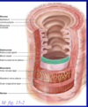
What are the layers of the alimentary canal?
- Mucosa (innermost later that faces the lumen)
- epithelium
- lamina propria
- muscularis mucosae
- Submucosa
- Meissner’s plexus
- Muscularis externa
- inner circular layer
- outer longitudinal layer
- Auerbach’s plexus
- Serosa/Adventitia
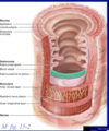
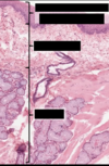
Identify the indicated layers of the alimentary canal

What is the lamina propria composed of?
What is its function?
loose connective tissue (filter tissue)
rich in immune system cells
prevent bugs from crossing epithelium and getting deep into the body
The submucosa is formed from what type of tissue?
Dense irregular connective tissue
Which layer of the alimentary canal provides most of the peristolsis movement to the intestine?
muscularis externa
What is the difference between serosa or adventitia?
Serosa is when the organ in interperitoneal (covered with outer epithelium)
Retroperotoneal, it does not have that lining, it is adventitia
What are the two major differences between the oral cavity and the rest of the alimentary canal?
- no muscularis externa
- no serosa/adventitia
Why can it be difficult to separate lamina propria from submucosa in the oral cavity?
What would you look for?
there is no muscularis mucosae
lamina propria: loose connective tissue
submucosa: dense connective tissue
What is located in the submucosa of the oral cavity?
- minor salivary glands
- intrinsc glands
- mucus-secreting
What is the difference between major and minor salivary glands?
Minor: fit within the wall of the organ; typically mucus-secreting
Major: extrinsic glands, too big to fit within the wall of the organ (parotid, submadibular, sublingual)
What is the shape of the minor salivary glands?
How do they drain into the oral cavity? Cell type?
Branched tubular gland
They have ducts that drain the products to the oral cavity through the mucosa. Lined with stratified cuboidal.
What are the layers of the oral cavity?
- mucosa
- non-keratinized stratified squamous epithelium
- lamina propria
- submucosa
- minor salivary glands

What type of epithelium is found on the tongue?
- Epithelium
- keratinized stratified squamous epithelium
What is the difference between extrinsic & intrinsic tonge musculature?
Intrinsic: both insertion and origin are within the tongue
Extrinsic: only insertion is within the tongue
What are the four major types of papillae found on the tongue? Major characteristics?
- Filiform
- most abundant
- throughout the tongue
- keratinized, no taste buds
- mechanical role
- Fungiform
- look like mushroom
- anterior
- taste buds on the apical surface
- Circumvallate
- v shaped row of very large papillae (8-12)
- dome shaped
- moat-like groove with taste buds
- Von Ebner’s glands
- serous lingual salivary glands
- flush substances out of the “most”
- Foliate
- parallel ridges on the lateral aspect of the tongue
- taste buds on lateral surfaces
- not very well developed in humans
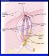
How do molecules enter the taste buds?
What two types of cells are located in the taste bud?
Where are taste buds located?
- taste pore (an opening on the surfaace)
- Types of cells
- Sensory cells with modified microvilli with chemoreceptors that project into the pore identify the different tastes
- neuroepithelial (no ACE2)
- support cells
- Sensory cells with modified microvilli with chemoreceptors that project into the pore identify the different tastes
- location
- lingual papillae
- oral cavity
- glossopharyngeal arch
- soft palate, epiglottis
The major function of the major salivary glands is to produce saliva. What is are the functions of saliva?
- lubrication
- moisten food
- digest carbohydrates
- amylase
- antimicrobial
- contains IgA
- source of Ca and phosphate
- for growing teeth and maintainace of teeth
What are the 3 components of the major salicary glands?
- secretory part
- ductal system
- stroma
- capsule adn septa, admipocytes
What are the 3 types of secretory portions that exist in the major salivary glands?
What do they secrete?
Cell structure?
Shape?
- Serous acini
- protein-secreting
- cell structure
- euchromatic nucleus
- abundandt RER
- Apical secretory granules
- spherical
- mucous acini
- mucus-secreting
- cell structure
- heterochromatic nucleus
- “frothy” appearance
- tubular
- seromucus acini: mixed

What are the 3 major categories of majori salivary gland ducts?
- Intralobular ducts
- within lobules
- interlobular ducts
- between lobules
- main (excretory) ducts
- from glands to oral cavity

What are the 2 types of intralobular ducts?
cell types?
characteristics?
- Intercalated duct
- cuboidal epithelium
- basally-placed nucleus
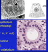
- Striated duct
- simple columnar epithelium
- basal membrane infoldings that push nucleus up into middle of cell
- more space for ion pumps
- mitochondria
- ion transport (Na+ in, K+ out)
- hypotonic saliva
What type of cells male up interlobular ducts?
stratified columnar epithelium
What are the differences between the 3 major salivary glands?
- Parotid
- largest
- serous gland
- long duct
- Submandibular
- floor of the mouth
- mixed gland
- mostly serous
- Sublingual
- flor of the mouth
- mixed gland
- mostly mucus
What are the layers of the esophagus including cell type?
- Epithelium
- non keratinized stratified squamous
- lamina propria
- thick and elastic
- muscularis mucosa
- thick
- longitudinal bundles

