GI III Flashcards
(35 cards)
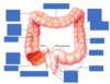
What are the divisions of the intestine?
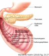
Small Intestine (bowel): duodenum, jejunum, ileum
Large intestine (colon): caecum, colon, rectum, anal canal
Describe the blood supply to the intestines
- superior mesenteric artery supplying most of small intestine and the large intestines (right ascending and part of transverse portions)
- inferior mesenteric supplies last 2/3 of large intestine

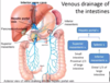
Describe venous drainage from the intestines
- inferior mesenteric vein drains distal 1/3 of large intestine
- superior mesenteric vein drains proximal 2/3 of large intestine
- all goes to hepatic portal vein
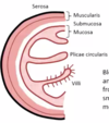
What are alterations in the small intestine?
- plicae circularis (increase SA- lots of enzymes here to breakdown food so increased SA enhances their ability to do that, also doing absorption here)
- villi (increase SA for digestion and absorption)
- microvilli (each epithelial cell in plasma membrane has invaginations to increase apical surface of absorptive cells)
- BVs, lymphatics, and nerves travel to and from the majority of the small intestine by the mesentery
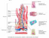
What is the function of the capillary bed and the lacteal in the villi?
- capillary bed absorbs water soluble nutrients
- lacteal is a lymphatic vessel; immune cells can use this or can use it to absorb dietary fats
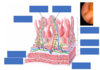

What is the most abundant cell of the villi? What other cells are around here?
- absorptive cell is most abundant; has microvilli
- goblet cells interspersed between these secreting mucous
- enteroendocrine cells releasing hormones
- paneth cell (specialized epithelial cell that talks to immune cell); constantly samples what you are ingesting and communicates that it is okay so immune system doesn’t activate inappropriately
What is the role of the duodenum?
- name means 12 (about 25cm)
- most dense plicae circularis
- major site of calcium and iron absorption (calcium absorption is regulated by vitamin D)
- in submucosa, there are submucosal glands which send mucous out to surface of duodenum (duodenal or Brunner’s glands); only exist in the first 10cm of duodenum
- this mucous is bicarb rich because this part of the small intestine is receiving the acidic chyme from the stomach

What are hepatopancreatic ampulla?
- tube
- accepts secretions from hepatocytes and pancreas and they will also produce bicarb rich mucous to finalize neutralization of the acidic chyme
What are the features of the jejunum?
- name means empty; in an autopsy you would never find residual matter here
- represents 40% of length of small intestine
- plicae circularis not as dense as you would find in the duodenum
- most of glucose and amino acid absorption (including water)
What are the features of the ileum?
- name means twisted
- represents 60% of length of small intestine
- contains abundant amount of lymphoid nodules (MALT/GALT)- Payer’s patches; where B and T cells hang out (no capsule around it so it is not a true lymph node)
- ileum is the last segment of the small intestine before the large intestine so need to make sure nothing pathogenic gets back into small intestine from bacteria in feces of large intestine
- most products of fat digestion (including bile salts) and vit B12 absorbed here
- lumen gets smaller because as you break down food you need less room
- ends in ileocaecal valve
- gastroileal reflex sends neural signals to spinal cord and enteric nervous system to stimulate distal end of ileum to pass undigested material into large intestines

How is digestion regulated in the intestines?
- during cephalic and gastric phases, stimulation by vagus nerve fibres releases pancreatic juice and stimulates smooth muscle within small intestine to contract
- acidic chyme entering duodenum causes the enteroendocrine cells of the duodenal wall to release secretin whereas fatty protein-rich chyme induces the release of cholecystokinin
- CCK and secretin enter bloodstream and tell pancreas and liver to release enzymes and bicarb-rich mucous into duodenum
What are the 2 patterns of intestinal contractions?
- fed pattern: during feeding, irregular contractions mix contents with digestive juices (segmentation)
- fasting pattern: migrating motor complex (peristalsis) begins at duodenum and propels residual material into colon (prevents bacterial overgrowth)

What enzymes break down carbohydrates?
- amylase
- lactase
- maltase
- sucrase
- begins with enzymes released by parotid gland (salivary glands) and by amylases secreted by pancreas
- intestinal absorptive cells lining small intestine have some of these enzymes in plasma membrane (brush border enzymes)

What enzymes break down protein?
- pepsin (released by stomach)
- trypsin, chymotrypsin, carboxypeptidase (released by pancreas)
- also comes from brush border enzymes in small intestine

What enzymes break down fats?
- bile salts (liver)
- lipases (lingual, gastric, and pancreatic)
How are nucleic acids broken down?
-nucleases

How does pancreas release enzymes to break down food stuff without digesting itself?
- when pancreas releases enzymes, they are in precursor form
- trypsin is key protease to activate the rest of the proteases released by pancreas
- trypsinogen is activated by membrane-bound enteropeptidase (brush border enzyme)
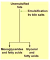
How is fat digested?
- primarily occurs in small intestine
1. Large fat globules are emulsified (broken up into smaller fat droplets) by bile salts in the duodenum
2. Digestion of fat by pancreatic enzyme lipase yields FFAs and monoglycerides. These then associate with bile salts to form micelles which “ferry” them to the intestinal mucosa.
3. FFAs and monoglycerides leave micelles and diffuse into epithelial cells. There they are recombined and packaged with other lipoid substances and proteins from chylomicrons.
4. Chylomicrons are extruded from the epithelial cells by exocytosis. The chylomicrons enter lacteals. They are carried away from the intestine by lymph. - from left thoracic duct, fluid ends up in left subclavian vein where it returns fluid back to circulation

How does absorption occur in the small intestine?
- active transport (needs energy to bring nutrient in)
- secondary active (using concentration gradient to power entry of another solute)
- facilitated diffusion (providing a carrier protein to let something walk down its concentration gradient)
- diffusion
*move into blood or lymphatics then end up in the liver

How is glucose absorbed?
- using 2 sodium travelling down its concentration gradient
- pulls glucose in
- secondary active transport
- facilitated transporter on basolateral side to get into the bloodstream

How are amino acids absorbed?
- secondary active transport using sodium
- uses simple diffusion on basolateral side
- if it’s an essential amino acid (from diet), energy might be put into this process to trap it in the absorptive cell; this uses ATP to pull amino acid in

How do fats get absorbed?
- short chain FAs diffuse across
- micelles are reassembled by absorptive cell, put onto protein called chylomicron
- extruded from absorptive cell and gets back to circulation through the lymphatics
- fat soluble vitamins (A,D,E,K) absorbed via micelles
- water soluble vitamins absorbed via diffusion