GI path Flashcards

1
Q

Histo of normal esophagus

A
  • stratified nonkeratinizing squamous epithelium
  • proximal 1/3=striated muscle
    distal 2/3=smooth muscle
  • GE junction (where rugal folds begin)
  • Z-line (squamocolumnar junction): mucosal junction of squamous and columnar epithelium, may not correspond to GE junction when columnar metaplasia has occured
2
Q

esophageal atresia and tracheoesophageal fistula

A

Def: Embryologic failure of tubal esophagus to connect mouth to stomach, ending in a blind pouch; fistula may connect segment to trachea

Clinical:

  • 50% have associated congenital anomalies–VATER syndrome (Vertebral defects, Anal atresia, TrachEoesophageal fistula, and Renal dysplasia)
  • Respiratory symptoms (aspiration pneumonia)

abnormal hedgehog signaling

3
Q
A

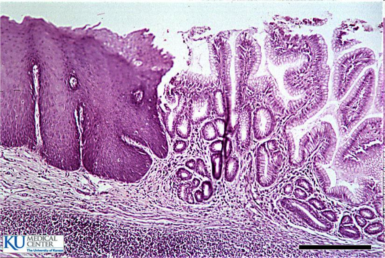
normal gastroesophageal junction

stratified sqaumous of esophagus

simple columnar of gastric cardia

4
Q

esophageal ring

A

Concentric, thin diaphragm of tissue in the distal esophagus, most commonly at GE junction (Schatzki’s ring

5
Q

esophageal webs

A
  • Eccentric, thin membranes of tissue in the esophagus, most commonly proximal region
  • Plummer-Vinson syndrome: webs + iron deficiency anemia + glossitis; women; responds to iron supplementation
6
Q

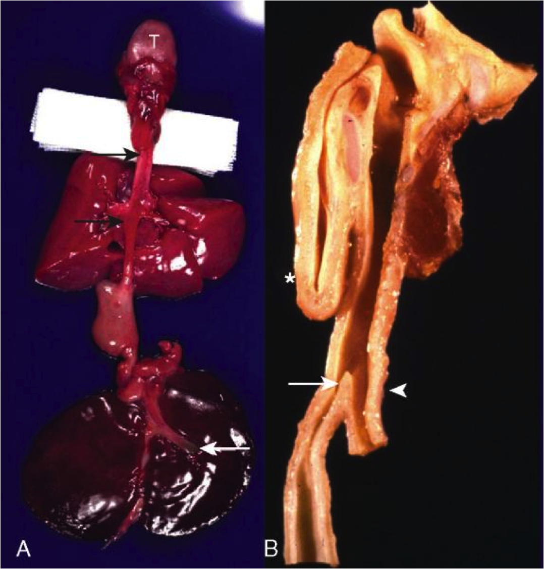
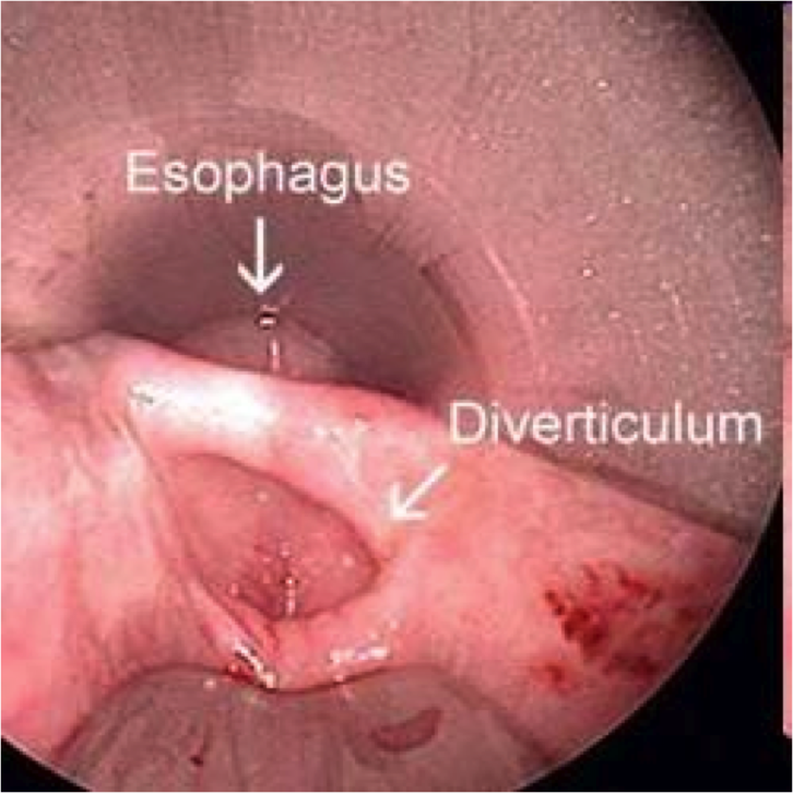
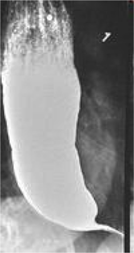
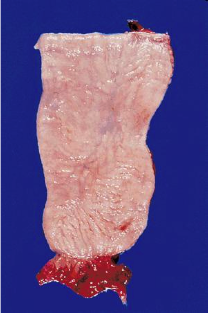
Esophageal diverticula

A

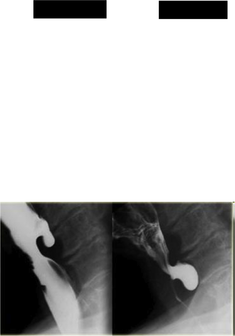
Outpouchings of the esophageal wall

  • True = all layers, including muscle
  • False = mucosa & submucosa only
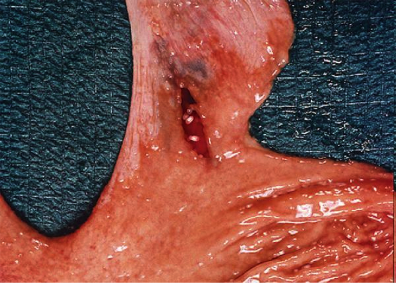
  • Zenker’s diverticulum = false, cervical esophagus, elderly (motor dysfx)
  • Epiphrenic diverticulum = true, any age, just above diaphragm

Reflect underlying motor dysfunction

image=Zenker’s diverticulum

7
Q

achalasia

def?

clinical?

microscopic?

A

Definition
Inability of the LES (lower esophageal sphincter) to relax after swallowing, resulting in periodic esophageal obstruction

Clinical
Dysphagia, odynophagia, regurgitation
inc. risk for squamous cell carcinoma

Microscopic Reduction or absence of myenteric ganglion cells

8
Q

Symptoms: heartburn, acid regurg, dysphagia, globus sensation, chronic sore throat

Dx?

risk factors?

contributing factors?

A

Dx: GERD (Reflux of gastric acid into the esophagus with mucosal damage)

Risk Factors: Age, EtOH, tobacco (LES relaxation), chocolate

Contributing factors: hiatal hernia, weak LES, impaired esophageal peristalsis, delayed gastric emptying, inc gastric acid production
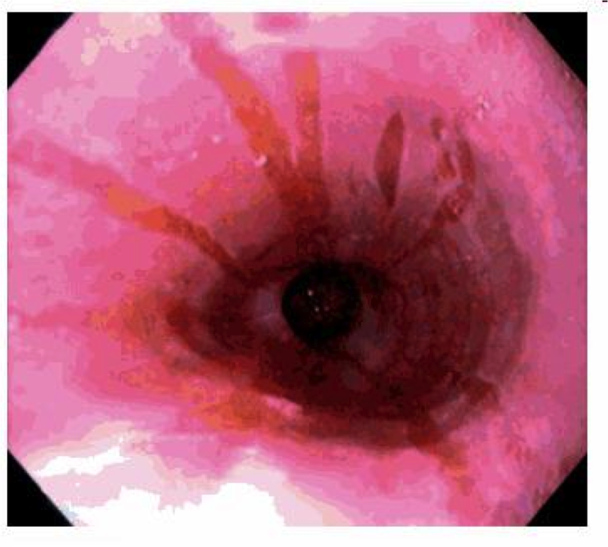
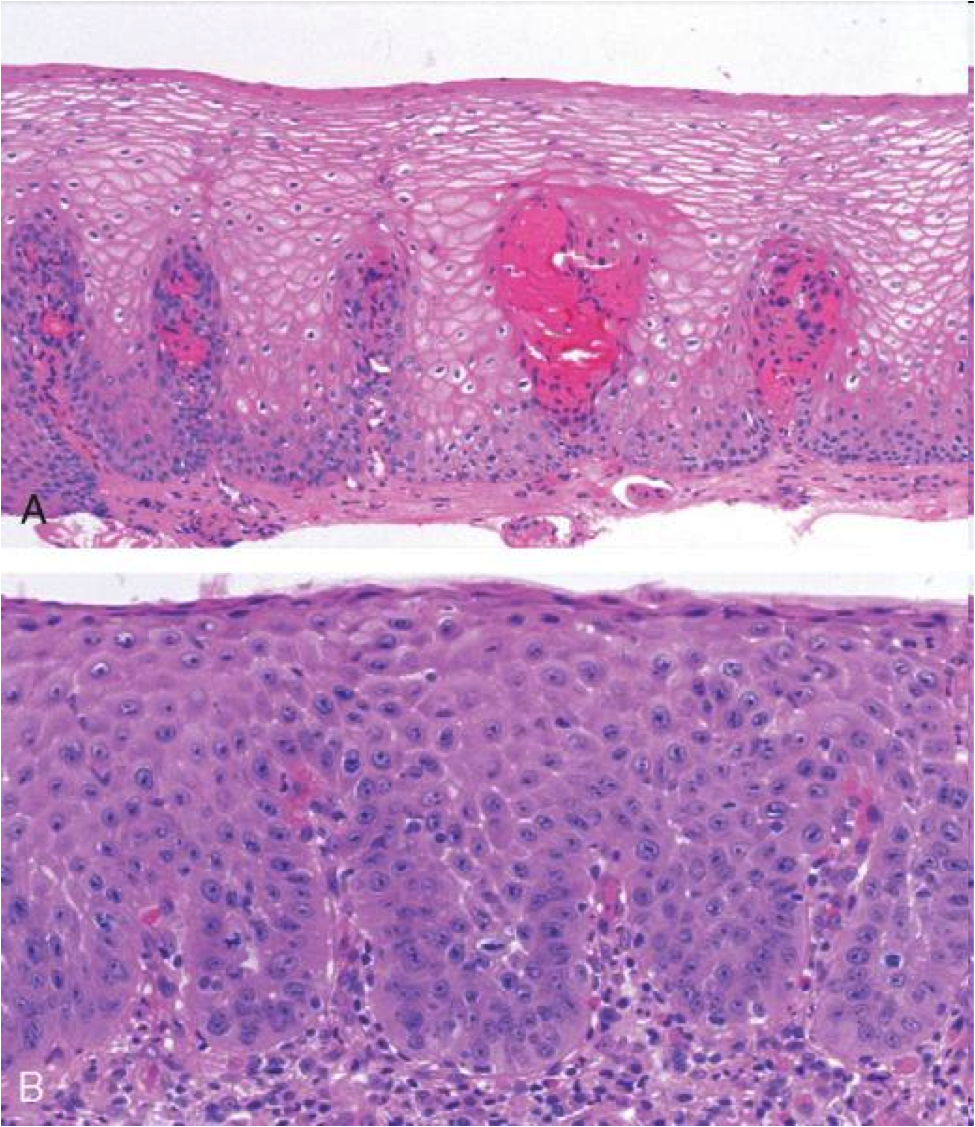
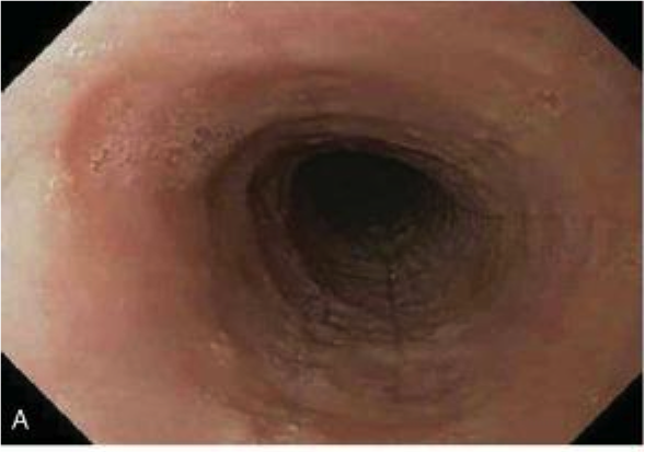
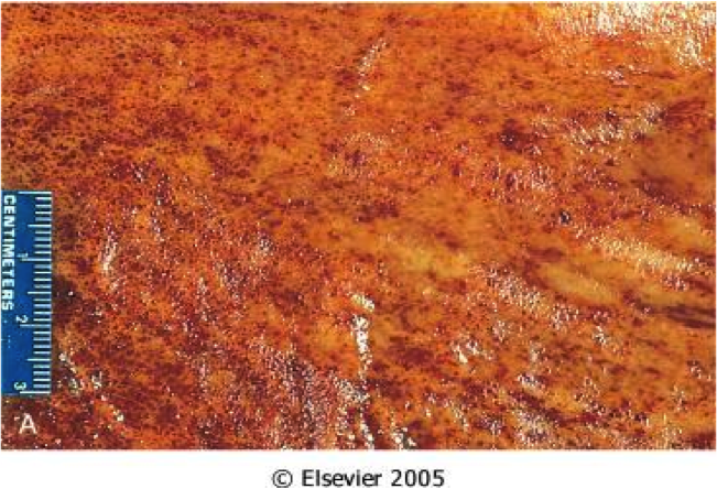
9
Q
A

GERD

Hyperemia, vertical linear streaks represent superficial mucosal erosions/ulcers

10
Q
A

GERD on bottom

squamous proliferation, papillae elongation, basal cell hyperplasia, inc. inflammation in lamina propria, decreased surface maturation

Note: biopsy findings are not specific! cannot make diagnosis w/ biopsy alone must have clinical info as well
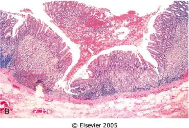
11
Q

Barrett’s esophagus

Def

A

Definition
Endoscopically recognizable columnar metaplasia of the esophageal mucosa that is confirmed pathologically to have intestinal metaplasia, the latter defined by goblet cells

Both the endoscopic and pathologic components should be present to establish BE.

12
Q
A

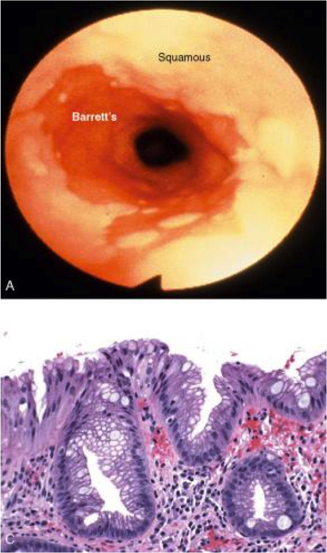
Barrett’s esophagus: columnar metaplasia (intestinal metaplasia)+goblet cells

13
Q
A

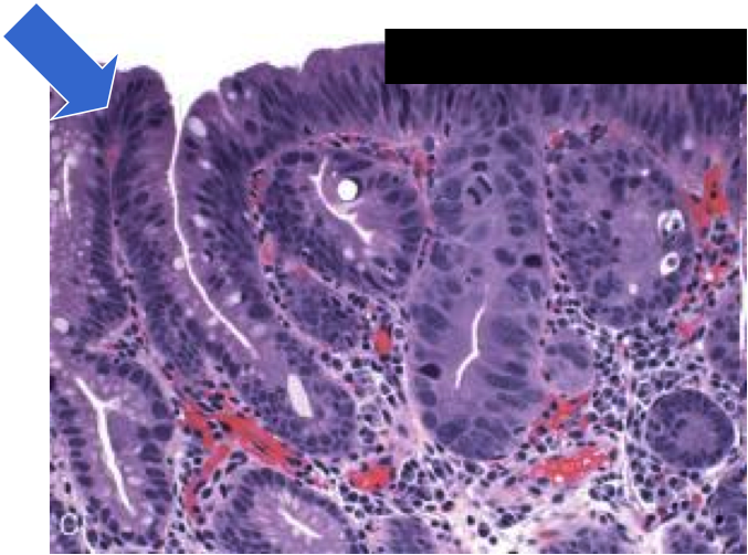
dysplasia in barrett’s

sequence of adenocarcinoma is metaplasia–>dysplasia–>carcinoma

14
Q

Sx: dysphagia, food impaction

Hx: allergies, failed antireflux therapy

Tests: normal pH monitoring

Dx?

A

primary eosinophilic esophagitis (>15 eos/hpf from pts who lack a positive response to proton-pump inhibitors, have normal pH monitoring, or both)
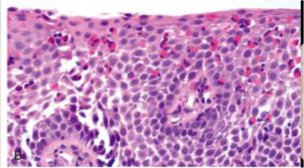
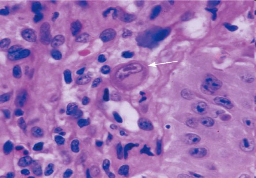
15
Q
A

herpes esophagitis

lateral margin of ulcer

cowdry A intranuclear inclusions (Multinucleation, chromatin Margination, nuclear Molding)
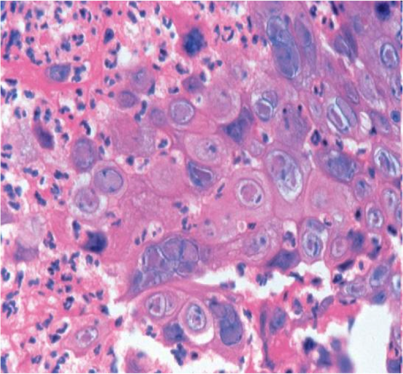
16
Q
A

CMV esophagitis

  • Base of ulcer
  • Lg intranuclear inclusion with granular cytoplasmic inclusions
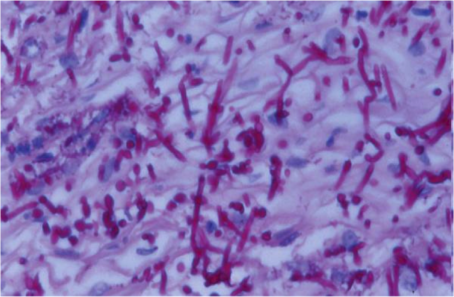
17
Q
A

Candida esophagitis

  • Immunosuppressed, Diabetics, recent Abx
  • Pseudohyphae
18
Q
A

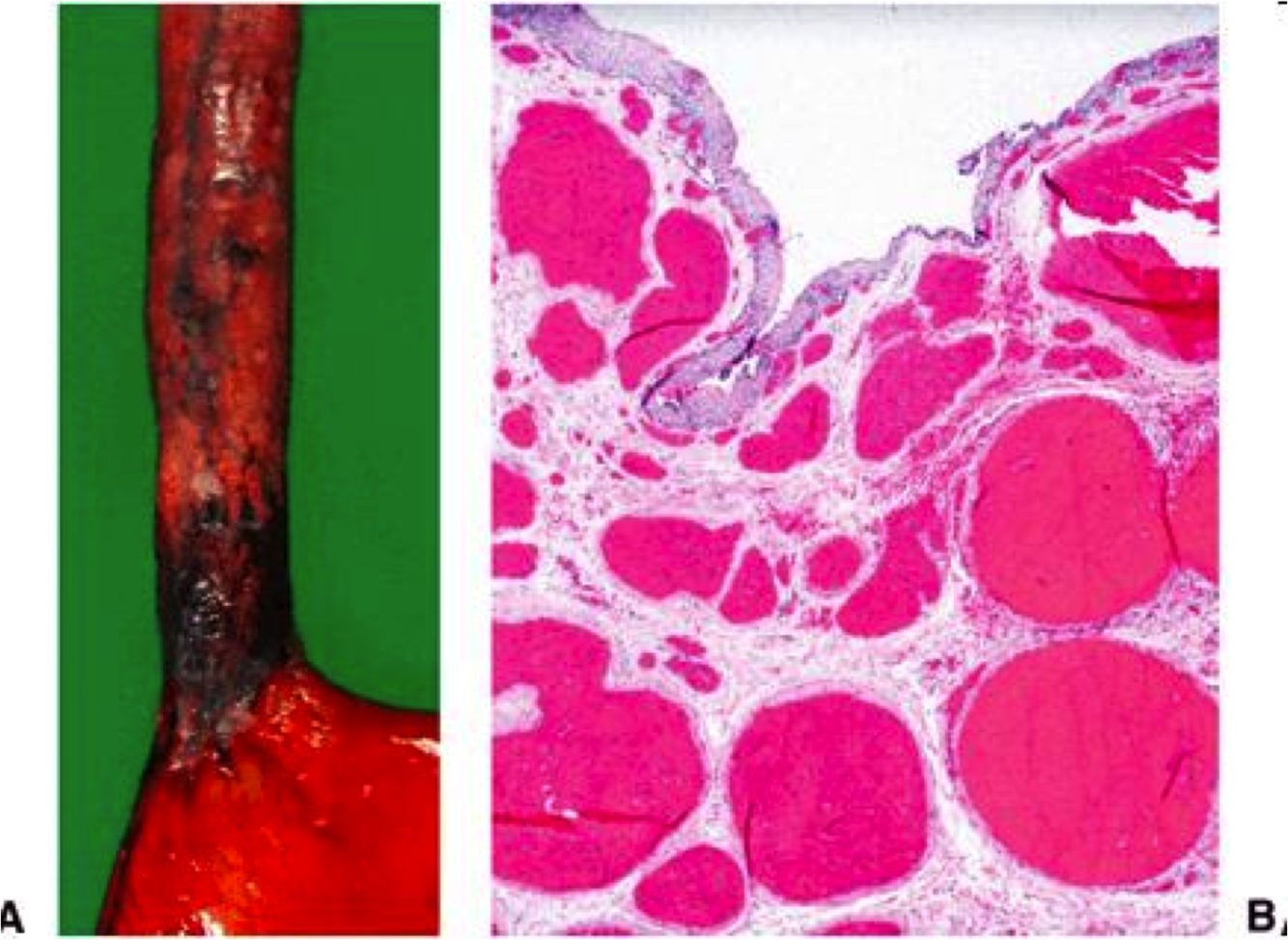
esophageal varices: Dilatation of submucosal esophageal veins

most often due to portal hypertension secondary to cirrhosis

19
Q
A
  • *Mallory-Weiss laceration**
  • at GE junction (usually on gastric side)
  • Forceful vomiting/retching forces prox stomach through diaphragm
  • Laceration may bleed profusely
  • Acute esophageal rupture –> Boerhaave’s syndrome
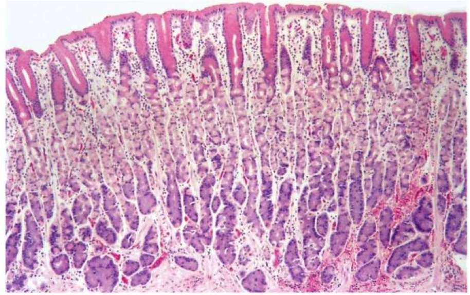
20
Q
A

Fundus, body
Parietal (oxyntic) glands
Pink = parietal cell
Blue/purple = chief cell
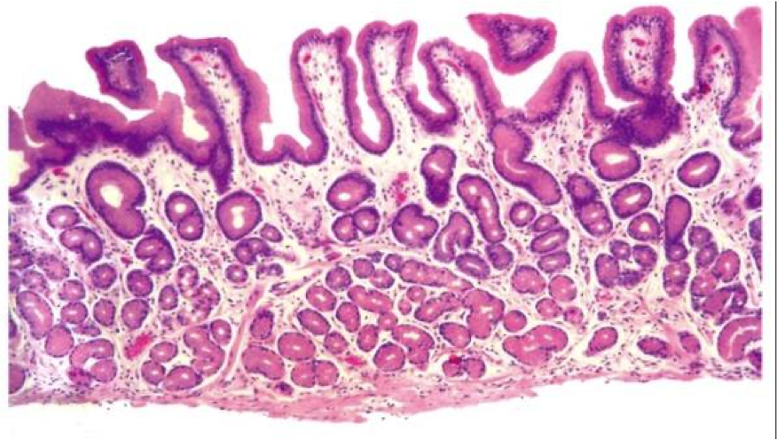
21
Q
A

Cardia and Antrum
Cardiac/antral glands
Mucus cells

22
Q

Congenital hypertrophic pyloric stenosis

A
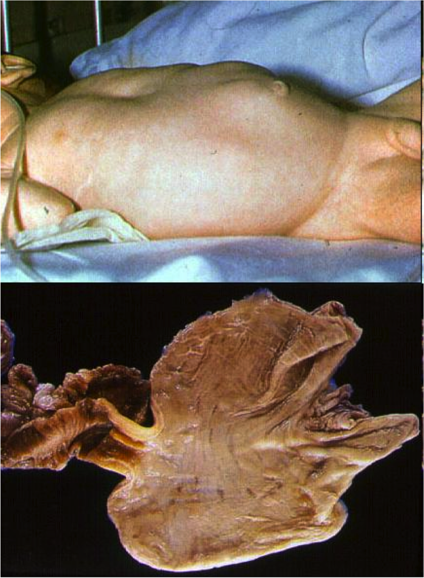
  • Concentric enlargement of the pyloric sphincter and narrowing of the pyloric canal that obstructs the gastric outlet
  • palpable epigastric mass (“olive”)

-M:F 4:1
Mostly Caucasian; rare in blacks & Asians

-Possible genetic as well as environmental (drugs, infection in utero) pathogenesis

23
Q

gastritis:

acute vs chronic vs reactive

A

Acute gastritis
Erosive/hemorrhagic

Chronic gastritis
Autoimmune
H. pylori associated
Lymphocytic (see text)
Granulomatous (see text)
Eosinophilic (see text)
Collagenous (see text)

Reactive gastropathy
Chemical (see text)

24
Q

Sx: abrupt onset abdominal pain and bleeding

Hx: alchohol use, NSAIDS

Dx?

Most common etiology?

Path findings?

Therapy?

A

Dx: Acute erosive/hemorrhagic gastritis (stress gastritis) (Abrupt onset of ab pain & bleeding a/w ETOH, NSAIDs, or low hemodynamic state following trauma)

Due to Breakdown of mucosal barrier (Direct irritant action, Drug mechanism of action, Hypoperfusion)

Most common etiologies: NSAIDS, post-op state

Gross: Petechiae, erosions, ulcers

Microscopic: Limited to mucosa: superficial lamina propria hemorrhage, mucosal sloughing/necrosis, neutrophils

Therapy: Acid-supression (histamine blockers, proton-pump inhibitors)

25
30yo female presents with fatigue and dyspepsia. Labs: megaloblastic anemia, B12 deficiency, increased gastrin Hx: SLE Biopsy of body of stomach Dx? Path?
Dx: autoimmune gastritis (Immune-mediated form of chronic gastritis, results in **loss of parietal cells**, hypo- or achlorhydria, vit B12 deficiency) Clinical: Autosomal dominant, \<5% of chronic gastritis, Women, Pernicious anemia (megaloblastic anemia d/t ↓vit B12), Other autoimmune diseases Labs: inc. gastrin, decreased vit B12 Path: Lymphoplasmacytic inflammation in deep, glandular lamina propria; limited to body/fundus
26
Fundic gland damage by patchy lymphocytic infiltrates Loss of glands over time--\> atrophy (atrophic gastritis)
27
autoimmune gastritis \*intestinal metaplasia is precursor to dysplasia and gastric carcinoma \*ECL cell hyperplasia is precursor to neuroendocrine tumors
28
Helicobacter pylori gastritis def? clinical?
Definition: Chronic antral-predominant gastritis caused by H. pylori Clinical: A/w peptic ulcer disease (stomach & duodenum), gastric lymphoma & carcinoma
29
H. pylori gastritis Lymphoplasmacytic inflammation admixed with neutrophils in superficial mucosa
30
Pititis, cryptitis, crypt abscesses (neutrophils = “activity”) due to H. pylori gastritis
31
H. pylori gastritis Line epithelium in mucus layer (do NOT invade, but secrete toxins)
32
Menetrier's disease **Definition:** Body & fundus-restricted hyperplasia of foveolar (mucous cell) epithelium with hypoproteinemia **Clinical:** dec plasma proteins (albumin) from gastric mucosa, dec acid production **Pathogenesis:** TFG-alpha overexpression **Gross:** Markedly thickened rugal folds in fundus & body (resembling brain) **Microscopic:** Foveolar (mucous cell) hyperplasia --\>corkscrewing
33
histo features of normal small bowel mucosa
-villous height-to-crypt depth ratio of 3:1 to 5:1 - -lamina propria contains mild physiologic mix of plasma cells, lymphocytes, and occasional eosinophils
34
Brunner's glands are in the duodenum. They are in the submucosa produce mucin rich alkaline (bicarbonate) secretion that protects duodenum from stomach acid and lubricates
35
normal terminal ileum w/ peyer's patches
36
endoscopic findings celiac disease
scalloping and loss of folds
37
histologic features of celiac disease
- increased intraepithelial lymphocytes with loss of so-called decrescendo pattern - blunted villi and crypt elongation - inc. plasma cells and lymphocytes in lamina propria (back is more "blue" and expanded from chronic inflammation)
38
Marsh classification
Used for classifying Celiac disease. Most symptomatic celiac pts are Marsh3a-c. classifies degree of villous blunting and crypt elongation
39
serologic tests for Celiac Disease
1) Anti-tTG IgA: highly sensitive and specific. Best test! 2) Serium IgA levels simultaneously tested to exclude IgA deficiency which would give false negative True gold standard diagnosis of CD is clinical/pathological response to gluten-free diet. Other less good tests: * anti-endomysial IgA * Anti-gliadin (used to monitor response to gluten free diet)
40
Genetics of Celiac Disease
almost all people w/ CD have MCH class II HLA-DQ2 or HLA-DQ8 absence makes diagnosis unlikely but 20% of normal population have this, so not used for screening
41
Other conditions besides Celiac that might have increased IEL and/or villous blunting
- immune (common variable immunodeficiency, HIV entropathy, autoimmune disease) - Infection (H. pylori, cryptosporidium, bacterial overgrowth, ) - NSAID injury \*NOTE: CVID mimics but has fewer plasma cells!
42
Diseases assoc. with celiac disease
* dermatitis herpetiform\* (responds to GF diet) * lymphocytic collagenous colitis * lymphocytic gastritis * refractory sprue * collagenous sprue * enteropathy-assoc. T cell lymphoma\* * small intestinal adenocarcinoma
43
- "test tubes in a rack" bases of crypts abut the muscularis mucosa - mild degree of chronic inflammation in lamina propria is normal (lymphocytes, plasma cells, eosinophils, mast cells)
44
characteristic triangular folds of the colon
45
microscopic colitis
lymphocytic and collagenous colitis pts have chronic watery (non-bloody) diarrhea and normal endoscopy middle aged-elderly assoc. with certain drugs (long-term NSAIDS) and celiac disease
46
lymphocytic vs collagenous colitis
**Lymphocytic colitis:** * \*F:M ratio of 3:1 * \*increased IEL * no inc. subepithelial collagen * inc. lymphocytes, plasma cells and eosinophils in LP * no architectural changes **Collagenous colitis:** * \*F:M of 8:1 * \*increased IEL * \*thickened subepithelial collagen * inc. lymphocytes, plasma cells and eosinophils in LP * no architectural changes
47
Inflammatory bowel diseases (3)
1) Ulcerative colitis 2) Crohn's disease 3) Intermittent colitis
48
Gross pathology of IBD
active disease: erythematous, friable, edematous, ahaustral w/ or w/o ulcerations, pseudopolyps, cobblestoning \*biopsy alone can't distinguish b/t Crohn or UC
49
Pathologic findings in UC
-distribution: continuous colonic involvement of mucosa beginning in rectum Gross findings: * inflammatory polyps * lead pipe colon (loss of haustral folds) * toxic megacolon Microscopic changes: limited to mucosa
50
Pathologic findings in Crohn's disease
- distribution: rectum usually spared, skip lesions, patchy disease both grossly and microscopically, disease can affect any part of columnar GI tract (classically terminal ileum) - Gross findings: * Long linear (bear claw-like) ulcers * cobblestone mucosa * creeping fat (mesenteric fat wrapping around serosal surface) * strictures (muscularis hypertrophy and hyperplasia) Microscopic changes: * inflammatory change involving entire wall of bowel (Vs UC only in mucosa) * "transmural inflammation": chronic inflammation and lymphoid aggregates * knife-like fissures * granulomata * combing fat
51
Mucosal changes in IBD chronicity activity
**chronicity:** - architectural distortion - metaplasia * paneth cell metaplasia of left colon * pyloric metaplasia (terminal ileum in Crohn disease) -muscularis hypertrophy and hyperplasia **Activity:** how much acute inflammation - cryptitis: neutrophils in epithelium - crypt abscess: collection of neutrophils in lumen of a crypt
52
chronicity in Crohn disease
- architectural distortion (villous blunting) - pyloric metaplasia - muscularis hypertrophy and hyperplasia (both muscularis mucosa and muscularis propria)
53
Acute self-limited colitis
- typically transient (2-4wks) colitis w/ diarrhea (sometimes bloody) - most commonly assoc. w/ bacterial enterocolitis, can be viral or parasitic - usually not biopsied but when done: biopsy has acute inflammation but no features of chronicity (distinguish from IBD, no architectural distortion, no paneth cell metaplasia, no MM hypertrophy)
54
C. difficile colitis
- related to previous antibiotic exposure - "pseudomembranous colitis": * not diagnostic: DDx includes C. diff and ischemia - PCR for C. diff toxins - pseudomembranes composed of fibrin, mucin and neutrophils. glands typically lose epithelial cells (C. diff toxin affects cell adhesion)
55
Strictures in bowel DDx?
- chronic ischemia (d/t fibrosis) - crohn disease (d/t muscularis mucosae and propria hypertrophy and hyperplasia) - tumor - recurrent bouts diverticulitis
56
celiac disease tip of villi w/ inc. IEL. architecture preserved so Marsh 1 lesion IEL are dark blue dots, enterocytes are lighter and more elongated
57
celiac disease small intestine is severely blunted so it looks completely flat
58
common variable immune deficiency mimics celiac diseae (but no plasma cells)
59
cryptosporidium mimics celiac disease
60
lymphocytic colitis. lamina propria filled with chronic inflammatory cells. No crypt distortion. No thickened subepithelial collagen band
61
lymphocytic colitis with surface damage (mucin depletion and reactive changes)
62
collagenous colitis. pink collagen bands under surface. No architectural distortion.
63
collagenous colitis. capilarries are entrapped in thickened subepithelial collagen band. lots of chronic inflammation in lamina propria and mildly inc. IEL and eosinophils.
64
IBD w/ features of chronicity. cyrpts have lifted off muscularis mucosa and are bifurcated--\>architectural distortion Muscularis mucosae is thickened (MM hypertophy and hyerplasia)
65
terminal ileum w/ pyloric metaplasia Pyloric metaplasia is feature of chronicity. glands look like pyloric glands of gastric antrum/cardia. pale/neutral in color w/ well-definied borders b/t cells pyloric metaplasia classically occurs in terminal ileum in Crohn disease Paneth cell metaplasia may rarely be seen in "backwash ileitis" of severe ulcerative colitis
66
activity (acute inflammaiton) w/ cryptitis and crypt abscess seen in acute self-limited colitis. neutrophils have faint pink cytoplasmic halo
67
crohn disease: transmural inflammation with lymphoid aggregates
68
granuloma in crohn disease
69
combing fat crohns disease (mesenteric fat extends to serosal surface)
70
acute self-limited colitis. No features of chronicity (no architectural distortion, no paneth cell metaplasia, no MM hypertorohy) to distinguish from IBD. Presence of chronic infammatory cells is not a feature of chronicity correlate w/ stool culture to exclude infection
71
C. diff colitis clue: epithelial cells fall of into lumen (due to C. diff toxin) Gross path: pseudomembranes (fibrin, mucin, neutrophils)
72
ischemia involving mucosa: hyalinization of lamina propria (pink), withered cypts, hemorrhage best way to asses hemorrhage is presence of hemosiderin laden macrophages!
73
pseudomembranes (fibrin, inflammatory cells) in ischemia
74
chronic ischmiea w/ inc. fibrosis (trichrome stain)
75
congenital anamolies of pancreas
o **Accessory (ectopic pancreas)**: pancreatic tissue outside it’s normal location (often in stomach, small intestine, meckel’s diverticulum) o Annular pancreas: abnormal ring of pancreas that encircles the duodenum; assoc w/ down’s syndrome o Pancreas divisum: failure of dorsal and ventral buds/ducts to fuse leading to retention of 2 separate duct systems; duct of santorinin provides main drainagel may cause acute pancreatitis