GU Eval & Congenital Anomalies Flashcards
(116 cards)
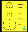
What would the indications for a renal eval be?
UTI’s – urinary tract infection
–
Increased BUN and or Creatinine
–
Hematuria
–
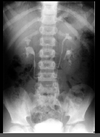
Mass seen on X-ray
–
Flank Pain, CVA tenderness
–
Decreased urine output – Oliguria
–
Azotemia - ↑ levels of nitrogen in the blood
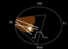
Transducer frequency used for kideny US (adult, ped, newborn)
types.
3-5 Mhz. (adult)
–
4-5 Mhz. (peds)
–
6-8 Mhz. (newborn)
–
Vector
–
Sector
–
Curved Linear
•
Rib artifacts
What is renal ultrasound able to demonstrate?
The acoustic properties of a mass, delineate an abnormal lie of the kidney resulting from extrarenal mass, or determine whether hydronephrosis is secondary to renal stones
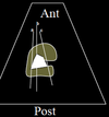
Patient postion Rt vs Lt
Right
–
Supine or Lt. lateral oblique or decubitus
•
Using liver as acoustic window
•
Imaging through muscles
–
posterior approach
•
Left
–
Supine or Rt. lateral oblique or decubitus
•
Spleen may be used as a window, generally upper pole only
•
Imaging through muscles
–
posterior approach
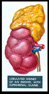
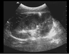
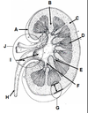
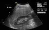
What are the Renal contours when imaged?
Smooth outer contours surrounded by reflected echoes of Perirenal fat. Renal parenchyma surrounds the fatty central renal sinus which contains the calyces infundibula pelvis vessels and lymphatics
US Appearance (adults vs infants)
ADULTS
Cortex; homogeneous, slightly less than liver echogenicity
•
Medulla-Pyramids; hypoechoic compared to cortex
•
Sinus; echogenic secondary to fat and fibrous tissue
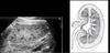
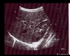
INFANTS
Cortex; more echogenic than in adults
•
Medulla-Pyramids; more prominent than adults and often mistaken for cysts
•
Sinus; not well developed and almost absent – lack of adipose tissue
Why is the renal sinus imaged as an area of intense echoes with variable contours?
Because of the fat interface
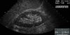
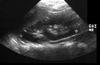
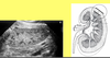
WHAT positon was this Td for this rt kidney?

long
What does dehydration do to the kidney?
Causes the infundibula and Renal pelvis to be collapsed making them indistinguishable from the echo dense renal sinus fat
what are the arrows pointing to?


What can you see if the bladder is hydrated?
The extrarenal pelvis medial to the kidney on transverse scans
how thick is the parenchyma? how about in the cortex?
Parenchymal thickness 1.5 cm or >
–
Even thickness throughout
–
Thin areas of cortex (< 1.5cm is pathologic)
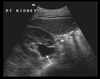
What window is used to image the right kidney?
The liver
is the cortex or sinus more echogenic?
cortex
What window do you use to see the left kidney?
The liver or spleen
how much can a kidney move during inspiration?
~2.5 cm
What should the renal cortical echo amplitude be compared to with regards to amplitude and depth to set appropriate TGC and sensitivity?
Liver parenchymal echo amplitude
what poles does the kidney have?
superior and inferior.
What two parts of the kidney make up the renal Parenchyma?
The renal sinus to the outer renal surface
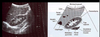
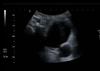
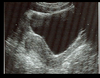
how can you tell this is a pediatric kidney?

the prominence of the medullary pyramids and the lack of prominence of the renal sinus as compared to the adult kidneys. (adult below)
What are the columns number Bertin?
The band of cortical tissue separating the hypo echoing medullary pyramids from the echogenic cortex
what can you see?

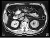
What normal structure could be confused for a renal artery and transverse?
The inferior vina cava. both will be tubular in transverse
what do you see?