Hand Fingertip Amputations, Dupuytren's, Vascular Flashcards

1
Q

A 50-year-old man comes to the emergency department after sustaining amputation of the right long finger involving an avulsion mechanism. The patient is taken to surgery for replantation. During surgery, extensive vascular injury is seen, and an approximately 2-cm vascular gap of the digital arteries and veins results following excision of injured vessels. Which of the following interventions is most likely to increase the probability of functional digit replantation?

A) Bone shortening
B) Medicinal leech therapy
C) Postoperative warming
D) Systemic heparin
E) Vein grafts

A

The correct response is Option E.

In patients who sustain digital amputation as a result of an avulsion mechanism, there is often an extensive zone of injury that precludes primary vascular anastomosis. Vein grafts permit vascular anastomosis outside of the zone of injury.

Bone shortening can sometimes allow excision of the injured vasculature and primary anastomosis. However, in this case, bone shortening is unlikely to make up for a 2-cm vascular gap.

Longer vascular gaps can be addressed with vein grafts. Despite the fact that vein grafts involve an additional anastomosis per vessel compared to primary anastomosis, they have been found to exhibit similar rates of thrombosis and replantation survival.

Medicinal leech therapy can help address venous congestion, but is typically considered when venous congestion occurs after attempt at surgical replantation, or if no suitable veins can be found for anastomosis.

While postoperative warming and systemic heparin are often used adjunctively in patients undergoing replantation, they have not been demonstrated to increase the likelihood of survival of the replanted part, and would most likely not have as significant an effect as restoring perfusion to the amputated part using vein grafts.

2018

2
Q

An otherwise healthy, nonsmoking 30-year-old mechanic has the long, ring, and little fingers amputated sharply through Zone II of the right hand. The amputated digits are stored appropriately, and he is rushed to surgery within 2 hours of the accident. Which of the following sequences is the best method of replantation?

A) Digit by digit: bone, tendons, arteries, nerves, veins
B) Digit by digit: bone, tendons, arteries, veins, nerves
C) Structure by structure: bone, nerves, tendons, arteries, veins
D) Structure by structure: bone, tendons, arteries, nerves, veins
E) Structure by structure: tendons, bones, veins, arteries, nerves

A

The correct response is Option D.

The most efficient sequence to perform the replantation is structure by structure: bone, tendons, arteries, nerves, and then veins. It has been shown that the time to complete the procedure is significantly shorter if the same anatomic structure on each severed digit is fixed before repairing the next structures, as opposed to completing all aspects of the replantation one digit at a time. With respect to the sequence of repair of the severed structures, the general thought is to have a stable construct prior to starting the delicate microscopic repairs. However, the technical sequence used by microsurgeons varies greatly.

The only consistent agreement is starting with bony shortening and fixation. The traditional sequence that follows is extensor and flexor tendon repair, and then vessel/nerve repair. However, individual surgeon preference and patient circumstances dictate the usual sequence thereafter. Some surgeons like to start dorsally and complete the extensor tendon, venous, and skin repair first, and then complete the volar structures next. On the volar side, some surgeons repair the tendon first, followed by the artery and nerve, while others fix the artery and nerve first, followed by the tendon. There are those who believe that the nerve is better repaired in a bloodless field, so that should be done first. Others feel that repairing the vein first reduces blood loss and keeps a bloodless field more reliably for better vision. In patients who present with long ischemia time, it may be beneficial to anastomose the artery first, because this provides the advantages of earlier revascularization and allows easier detection of the most functional veins by their spurting backflow. In short, any of these sequences is fine, as long as it follows the bony fixation.

The other options are incorrect sequences for the above reasons.

2018

3
Q

A 56-year-old woman with a history of systemic sclerosis (scleroderma) is evaluated for intractable pain and progressive ulceration to the right index and middle fingers despite medical management. Duplex ultrasonography shows no identifiable vascular occlusion in the affected digits. Which of the following is the most appropriate surgical management?

A) Interposition bypass grafting
B) Intra-arterial TPA
C) Sympathectomy
D) Thrombectomy
E) Venous arterialization

A

The correct response is Option C.

For patients who have patent arterial inflow on imaging, spasm is likely to be responsible for their ischemia. Spasm is most common in those with autoimmune disease. Digital sympathectomy involves stripping the adventitia from the radial, ulnar, and digital arteries in an effort to decrease sympathetic input that is the presumed cause of pathologic vasoconstriction. Vascular occlusion with a satisfactory distal target may require an interposition bypass. Occlusion without a distal target for bypass may require venous arterialization. In the absence of evidence of occlusion, there is no indication for thrombolytic therapy.

2018

4
Q

A 25-year-old man sustained traumatic amputation of the nondominant index finger 3 hours ago and requests replantation. Which of the following factors has the greatest influence on survival of the injured digit after replantation?

A) Mechanism of injury
B) Number of vessels repaired
C) Patient’s smoking status
D) Time from injury to replantation
E) Use of anticoagulation

A

The correct response is Option A.

The mechanism of injury has the greatest influence on survival of replanted digits. Injuries from sharp devices that leave a clean cut with little or no crush component are the most amenable to replantation. The more the tissue is crushed or avulsed, resulting in greater vessel injury, the less likely the digit will survive. No studies have shown that the use of anticoagulants changes survival rates. Smoking decreases blood flow in digits, but has not been widely studied in replantation. Fingers have no muscle, which is the tissue most susceptible to ischemia, so digits can tolerate long delays as long as they are treated correctly. At least two veins per artery have been shown to help prevent venous congestion.

2018

5
Q

A 58-year-old man is to undergo excision of a painful ulnar artery aneurysm of the palm, which has been causing ulnar nerve compression. Preoperative examination shows a digital/brachial index (DBI) of 0.5 in the small finger. After excision of the diseased segment, which of the following is the most appropriate next step in management?

A) Arterial reconstruction
B) Botulinum toxin type A injection
C) Extended periarterial sympathectomy
D) Periarterial injection of 2% lidocaine
E) Postoperative anticoagulation

A

The correct response is Option A.

Ulnar artery aneurysms may cause symptoms because of local mass effect, distal embolization, and/or episodic vasospasm. Ligation of the ulnar artery to exclude the aneurysm from hand circulation can effectively eliminate risk for embolism, but may rob the digits of necessary blood flow if there is not enough collateral circulation from the deep arch or other sources. Measuring the digital-brachial index (DBI) is an effective way to assess whether or not there is sufficient blood flow to the digits. A normal DBI is between 0.75 and 0.97. Values equal to or less than 0.7 indicate inadequate perfusion. Below a DBI of 0.5, tissue loss is inevitable. Following ulnar artery ligation, if the DBI is below 0.7, then reconstruction of the ulnar artery is recommended rather than simple aneurysm excision or ligation. This is typically accomplished with a reversed vein graft or an arterial graft (e.g., from the lateral femoral circumflex system).

Anticoagulation alone, or anti-vasospastic drugs, such as botulinum toxin type A or lidocaine, are not sufficient in this clinical situation, where blood flow is limited because of blockage of flow. While sympathectomy could improve circulation in cases of vasospasm, this patient had no history of this, and sympathectomy alone would not be a substitute for arterial reconstruction.

2018

6
Q

A 53-year-old man comes to the emergency department because of an avulsion degloving injury to the left nondominant thumb sustained 3 hours ago. The amputated part is not retrievable. Physical examination shows loss of skin from the interphalangeal joint distally on both volar and dorsal surfaces. The distal phalanx and flexor pollicis longus and extensor pollicis longus tendons are intact. He has no other associated injuries. Which of the following is the most appropriate method of reconstruction of the thumb?

A) Amputation revision at the mid-proximal phalanx
B) Great toe wraparound flap
C) Radial forearm osteocutaneous flap
D) Second toe-to-thumb transfer
E) Volar neurovascular advancement flap

A

The correct response is Option B.

Thumb reconstruction remains a difficult challenge for hand surgeons. Amputations of the skin distally may be covered with palmar advancement flaps; however, this technique is only limited to wounds less than 50% of the palmar surface of the thumb distal to the interphalangeal joint. In order to preserve length and function in more proximal amputations, either a regional or distant flap is required. The toe-to-thumb wraparound flap requires a microvascular anastomosis of digital vessels and nerves, providing excellent sensation and cosmetic results. The toe donor site can be covered with a skin graft in order to preserve length.

The volar neurovascular advancement flap would not adequately cover a defect this size. Amputation at the mid-proximal phalanx would result in a very short thumb with loss of function. The radial forearm flap may be utilized to cover the above defect; however, it would lack adequate sensation. Any osteocutaneous radial forearm flap would not be indicated since there is preservation of the bone. Similarly, a second toe-to-thumb transfer would not be indicated since there is preservation of bone in this patient.

2018

7
Q

A 21-year-old man sustains traumatic amputation of the right thumb at the level of the metacarpal base. Pollicization should include osteosynthesis of which of the following?

A) Index metacarpal base to trapezium
B) Index metacarpal to thumb metacarpal
C) Index middle phalanx to thumb metacarpal
D) Index proximal phalanx to thumb metacarpal
E) Index proximal phalanx to trapezium

A

The correct response is Option D.

Transfer of the index finger to the thumb position on the hand (pollicization) typically transfers the proximal phalanx to the thumb metacarpal, as long as the base of the thumb metacarpal is preserved. Transfer of the middle phalanx or metacarpal of the index would create a neo-thumb that is too short or too large, respectively. Obliterating an intact carpometacarpal joint by transferring the index metacarpal to the trapezium would eliminate palmar and ulnar abduction of the thumb and compromise global hand function.

2018

8
Q

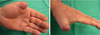
A 27-year-old man is brought to emergency department because of a thumb avulsion injury measuring 3 × 3 cm. A photograph is shown. Which of the following is the best option for sensate, soft-tissue coverage?

A) Cross-finger flap
B) First dorsal metacarpal artery flap
C) Moberg flap
D) Pedicled groin flap
E) Skin grafting

A

The correct response is Option B.

There are several sensate options for thumb pulp deformities. These include neurovascular island flaps, Moberg flaps, free toe pulp flaps, and the first dorsal metacarpal artery flap (FDMA). Cross finger flaps, skin grafts, and pedicle groin flaps do not have innate innervation. The defect in the question involves the entire pulp of the thumb overlying the distal phalanx and is too large for a Moberg advancement flap.

The FDMA flap is supplied by its eponymous artery, which travels in the fascia overlying the index metacarpal and supplies the skin overlying the dorsum of the proximal phalanx. The vessel is accompanied superficially by a branch of the radial nerve that provides neural activation to the skin overlying the proximal phalanx of the index finger. The flap can be transposed to provide sensate coverage of the tip of the thumb, and can provide sufficient size to resurface relatively large defects.

Cortical reorientation is the fact that the brain recognizes a stimulus from the flap area as a stimulus from the thumb, and not from the index finger. This process takes some time, but is usually complete after 2 years. Average static two-point discrimination in these flaps utilized for thumb resurfacing is 10 to 11 mm.

Use of the FDMA flap for resurfacing of the thumb pulp has been compared to heterodigital island flaps in several studies. Both remain options to be considered, but the ease of elevation, limited dissection, and acceptable donor site morbidity make the FDMA flap a common primary option for thumb tip resurfacing.

2018

9
Q

A 45-year-old woman with systemic sclerosis (scleroderma) has severe Raynaud phenomenon. A photograph is shown. Periarterial injection of botulinum toxin type A is being considered for treatment in this patient. This treatment is believed to relieve vasospasm in Raynaud phenomenon by which of the following mechanisms?

A) Blocking fast sodium channels in axonal gap junctions
B) Increasing the activity of chronically down-regulated group C nerve fiber nociceptors
C) Inhibiting Rho/Rho kinase activity
D) Obstructing myofibroblast contractile activity in vascular smooth muscle
E) Promoting substance P secretion/receptor sensitivity

A

The correct response is Option C.

Several mechanisms have been proposed to explain the effect of botulinum toxin type A (Botox) to inhibit Raynaud phenomenon in patients with scleroderma. Studies have demonstrated inhibition of Rho/Rho kinase activity, inhibition of substance P secretion and receptor sensitivity, and decreasing the activity of chronically up-regulated C-fiber nociceptors all to occur in models of Raynaud phenomenon that responded to Botox treatment. Fast sodium channels conduct axonal signals AT in gap junctions, but have not been shown to be affected by Botox. Myofibroblasts may be involved in late fibrosis of scleroderma patients but do not exist within the vascular smooth muscle.

2018

10
Q

A 23-year-old woodworker sustains an injury to the dominant left thumb that involves the loss of less than 2 cm of the distal pulp with exposed bone from a planing machine. Which of the following reconstruction methods is most likely to provide normal sensation to the volar pulp of this patient’s thumb?

A) Cross-finger flap from the long finger
B) Islandized Moberg flap
C) Flag flap
D) Thenar flap
E) Venous flow-through flap

A

The correct response is Option B.

The venous flow-through flap was described for small defects of the dorsum of a digit or hand where end-to-end anastomoses of the included veins on the proximal and distal edges of the flap can provide venous outflow for the digit and supply the flap. A defect from the distal, volar surface of the thumb would not have any veins large enough to use. A cross-finger flap is a classical solution to cover the volar aspect of a digit. The other mentioned flaps are also excellent options for volar thumb coverage, except for the thenar flap. The thenar flap is used for distal pulp defects of the fingers in children. The only flap that maintains the normal sensation of the thumb pulp is the Moberg flap, which advances the volar aspect of the thumb on its neurovascular pedicles. The islandized (O’Brien) modification was to make a transverse incision at the base of the thumb and dissect the neurovascular bundles to allow the flap to travel further distally, and then placing a skin graft over the proximal defect.

2018

11
Q

A 30-year-old man is scheduled to undergo transhumeral amputation after unsalvageable brachial artery occlusion. A photograph is shown. Use of targeted muscle reinnervation may allow improved functional recovery by which of the following means?

A) Better bulk and durability by preventing denervation atrophy of muscles at the amputation stump
B) Better prosthesis control by input from median and ulnar nerve signals
C) Better sensory detection in the prosthesis by positioning amputated nerve stumps closer to the skin closure
D) More precise control of an osseointegrated body-powered prosthesis
E) Preservation of greater bony length in the amputation stump

A

The correct response is Option B.

A body-powered prosthesis uses motion of remaining joints, such as the gleno-humeral and scapulo-thoracic joints, to control an upper extremity prosthesis.

Targeted muscle reinnervation (TMR) would not affect function of a body-powered prosthesis. TMR positions amputated nerve stumps well within the remaining muscle and far from the cutaneous closure. Current prosthetics are not yet able to detect sensation and transmit this to the patient. Having nerve stumps near the amputation closure site increases the risk for neuroma pain.

TMR has not been shown to decrease denervation atrophy of residual upper extremity muscles. TMR has gained increasing acceptance in the treatment of patients who have undergone or will undergo upper extremity amputation. Resected nerves, such as the median and ulnar nerves, can be coapted to nerve branches to remaining muscles, such as the pectoralis and deltoid. Transcutaneous EMG detectors are positioned over these reinnervation sites to detect nerve signal, which a myoelectric prosthesis can then use to better control distal joints.

TMR does not affect the amount of bony length that can be preserved in an amputation.

2018

12
Q

A 54-year-old man comes to the office because of an injury to the long finger of the dominant right hand sustained when it was pinched in a machine at work. Physical examination shows a 1.3-cm loss of pulp tissue with no exposed bone. To preserve function and sensation in the digit, which of the following is the most appropriate intervention?

A) Amputation at the distal interphalangeal joint
B) Cross-finger flap
C) Moist dressings
D) Thenar flap
E) Volar V-to-Y advancement flap

A

The correct response is Option C.

Fingertip injuries are one of the most common problems encountered in hand surgery. The long finger is the most common finger involved. The patient’s age, occupation, and compliance with treatment should be considered when determining treatment. When possible, if the patient has no exposed bone or only a small area of exposed bone, treatment with dressing changes offers excellent results. There is no donor site morbidity, scarring is often minimal, and return of sensation is generally excellent. Patients, however, need to be cautioned that a prolonged period of dressing changes is required, often lasting 3 to 6 weeks.

Amputation at the distal interphalangeal joint would result in loss of function of the profundus tendon and grip weakness. Neurovascular island flaps and V-to-Y advancement flaps offer excellent closure options when digital length needs to be preserved and there is significant exposure of bone. However, with these flaps there is a donor defect and decreased sensation. Care must be taken when using a cross-finger flap or thenar flap in older patients to avoid contractures and stiffness of the digits.

2018

13
Q

A 45-year-old woman with scleroderma is evaluated because of a 2-year history of severe resting pain in both hands. She does not smoke cigarettes. Despite appropriate medication therapy, she has had no relief of her symptoms. Injection of botulinum toxin type A into which of the following locations is the most appropriate treatment for this patient’s Raynaud phenomenon?

A) Around the stellate ganglion
B) Intradermal at the wrist
C) Intradermal in the palm
D) Perivascular at the wrist
E) Perivascular in the palm

A

The correct response is Option E.

Injection of botulinum toxin around the digital vessels in the palm has been shown to decrease pain associated with vasospastic disorders like Raynaud phenomenon. This is a relatively quick, easy, and low-risk method of treating a patient with incapacitating ischemic pain of the hand. The exact mechanism by which botulinum toxin works in this clinical scenario is still under investigation, but some theories suggest an effect on the vessels and/or nerves of the hand through inhibition of sympathetic nerves, sensory nerves (c-fibers), substance P, and/or other signal transduction pathways. Studies show a 75 to 100% reduction in pain and up to 50% healing of chronic ulcers. Approximately 10 units of botulinum toxin is bathed around each of the digital neurovascular bundles in the palm. The most common side effect reported is temporary minor intrinsic hand weakness.

Injecting botulinum toxin in the skin or too proximally in the wrist has not shown the same response as around the digital neurovascular bundles in the palm. Surgical sympathectomies by stripping the adventitia of the digital and wrist vessels have also shown some success in symptom control. Stellate ganglion blocks have also been used for this purpose among others (complex regional pain syndrome); however, local anesthetics, not botulinum toxin, are used to block the ganglion.

2017

14
Q

A 27-year-old man who is right-hand–dominant and works as a manual laborer comes to the emergency department for evaluation 6 hours after inadvertently incurring a high-pressure latex paint injection to the volar aspect of his left index finger. Which of the following is the most appropriate management?

A) Admission to the hospital and intravenous administration of antibiotics
B) Operative exploration
C) Radial gutter splint with follow-up in 3 days
D) Topical application of acetone
E) Warm compresses, elevation, and observation

A

The correct response is Option B.

Emergent incision and drainage is mandatory for high-pressure paint gun injuries. Although clinically these may appear benign and/or superficial, there is often significant underlying injury. Even small amounts of material can lead to compartment syndrome, poor perfusion, and closed space infections resulting in tissue necrosis and ultimately, amputation. History is critical, but plain films may be used to confirm the diagnosis, as both latex and the less common oil-based paints are easily seen. Grease may be radiolucent or radiopaque, depending on lead content. The most commonly injected materials are paint and grease but can also include paint solvents and fuel oil.

Nearly all reported cases involved male occupational injuries and injury to the non-dominant second or third digit, as in this case. These machines can generate pressures of 2,000 to 12,000 pounds per square inch (psi), which far exceeds the 100 psi needed to break the skin. These extreme pressures can propel injected material through the skin and subcutaneous tissues down to the bone or along fascial planes, tendon sheaths, and neurovascular bundles.

The overall rate of amputation was 30% and particularly related to the location of injury and type of material injected. Optimal time for wide surgical debridement was within 6 hours of injury. Other studies have documented an amputation rate of approximately 40% when surgery is performed within 6 hours, and an amputation rate of 57% when surgery is delayed beyond 6 hours. The amputation risk is as high as 87% without treatment or if treatment is further delayed.

None of the other interventions listed are appropriate for this type of emergent injury.

2017

15
Q

A 28-year-old, right-hand–dominant woman is brought to the emergency department after sustaining a severe crush injury to the right upper extremity during a rollover motor vehicle collision. Examination shows multiple digit amputations and comminuted fractures of the distal radius and ulna. After multiple debridements, the limb is unsalvageable. Which of the following is the shortest stump length distal to the elbow that is required when fitting a prosthesis to maintain native elbow motion?

A) 3 cm
B) 8 cm
C) 13 cm
D) 18 cm
E) 23 cm

A

The correct response is Option B.

The minimum stump length required for prosthesis fitting is 5 to 10 cm distal to the elbow. Major upper extremity amputations are defined as amputations at or proximal to the wrist joint. Data from 2005 estimate that upper extremity amputations account for 34% of the 1.6 million people living in the US with limb loss, and 41,000 of these were considered major amputations. Limb salvage is always the goal of the initial surgical management; however, the decision to amputate is made when limb salvage will result in a less functional outcome for the patient.

The ideal stump has adequate length, durable soft tissue, minimal edema, and a tapered shape with minimal scar tissue that is not directly over the bony prominence. Muscle preservation is important for the potential use of a myoelectric prosthesis.

In order to preserve elbow function and allow for fitting of a prosthesis, at least 5 cm of a bony stump is required. Although shorter transradial stumps do not allow for pronation and supination, preservation of elbow function is felt to be worthwhile functionally. Transfer of the biceps tendon to the ulna should be considered in shorter transradial stumps to decrease the risk of developing a flexion contracture at the elbow.

Amputations at least 10 cm proximal to the wrist or at the junction of the middle and distal 1/3 are felt to be ideal in terms of muscle coverage, stability of prosthesis fit, and forearm rotation, but not required. More distal stumps can be problematic in terms of soft-tissue coverage over bone and limb-length discrepancy to accommodate the wrist unit of the prosthesis.

2017

16
Q

A 30-year-old man is brought to the emergency department because of ring avulsion of the right ring finger with complete amputation through the proximal phalanx. Which of the following factors is most likely to influence survival of the replanted finger in this patient?

A) Associated fracture of the middle phalanx
B) Level of amputation
C) Need for skin graft
D) Number of dorsal veins repaired/grafted
E) Patient history of cigarette smoking

A

The correct response is Option D.

The only factor that correlated with survival in the reported series and reviews is the repair and/or grafting of two or more dorsal veins. Reports that did not compare groups recommend repairing at least two dorsal veins of replanted digits.

Smoking, level of amputation, need for skin grafting, and associated fractures were not found to have any effect on survival of digits that had ring avulsion injuries.

2017

17
Q

A 45-year-old woman comes to the office because of a split in the nail plate following a previous crush injury to the left index finger. The patient desires improvement in the appearance of the nail. A photograph is shown. Which of the following is the most appropriate treatment?

A) Application of topical phenol
B) Excision/repair of the nail bed
C) Nail plate avulsion
D) Oral antifungal therapy
E) Split-thickness skin grafting

A

The correct response is Option B.

The most appropriate method of treatment is excision and repair of the nail bed.

The anatomy of the nail consists of a nail plate, nail fold, and a nail bed. The nail bed is the soft tissue beneath the nail plate, which is composed of the germinal matrix proximally and the sterile matrix distally. Most nail plate growth (90%) is provided by the germinal matrix. In cases of trauma, adherence between the nail fold dorsally and nail bed volarly can result in synechiae, interfering with nail growth and resulting in a longitudinal split in the nail. It is important to prevent adherence of dorsal and palmar elements by splinting the nail fold open during the healing phase. This can be accomplished by replacement of the nail plate if available, or using a piece of foil from the suture packet. This patient presents with a split nail deformity after previous trauma. There is scarring between the nail fold and the nail bed, resulting in a longitudinal split with inability to allow for growth of the nail plate in the central portion. Proper treatment consists of excision of the nail bed scar, with repair of the nail bed. Splinting of the nail fold during the healing period will prevent recurrent scarring of the dorsal fold to the palmar surface. In cases where there is significant scar tissue and inability to close the resultant defect after excision, grafting of the nail bed may be required. A split graft of the sterile matrix can be performed if the deficit is only present distally. If the germinal matrix is involved, a full-thickness graft is needed.

Avulsion of the nail plate alone will not eliminate the scarring at the proximal nail fold.

Oral antifungal therapy is useful in treatment of fungal onychomycosis.

Complete excision of the nail bed and split-thickness skin grafting can be used in nail ablation, but would result in absence of the nail and not yield a more cosmetic appearance.

Topical phenol application has been used for nail matricectomy, but can produce irregular tissue destruction and would result in loss of the nail.

2017

18
Q

A 32-year-old, right-hand–dominant man is brought to the emergency department 3 hours after sustaining an avulsion injury to the left thumb. The avulsed digit was immediately placed on ice and transported with the patient. Photographs are shown. Replantation fails; the necrotic digit is removed and the wound closed. The carpometacarpal (CMC) joint is disarticulated. Which of the following is the most appropriate method of reconstruction in this patient?

A) Great toe to thumb transfer
B) Metacarpal lengthening
C) Osteoplastic reconstruction
D) Pollicization of index finger
E) Web deepening

A

The correct response is Option D.

This patient has a proximal thumb avulsion with disruption of the carpometacarpal (CMC) joint. In this scenario, the best reconstructive option (besides successful replantation) is pollicization of the index finger. Reconstruction after thumb amputation, as with congenital deficiencies, depends largely on the length of the remaining skeletal structure. One can lose most of the distal phalanx and still retain good overall thumb function. Amputations that involve the proximal phalanx or the metacarpal suffer from deficient bone length and procedures that add length, like distraction, toe to thumb, or osteoplastic reconstruction. When the entire metacarpal is absent, the aforementioned procedures will not be effective. Pollicization will restore thumb length and provide very good function.

2017

19
Q

A 55-year-old, right-hand–dominant man who is a machinist comes to the office because of inability to fully extend the right ring finger. Photographs are shown. The patient reports that his symptom began 5 years ago and has worsened progressively. Examination shows a 45-degree flexion contracture of the right ring finger (PIP) joint during attempts at full extension. All other joints demonstrate full extension, and the patient can create a complete fist during flexion. Regarding treatment options for this patient, which of the following interventions is most likely to provide the longest relief of his symptom prior to recurrence?

A) Collagenase injection and manipulation
B) Limited fasciectomy
C) Percutaneous aponeurotomy with lipografting
D) Percutaneous needle fasciotomy
E) Radiation therapy and splinting

A

The correct response is Option B.

Radiotherapy has been proposed as a potential treatment to slow or stop progression of Dupuytren contractures (palmar fibromatosis). A prospective study of radiotherapy revealed no greater efficacy than observation as an intervention for slowing the disease process. There is no evidence to suggest radiotherapy for correction of an established contracture.

Rijssen and colleagues established quantitative criteria for recurrence, using an increase of total passive flexion contracture of 30 or greater, compared to the 6-week follow-up values in previously treated joints. After 5 years, their recurrence rate following percutaneous needle fasciotomy was 85%; 21% for limited fasciectomy; and 32% of joints successfully treated with Clostridial collagenase. Percutaneous aponeurotomy with lipografting is an experimental technique which has shown some promise with correction of contractures and prevention of recurrence, but the evidence is level 4, with no controlled studies looking at this technique, in comparison to other established techniques.

Although limited fasciectomy provides the greatest degree of initial correction for Dupuytren contractures, as well as the longest period prior to recurrence, the costs associated with the procedure are by far the highest. When comparing the QALY costs of three interventions (limited fasciectomy, percutaneous needle fasciotomy, and collagenase injection), limited fasciectomy yielded the highest cost per QALY. The authors emphasize that this does not indicate limited fasciectomy is an inappropriate intervention—only that it is relatively the most expensive.

2017

20
Q

A 19-year-old man is brought to the emergency department because of pain and swelling of the left lower extremity after it was pinned beneath a large granite stone for 2 hours. On physical examination, the left leg is swollen, tense, and erythematous; a palpable pulse is noted. X-ray studies are negative for fracture. The patient reports marked pain that is uncontrolled by increasing doses of narcotics. Pain on passive movement of the ankle and toes is noted. Which of the following is the most appropriate next step in management?

A) Angiography
B) Compression wrap
C) CT scan
D) Duplex ultrasonography
E) Fasciotomy

A

The correct response is Option E.

The most appropriate next step is fasciotomy.

The patient is exhibiting signs of compartment syndrome after sustaining a significant crush injury to the lower extremity. Signs and symptoms of compartment syndrome include pain with passive stretch, increased pressure on palpation, paresthesias, paralysis, pallor, and pulselessness.

Early recognition and treatment are necessary to prevent permanent damage. The pressure within the muscles increases, and prevents blood flow to the area and capillary exchange of nutrients. Fasciotomy is recommended if compartment pressure exceeds 30 mm Hg, or if the difference between intracompartmental pressure and diastolic blood pressure is less than 30 mm Hg. If left untreated, ischemic necrosis to the muscles can result, causing permanent disability.

Compartment pressures can be measured by a handheld manometer, or the method of Whitesides with an arterial line setup. Operative fasciotomy is indicated to release the compartment pressures and prevent tissue loss and muscle necrosis. Loss of pulse typically occurs later in the spectrum of findings.

Angiography would be useful in evaluating vasculature and blood flow to the lower extremity. Typically pain with passive stretch does not occur in cases of arterial insufficiency.

Duplex ultrasound is used to look for deep venous thrombosis, which can be a source of pain and swelling in the lower extremity. This is more typical in the postoperative period or after prolonged immobilization. In this case, the mechanism of injury would prompt urgent fasciotomy.

Compression wrap and elevation are used in treatment of venous stasis and lymphedema, which is unlikely to be the cause of swelling in this case of acute trauma.

CT scan can provide better detailed imaging, but would not be indicated in this situation and would delay treatment.

2017

21
Q

A 24-year-old, right-hand–dominant man is brought to the emergency department after sharp amputation of the index, long, and ring fingers of the left hand at the middle phalanx level sustained in a rollover motor vehicle collision. The digits are appropriately preserved. Before replantation surgery is performed, which of the following is the most appropriate next step in management?

A) Administer aspirin orally
B) Administer subcutaneous heparin
C) Obtain cervical spine x-ray
D) Obtain x-rays of the hand and digits
E) Predissection of the amputated digits

A

The correct response is Option C.

The NEXUS Criteria were developed to help physicians determine whether cervical spine imaging could be safely avoided in appropriate patients. The NEXUS literature defines a distracting injury as “a condition thought by the clinician to be producing pain sufficient to distract the patient from a second (neck) injury.” Similarly, the Canadian C-spine rule describes distracting injuries as “injuries […] that are so severely painful that the neck examination is unreliable.” It also must be recognized that the surgeon and ER staff can be “distracted” by what appears to be the overwhelming injury. Trauma evaluation algorithms strictly apply.

A patient involved in a rollover motor vehicle accident has significant mechanism of injury to warrant a complete trauma evaluation.

All other answers here are appropriate to prepare for the operating room AFTER the initial trauma clearance is obtained.

2017

22
Q

A 5-year-old boy is brought to the emergency department after sustaining a crush injury to the index finger of his right dominant hand, resulting in amputation through the distal interphalangeal (DIP) joint. X-ray study shows a comminuted fracture of the proximal phalanx. Which of the following is the most significant CONTRAINDICATION to replantation in this patient?

A) Children have a difficult time adapting to functional deficits
B) The index finger is expendable
C) The mechanism and multiple-level nature of the injury preclude a functional result
D) Microvascular anastomosis is unlikely to be successful in a child of this age
E) Replantation will adversely affect epiphyseal growth

A

The correct response is Option C.

In the patient described, the most significant contraindication to replantation is the mechanism (crush injury) and multiple-level nature of the injury. It is highly unlikely that replantation will be successful with a crush mechanism due to the zone of injury. In addition, the multiple-level injury including the proximal phalanx and distal interphalangeal (DIP) joint precludes a functional result.

Replantation in children does not adversely affect epiphyseal growth. Children adapt quite well to functional deficits of the hand. Microvascular surgery in children, while challenging, has been shown to have a very high success rate when performed by skilled microsurgeons. The index finger can be considered expendable; however, children tend to have more favorable results than adults when it comes to replantation and, therefore, whenever feasible, replantation should be attempted, even if it is an isolated index finger injury. The mechanism of injury plays a greater role than the type of digit in determining the feasibility of replantation in the pediatric population.

2016

23
Q

A 40-year-old woman has an extravasation injury following a CT scan. An automated power injector was used to inject 100 mL of nonionic contrast medium into an intravenous cannula on the dorsum of the hand approximately 60 minutes ago. On examination, the hand appears edematous with mild erythema and is moderately tender. The skin is intact with no blistering. Capillary refill time is normal, and there is no neurologic deficit. Which of the following is the most appropriate next step in management?

A) Administration of a corticosteroid
B) Bedside exploration of the intravenous cannula site
C) Elevation of the extremity
D) Surgical exploration with dorsal fasciotomy and carpal tunnel release
E) Surgical exploration with dorsal fasciotomy only

A

The correct response is Option C.

In a patient with an intravenous extravasation, if the symptoms are mild (pain, swelling, or erythema), elevation and cold compresses will usually lead to complete resolution. Patients who have more severe symptoms, such as neurovascular compromise, may need additional evaluation of compartment pressures and potentially surgical exploration for decompression; however, the first step in treatment is still elevation of the extremity.

Extravasation injury was once a difficult problem. The extravasation of ionic contrast at high osmolality had increased risk for soft-tissue complications, and plastic surgery consultation was often necessary. The switch to nonionic, low-osmolality contrast over the past decade has resulted in a significant decrease in complications. In a recent study of 69,657 patients undergoing a CT scan, the rate of extravasation was 0.7% (476 patients), and only one patient required operative intervention and decompressive fasciotomy.

Corticosteroids do not have a role in extravasation injuries.

2016

24
Q

A 67-year-old man with a history of Dupuytren contracture of the right small finger comes for evaluation one week after noticing numbness and paresthesias of the outer aspect of the right small finger. Two days prior to the onset of the numbness and paresthesias, he underwent injection of collagenase Clostridium histolyticum to the finger. On physical examination today, there is mild edema of the finger. Extension of the finger has significantly improved, and there is good flexor tendon function. However, there is no sensation in the ulnar digital nerve distribution; two-point discrimination is greater than 10 mm. Nerve function was intact prior to the injection. Which of the following is the most appropriate next step?

A) Electromyography and nerve conduction velocities
B) Immediate surgical exploration and direct repair of the ulnar digital nerve
C) Immediate surgical exploration and repair of the ulnar digital nerve with nerve conduit
D) Observation only

A

The correct response is Option D.

The use of collagenase Clostridium histolyticum for Duypuytren contracture has been well studied. Reports of its efficacy and safety have been published in numerous papers in peer-reviewed journals. Though postulated, there have been no cases reported in the literature of digital nerve rupture during cord rupture with collagenase Clostridium histolyticum. Pulley rupture and flexor tendon rupture have been reported. In this case, observation would be the most appropriate next step. It is more likely that there is a neuropraxia rather than a frank rupture of the nerve. Electromyography and nerve conduction velocities will not elucidate whether the nerve has been severed. Exploration and repair is not indicated only 1 week after injury; exploration of a neurapraxia injury is indicated 8 weeks after injury. The best option is to observe the patient’s injury.

2016

25
Q

An 18-year-old man comes for evaluation 4 days after he sustained an avulsion injury of the pad of the index finger of his dominant right hand. A photograph is shown. The part was never recovered, and the patient has been performing dressing changes. The defect is 2.5 cm2 and extends to the distal phalanx bone. Coverage with which of the following flaps is most likely to result in fingertip sensation closest to pre-injury?

A) Cross-finger flap
B) First dorsal metacarpal artery flap
C) Homodigital island flap
D) Reverse radial forearm flap
E) Thenar flap

A

The correct response is Option C.

The homodigital island flap raises skin and fat overlying one digital neurovascular bundle that can be advanced distally to cover a pulp defect. As long as both digital arteries are patent, the flap can be raised on either digital artery on any finger. Because the digital nerve is raised with the flap, the overlying skin retains its sensibility (figures 2b and 2c).

The first dorsal metacarpal artery flap raises skin and fat from the dorsum of the proximal phalanx of the index finger based on branches of the radial artery and superficial radial nerve. It is useful in providing sensate coverage of the thumb, but the pedicle is not long enough to allow the flap to reach the tip of a finger.

The thenar flap and cross-finger flap raise skin from the thenar eminence and dorsal middle phalanx of a finger, respectively. Innervation is not transferred in coverage with these flaps; sensation recovery must occur with growth of the tissue surrounding the original defect. Both of these flaps require two stages, making them less appealing options.

The reverse radial forearm flap is a large, robust flap that provides excellent coverage of the hand. Its pedicle allows it to reach the tip of a finger; however, when raised in a reverse pattern with retrograde vascular flow, the flap would not bring sensation with it. In addition, the flap is too large for the defect shown in the photograph.

2016

26
Q

A 50-year-old man comes to the emergency department after sustaining an injury to the right thumb. A photograph is shown. The patient declines revision amputation and states that he does not want to lose thumb length or the nail. Which of the following is the most appropriate next step in management?

A) Complete the amputation of the thumb proximal to the germinal matrix
B) Obliterate the germinal matrix and remaining nail bed, followed by skin grafting
C) Transplant germinal matrix from the great toe
D) Transplant sterile matrix from the great toe
E) Continue to observe

A

The correct response is Option D.

The patient described has lost the sterile matrix of the nail bed. The germinal matrix is intact, as the nail continues to grow. The nail bed consists of the germinal matrix proximally and the sterile matrix distally. The sterile matrix allows the nail to adhere to the bed. Observation, amputation, and obliteration of the nail bed do not seem to be good options for a patient who has stated that appearance is paramount. The issue here is that the nail continues to grow, but cannot adhere to the full-thickness skin graft. The best option would be to harvest a split-thickness sterile matrix graft from the big toe. No additional germinal matrix is necessary as the germinal matrix is intact and producing nail.

2016

27
Q

A 28-year-old man is brought to the emergency department 3 hours after sustaining complete amputation at the level of the right mid forearm during a paper mill accident. A tourniquet was placed at the scene of the accident. He is medically stable and has no additional injuries. Which of the following is the most appropriate management of the amputated segment until vascular anastomoses are completed?

A) Clamp distal vessels
B) Cool amputated extremity
C) Flush artery with thrombolytics
D) Immerse in saline
E) Perform fasciotomies

A

The correct response is Option B.

The correct response is that the limb should be cooled until vascular anastomoses are complete.

Replantation of an amputated extremity remains the primary option at initial presentation. Contraindications for immediate replantation include an unstable patient who cannot tolerate a prolonged operative procedure, and patients in whom amputation and prosthesis would provide a better functional result than reconstruction. Warm ischemia should not exceed 6 hours, but can be extended to 10 to 12 hours when a part is cooled. Therefore, keeping a replanted part cooled until reperfused is important to success.

Clamping vessels in an amputated segment damages vessels needed for microanastomoses.

Cold heparinized saline and University of Wisconsin solution are commonly used to flush amputated parts to reduce thromboses and cool the part. Systemic complications are not common with the doses used. Persistent cold intolerance is a complaint of the majority of replanted patients. Unless thrombi are present, it is not routine to flush with thrombolytics. Immersing in saline is not appropriate.

When performing vascular anastomoses on larger masses of tissue with a longer ischemia time, performing arterial anastomoses and allowing run-off of metabolites through an open venous system before venous anastomoses are performed is recommended. Arterial perfusion may also be established early, allowing more time for repair of bone and tendon without prolonging ischemia times.

Fasciotomies are recommended as periods of ischemia can lead to compartment syndrome. However, these do not need to occur before anastomoses and should not delay reperfusion. Elevation of a replanted part is helpful to reduce post-operative edema but has no role before anastomoses.

2016

28
Q

A 45-year-old electrician is evaluated because of a fingertip injury to the index finger. The wound is allowed to heal by secondary intention but eventually results in hook-nail deformity. This patient is most likely to have sustained an injury to which of the following parts of the nail?

A) Eponychium
B) Germinal matrix
C) Lunula
D) Nail groove
E) Sterile matrix

A

The correct response is Option E.

Loss of the distal fingertip and the associated soft-tissue defect can be treated using different methods, but the involvement of the nail influences the choice of surgical approach and makes reconstruction more difficult. If the sterile matrix is foreshortened and heals to the volar soft tissues, it can result in hook-nail deformity.

The germinal matrix is responsible for formation of nail plate elements and injury does not result in hook-nail. The nail groove is the lateral margin of the nail and the site of ingrown nails and paronychia. The eponychium is the cuticle covering the proximal nail plate. Loss of the eponychium results in a rough nail lacking the usual lustre. The lunula is similarly on the proximal nail and is under the plate.

2016

29
Q

A 28-year-old woman who works as a manicurist comes for left thumb reconstruction 1 year after sustaining an amputation injury through the metacarpophalangeal joint. She desires improved pinch and grip with the best possible aesthetic appearance. Which of the following reconstruction techniques is most likely to offer her improved function with the least aesthetic donor site deformity?

A) First webspace Z-plasty
B) Great toe flap
C) Great toe wraparound flap
D) Osteocutaneous radial forearm flap
E) Second toe flap

A

The correct response is Option E.

While amputations distal through the proximal phalanx may benefit from isolated webspace deepening, it is unlikely that adequate length would remain at this level to provide good pinch or grip function even with a deeper web.

Prior to the advent of free-tissue transfer, osteoplastic reconstructions, including the reversed radial forearm flap with inclusion of a radius bone segment, were the workhorses of thumb reconstruction. The resulting thumb, however, is insensate and lacks any motion except at the level of the remaining CMC joint. Aesthetically, this reconstruction looks the least like a thumb when compared to toe transfers.

The great toe wrap-around flap provides a cosmetically acceptable way to resurface a thumb when the skeletal support is already present, either due to retention from the trauma or from an osteoplastic reconstruction. In this case, the skeletal support is absent, and the wrap-around flap by itself would not allow recreation of a stable thumb.

The great toe free flap provides an excellent reconstructive choice for a sensate, mobile thumb. Functionally, it will be nearly identical to the second toe transfer, and the two have both been used extensively for functional restoration. Aesthetically, the great toe tends to be larger than the contralateral thumb, leading to the development of the “trimmed” great toe transfer. In addition, the cosmetic impact on the foot is greater than that of harvesting the second toe, making this a less aesthetically acceptable reconstruction than the second toe.

The second toe transfer offers functionality equivalent to the great toe transfer and creates a thumb closer in size to the contralateral thumb. In addition, the harvest of the second toe avoids sacrifice of the aesthetic unit of the great toe on the foot, a consideration for this patient as she may still wear open-toed footwear.

2016

30
Q

A 42-year-old man comes to the emergency department after sustaining an injury to the right hand when he caught his thumb in a table saw. Physical examination and x-ray studies show a 1.5-cm defect of the distal and volar aspect of the thumb with exposed bone. Which of the following flap and blood supply classifications is most appropriate for coverage of this defect?

A)
B)
C)
D)
E)
F)

A

The correct response is Option C.

The most appropriate coverage option for this 1.5-cm defect of the thumb is a Moberg flap. The Moberg flap has an axial pattern blood supply. It is indicated for defects as great as 1.5 cm on the volar surface of the thumb. It provides sensate soft-tissue coverage of thumb tip wounds.

The VY advancement flap will not cover defects as large as 1.5 cm; and the cross-finger flap, although it will cover defects as large as 1.5 cm, is inferior to the Moberg flap for defects of this type. It also does not carry innervated tissue, making the Moberg flap the most appropriate flap option for this defect.

2016

31
Q

A 30-year-old man who works as a carpenter comes for evaluation because of a full-length longitudinal ridge over the nail of his left index finger. He recalls hitting it with a hammer 6 months ago and saw “blue under the nail” at that time. He has not sought medical attention until now. An x-ray study shows no abnormalities. Which of the following is the most appropriate management?

A) Observation
B) Excision of scar and ablation of the nail matrix
C) Excision of scar and primary repair of the nail matrix
D) Excision of scar and wrapped second toe procedure
E) Trephination of the nail plate

A

The correct response is Option C.

The correct treatment for this deformity would be excision of scar and repair of the nail germinal and sterile matrices, which had not been done initially. Usually, removal of scar tissue and undermining will allow for delayed primary closure. Grafting of the nail bed should not be necessary.

Trephination is done only in the acute setting to evacuate hematoma, if the subungual hematoma is symptomatic.

The nail plate grows at 3 mm per month; therefore, full-length nail growth would require 3 to 4 months. Because this deformity has occurred 6 months after the injury, the nail plate has completed a full growth cycle. Nonsurgical treatment would therefore be incorrect.

Nail bed ablation, also called matricectomy, is complete chemical or surgical ablation of the nail matrix and is used for recalcitrant and recurrent fungal infections. Matricectomy is also used in painful post traumatic conditions such as pincer or split nail deformities.

A wrapped toe procedure is a microsurgical replacement of soft tissue to an amputated finger, and can include nail matrix and surrounding soft tissue replacement, but is not indicated in this setting where only the nail matrix is involved.

2016

32
Q

In a transplanted hand, which of the following tissue components is most likely to elicit an IgM and IgG immune response that results in cellular destruction?

A) Bone marrow
B) Endothelium
C) Muscle
D) Nerve
E) Skin

A

The correct response is Option E.

Skin is the most reactive component of vascularized tissue allograft. Initial response is an acute rejection, which is modulated by type II hypersensitivity. This is when IgG and IgM antibodies bind to the offending cell, targeting it for destruction by the immune response. Dr. Joseph Edward Murray, plastic surgeon and Nobel Laureate who performed the first human kidney transplant in 1954, elucidated the difficulty in suppressing skin immunogenicity. In fact, the first hand transplant was performed in Ecuador in 1964; however, it quickly failed because of acute rejection. This demonstrated that although the technical ability of vascularized allografting was possible, the medical knowledge of transplant immunology was not yet advanced enough for success.

Since it contains antigen-presenting cells such as Langerhans cells, skin is quite immunogenic and current therapy and monitoring of upper extremity vascularized composite allograft relies on skin monitoring to detect for acute rejection. Interestingly, although the skin may be in the process of being rejected, other tissue components of the vascularized composite allograft do not appear to suffer the same amount of immune-modulated damage.

2016

33
Q

A critically ill 85-year-old man requires arterial catheterization for serial arterial blood testing and continuous blood pressure monitoring. Which of the following is the most likely complication of brachial artery catheterization in this patient?

A) Increased risk of catheter thrombosis compared with radial artery catheterization
B) Increased risk of infection compared with femoral artery catheterization
C) Paresthesia to the radial three digits
D) Progressive clawing of the ring and little fingers
E) Transient loss of wrist and finger extension

A

The correct response is Option C.

Of the choices given, the most likely complication of brachial artery catheterization is paresthesia to the lateral three digits due to median nerve injury. The median nerve travels adjacent to the brachial artery along its length of the arm until the two separate deep to the bicipital aponeurosis and is thus subject to unintended injury in the course of attempted cannulation in the distal arm.

Progressive clawing of the ring and little fingers, and transient loss of wrist and finger extension, manifesting ulnar and high radial nerve injuries, respectively, have not been described as complications of brachial artery catheterization.

Common sites of arterial catheterization in adults include the radial, femoral, brachial, dorsalis pedis, and axillary arteries. Complications common to all sites include local and systemic infection, catheter thrombosis, bleeding, hematoma, and pain. One prospective cohort study involving almost 2500 patients found that femoral artery catheters were associated with increased incidence of both local infection and bloodstream infection when compared with radial arterial catheters. A significant relationship between other anatomical sites of arterial catheterization and increased incidence of infection has not been shown in other large studies.

There is a decreased risk of catheter thrombosis in larger caliber vessels like the brachial artery compared with smaller vessels such as the radial artery. Other risk factors for catheter thrombosis include duration of catheterization greater than 72 hours, larger gauge catheters, low blood flow states, peripheral artery disease, and vasospastic disorders.

2016

34
Q

A 45-year-old man comes to the office for follow-up evaluation 3 months after undergoing amputation through the distal third of the middle phalanx of the long finger. He underwent 6 weeks of occupational therapy, but he demonstrates 45 degrees of active proximal interphalangeal (PIP) joint flexion with the initiation of flexion, followed by paradoxical extension with continued attempted flexion. Passive flexion at the PIP joint is 100 degrees. The unaffected digits have full 100 degrees of active flexion at the PIP joint. Which of the following surgical interventions performed on the long finger is most appropriate to achieve full flexion at the PIP joint of the long finger?

A) Division of the flexor digitorum profundus tendon
B) Extensor tenolysis
C) Flexor tenolysis
D) Release of PIP joint contracture
E) Release of the lumbrical
F) Revision amputation

A

The correct response is Option E.

This is a lumbrical plus finger as a result of amputation of the long finger at the distal third of the middle phalanx. The flexor digitorum profundus (FDP) tendon has retracted, increasing tension on the lumbricals during flexion through the FDP tendon. This leads to paradoxical extension of the proximal interphalangeal (PIP) joints with attempted flexion of the digit. Division of the lumbrical to the long finger will solve this problem.

PIP joint arthrotomy is not necessary, as this is not a PIP joint flexion or extension contracture. The scenario described insinuates this by providing the information about the passive range of motion as compared with the active range of motion at the PIP joint.

Flexor tendon adhesions do not lead to paradoxical extension with attempted flexion. Therefore, flexor tenolysis will not improve the range of motion of the PIP joint.

The PIP joint has full passive flexion and an ability to extend at the PIP joint, making significant extensor adhesion less likely, and therefore, extensor tenolysis a less worthwhile procedure than lumbrical release to address this issue.

Division of the FDP tendon will not improve flexion of the long finger PIP joint or address the paradoxical extension at the PIP joint.

Revision amputation of the long finger will not address the paradoxical extension at the PIP joint.

2015

35
Q

A 48-year-old right-hand–dominant man who is a carpenter is evaluated because of progressive intermittent discomfort in his dominant hand. Symptoms include hand cramping during work activities, sensitivity to cold, tingling of the ulnar fingers, and difficulty holding heavy objects. He does not smoke cigarettes. Physical examination shows decreased sensation, pallor, and decreased capillary refill time in the ring and little fingers. Which of the following is the most likely diagnosis?

A) Hypothenar hammer syndrome
B) Raynaud disease
C) Thoracic outlet syndrome
D) Thromboangiitis obliterans
E) Ulnar tunnel syndrome

A

The correct response is Option A.

The patient’s presentation is classic for hypothenar hammer syndrome, or trauma-induced thrombosis of the ulnar artery. This condition is significantly more common in men than in women (9:1), peaks in incidence between 40 and 60 years of age, and is often associated with vocational (e.g., carpenter, machinist, mechanic) or recreational activities (e.g., golf, baseball catchers) that subject the ulnar base of the palm to repeated vibration or blunt trauma. Unlike thromboangiitis obliterans (Buerger disease) or Raynaud disease, the presenting signs and symptoms are almost always unilateral and localized to the ulnar side of the hand and can include pain over the hypothenar eminence, cold sensitivity, paresthesias in the ring and little fingers, blanching and slow capillary refill in the ring and little fingers, and possible positive Allen test. Occasionally, there may be an aneurysm (pulsatile mass) in the ulnar tunnel. Nonoperative treatment, such as activity modification, is effective in many cases, but persistent symptoms or the presence of an aneurysm may warrant resection of the involved area with vein grafting.

Buerger disease is an acquired vasculitis that occurs almost exclusively in smokers. It is typically bilateral, not localized to the ulnar side of the hand, more common in males (3:1) between the ages of 30 and 45 years, and progresses from intermittent claudication to severe ischemia with ulceration and necrosis of the affected fingers. Raynaud disease is an idiopathic vasospastic disorder that is more common in females under the age of 40 years. This typically affects all fingers and is often bilateral. Symptoms include acute episodes of blanching and lack of blood flow (white fingers), followed by cyanosis (blue finger discoloration), and eventual rapid reperfusion and hyperemia (fingers turn bright red). Ulnar tunnel syndrome refers to compression of the ulnar nerve in the ulnar tunnel, often by a ganglion cyst. While ulnar nerve compression can be a component of hypothenar hammer syndrome, this diagnosis does not include an ischemic presentation as portrayed in the case. Thoracic outlet syndrome is neurovascular compression of the subclavian vessels and brachial plexus. This condition can present with upper extremity cold intolerance and sensory symptoms localized to the ulnar nerve, but the symptoms are usually more diffuse, and vascular compromise is rare and not specific to the ulnar hand.

2015

36
Q

A 9-year-old boy is evaluated because of severe, worsening arm pain, finger swelling, and numbness 2 days after undergoing cast treatment for a fracture. The cast is removed, and a fasciotomy is performed. Which of the following muscles is most likely to have sustained damage?

A) Extensor carpi radialis brevis
B) Extensor digitorum communis
C) Extensor pollicis longus
D) Flexor digitorum profundus
E) Flexor digitorum superficialis

A

The correct response is Option D.

The most likely muscle to sustain damage is the flexor digitorum profundus.

The patient described has signs of compartment syndrome, resulting from a tight cast and swelling due to the fracture. Early recognition and treatment is necessary to prevent permanent damage. The pressure within the muscles increases and prevents blood flow to the area and capillary exchange of nutrients. Fasciotomy is recommended if compartment pressure exceeds 30 mmHg, or if the difference between intracompartmental pressure and diastolic blood pressure is less than 30 mmHg. If left untreated, ischemic necrosis to the muscles can result, causing Volkmann ischemic contracture. This gives rise to scarring and permanent shortening of the muscles. The deep muscles of the forearm, the flexor digitorum profundus, and flexor pollicis longus are the first to sustain damage. If the condition continues, all muscles of the forearm can be involved. The hand is left in a contracted intrinsic minus configuration, with severe disability.

The more superficial muscles (flexor digitorum superficialis) are less likely to be affected than the deep muscles. The extensor muscles (extensor digitorum communis, extensor pollicis longus, and extensor carpi radialis brevis) are the last to be involved in Volkmann ischemic contracture.

2015

37
Q

A 10-year-old boy who underwent surgical repair of near-complete avulsion of the fingertip dorsally at the level of the mid nail bed 8 months ago is evaluated because of hook-nail deformity of the long finger. Which of the following structures is most likely to be deficient when considered for surgical reconstruction?

A) Distal tuft
B) Germinal matrix
C) Sterile matrix
D) Volar epidermis
E) Volar pulp

A

The correct response is Option A.

Hook-nail deformity is a volar curvature of the nail that occurs because of lack of bony support to the sterile matrix. Hook-nails most commonly occur after trauma. The best means of prevention is ensuring that the distal limit of sterile matrix is a minimum of 2 mm proximal to the distal phalanx tip. Reconstruction includes grafting, local and regional flaps, and distal flaps. Bone grafting (free, vascularized or phalanx osteotomy) is another option. Bone grafting has the highest failure rate due to reabsorption. Free vascularized nail flaps, arterialized venous nail flaps, and osteo-onychocutaneous nail flaps are another option.

2015

38
Q

A 56-year-old man is evaluated because of Dupuytren contractures of the hand with palpable cords. Collagenase injection of which of the following joint contractures is most likely to result in serious complications?

A) Index metacarpophalangeal (MCP) contracture of 50 degrees
B) Long proximal interphalangeal (PIP) contracture of 30 degrees
C) Ring MCP contracture of 60 degrees
D) Little PIP contracture of 20 degrees
E) Thumb MCP contracture of 40 degrees

A

The correct response is Option D.

Collagenase injection has been FDA-approved for the treatment of Dupuytren contractures with palpable cords and works by dissolving collagen contained in the cord. Injection is typically performed with placement of collagenase along several areas along the Dupuytren cord using a hubless 1-mL syringe, followed by a finger extension procedure approximately 24 hours after injection. Care should be taken to avoid injecting into tendons, nerves, blood vessels, or other collagen-containing structures.

Injection of the 20-degree contracture of the little finger proximal interphalangeal (PIP) joint is most likely to result in serious complications.

Dupuytren contracture is a disease resulting in progressive contracture of the palmar fascia. The disease involves activity of myofibroblasts and has a genetic component, being more common in people of northern European descent.

Treatment of PIP contractures of the little finger is most likely to result in serious complications. There have been a few incidents of flexor tendon rupture occurring from collagenase injections, and these are thought to occur due to the proximity of the flexor tendon to the Dupuytren cord. Care must be taken to keep the collagenase injection away from the flexor tendon in this finger. Recommendations for collagenase injections for PIP contractures in the little finger include keeping the injection no more than 2 to 3 mm deep and as close to the palmar digital crease as possible, staying no more than 4 mm distal to the palmar digital crease.

Serious complications are not found to be more frequent in other digits or in the metacarpophalangeal (MCP) vs. PIP joints (other than in the little finger PIP joint).

The degree of contracture does not have a bearing on the incidence of serious complications.

2015

39
Q

A 25-year-old man is evaluated for thumb reconstruction after failed replantation just distal to the metacarpophalangeal (MCP) joint. The amputation was a sharp injury with no avulsion component. A photograph is shown. Which of the following is the dominant arterial pedicle for the most appropriate flap for reconstruction?

A) First dorsal metatarsal artery
B) Lateral plantar artery
C) Peroneal artery
D) Radial artery
E) Superficial circumflex iliac artery

A

The correct response is Option A.

The microvascular reconstruction of choice is a toe-to-thumb transfer involving the great toe. The most common arterial pedicle for this composite flap is the first dorsal metatarsal artery (~70%), a branch of the dorsalis pedis artery. The great toe can be harvested en bloc, as a trimmed flap to improve size match, or as a wrap-around soft-tissue flap for more distal or soft-tissue–only reconstructions. Less commonly, there is a dominant plantar arterial system from the plantar digital arteries via the lateral plantar artery. Communications between the dorsal and plantar systems exist between the metatarsals, and a flap with plantar-dominant inflow can be traced back to the dorsalis pedis in most cases, although the dissection is tedious. Many authors recommend vein grafting if more length is needed in a plantar-dominant flap.

This patient requires mostly restoration of length and sensibility. Reconstruction with a toe-to-thumb transfer has the advantages of transferring similar glabrous tissue with good cosmetic match. Flexion, extension, and sensation can be restored with good outcomes, according to the literature. This patient has an intact carpometacarpal joint and adequate range of motion should be maintained.

The lateral plantar artery is the arterial pedicle for the medial plantar artery fasciocutaneous flap. It does not contribute to the plantar arch. This flap is used mostly as a pedicle flap for coverage of defects on the forefoot and heel. It can be used for free tissue transfer. The deep plantar arch is an anastomotic network between the lateral plantar and dorsalis pedis arteries.

The peroneal artery is the pedicle for the free fibula osteocutaneous flap; however, this is not described for use in thumb reconstruction.

Osteoplastic reconstruction for thumb defects not involving the basilar joint have been described as both pedicle and microvascular free flaps. These involve a vascularized soft-tissue flap surrounding a nonvascularized bone graft. The radial artery is the pedicle for the radial forearm flap, which can be harvested as a soft-tissue flap around an iliac crest bone graft, or as an osteocutaneous flap. The superficial circumflex iliac artery is the pedicle for the groin flap. This is mostly described as a staged pedicled flap for osteoplastic reconstruction around an iliac crest bone graft. Both of these techniques are complicated by poor return of sensation and bone resorption. They should be used as second-line options when free toe transfer or pollicization is not available due to severity of injury or other patient-related concerns.

40
Q

A 21-year-old man with a crush injury to the right forearm is evaluated because of severe pain at rest and with passive range of motion 24 hours after he was released by the emergency department. The patient reports no paresthesias. Which of the following assessments is the most appropriate next step in management?

A) Compartment pressures
B) CT scan
C) MRI
D) Ultrasonography
E) Urinalysis

A

The correct response is Option A.

The patient described sustained a crush injury, and subsequently experienced increased pain in the affected extremity. He ultimately experienced pain upon passive motion as well. This scenario should elevate suspicion for compartment syndrome. Other signs and symptoms of compartment syndrome include paresthesias, which this patient did not have, as well as poikilothermia, and pulselessness, which is a late finding. Compartment syndrome can be the result of high-impact trauma or low-impact injury, and it should be identified as early as possible to prevent permanent disability. Therefore, the next best step in the care of this patient is measurement of compartment pressures to determine if fasciotomy is necessary to release the build-up of pressure in the forearm. Radiological imaging will waste time and possibly worsen the patient’s prognosis. Urinalysis should be performed as part of the patient’s overall workup in order to assess the potential for acute tubular necrosis, but it is not the definitive next step.

2015

41
Q

A 60-year-old woman undergoes bilateral hand transplantation. Within 12 hours of the procedure, the transplanted tissues show evidence of rejection. Despite aggressive medical and surgical management, the transplants fail. Which of the following is the most likely type of tissue rejection in this patient?

A) Acute cellular
B) Acute humoral
C) Chronic
D) Hyperacute

A

The correct response is Option D.

The most consistent clinical stage of rejection in this case is hyperacute rejection. In hyperacute rejection, the transplanted tissue is rejected within minutes to hours because of preformed antibodies in the recipient. These antibodies are usually induced by previous blood transfusions, multiple pregnancies, or previous transplantation. The antigen-antibody complexes activate the complement system, causing massive thrombosis in the capillaries, which prevent the vascularization of the graft. If the graft is not removed, severe systemic complications such as systemic inflammatory response syndrome will result.

Acute humoral rejection is also primarily mediated by antibody and complement, similar to the hyperacute form of rejection. However, these antibodies are not preexisting, but rather are rapidly induced after exposure to the graft. This usually takes a few days, and the rejection appears in about 3 to 7 days. Another important difference between the hyperacute and acute form of rejection is that there is no known treatment for the former, while the latter may be reversed by plasmapheresis and treatment with anti–B-cell reagents.

Acute cellular rejection is mediated by T cells that have been activated against donor antigens, primarily in the lymphoid tissues of the recipient. This is the most common form of rejection treated by clinicians and usually occurs in the first 3 to 6 months of the transplant. Acute cellular rejection is usually treated with increased doses of standard immunosuppressive drugs or anti-lymphocytic antibodies.

Chronic rejection develops months to years after acute rejection episodes have subsided. Chronic rejections are both antibody- and cell-mediated. The use of immunosuppressive drugs and tissue-typing methods has increased the survival of allografts in the first year, but chronic rejection is not prevented in most cases.

2014

42
Q

A 38-year-old right-hand–dominant man is evaluated in the emergency department 4 hours after amputating the left thumb and index finger with a circular saw. Microvascular replantation surgery is planned. Which of the following is first in the sequence of repair?

A) Artery
B) Bone
C) Nerve
D) Tendon
E) Vein

A

The correct response is Option B.

When multiple digits are amputated, thumb replantation takes priority. If the amputated thumb is not suitable for replantation, the best available finger is replanted in its position. If there are injuries to other fingers or parts of the hand, they should be repaired first before replantation. In a mutilated hand, functional preservation takes priority. Procedures that ensure maximal function must be done first because the hand should not be disturbed after replantation. The sequence of repair of structures in multi-digit replants can be performed either digit-by-digit or structure-by-structure. While structure-by-structure is more efficient, warm ischemia time tends to be longer. If the thumb is involved, it is preferred that the thumb be replanted first followed by the remaining digits.

After debridement, vessels and nerves should be identified and tagged because they may be more difficult to locate after bone fixation. The sequence usually is bone fixation, tendon repair, and then vessel and nerve repair. Bone shortening facilitates repair of structures without tension. Bone fixation is performed first and should be stable enough to undergo the rigors of hand therapy. After bone fixation, the volar structures are repaired by structure type (i.e., flexor tendon, then the artery and nerves) followed by the dorsal structures (extensor tendon and veins).

2014

43
Q

A 21-year-old man is brought to the emergency department 6 hours after he reportedly fell asleep on his right arm after ingesting a large amount of narcotics and alcohol. On examination, the arm is warm, swollen, and tense to palpation. Physical examination shows no sensation or movement of the fingers, wrist, or forearm. After initiating resuscitation, which of the following is the most appropriate next step in management?

A) Angiography
B) Decompression fasciotomies
C) Discharge with follow-up
D) MRI
E) Observation and elevation

A

The correct response is Option B.

The patient described is presenting with the signs and symptoms consistent with compartment syndrome of the upper extremity. It is still early in the process but time is running out on being able to save muscle and function. The patient must be resuscitated and is likely intravascularly depleted. Of the options presented, the next best option would be to take the patient to the operating room for exploration and decompression of the arm, forearm, and possibly hand. While compartment pressures could be helpful, with this clinical picture, it is important to make a clinical diagnosis and move forward with treatment. Observation might be indicated if there were a delayed presentation in which there is the theoretical risk of increasing infection without restoring function. However, one should typically err on the side of decompression in the hope of saving muscle and function.

Imaging studies are not indicated for compartment syndrome.

2014

44
Q

A 45-year-old woman is evaluated for a dorsal oblique amputation of the tip of her index finger sustained when she was cutting vegetables with a sharp knife. X-ray studies and physical examination show tuft exposure. Which of the following is the most appropriate management?

A) Cross-finger flap
B) Groin flap
C) Moberg flap
D) Split-thickness skin grafting
E) Volar V-Y advancement flap

A

The correct response is Option E.

Although daily dressing changes are appropriate for fingertip injuries with one dimension measuring 1 cm or less, the exposed bone makes this less appropriate due to the increased risk of infection with prolonged bone exposure. A split-thickness skin graft would adequately cover the defect, but padding over the bone may not be sufficient and sensory recovery would not be as good as with a flap. Use of an Atasoy volar V-Y advancement flap is ideal in transverse and dorsal oblique fingertip amputations, particularly those with bone exposure where durability and padding might be a consideration. In addition, Atasoy flaps provide excellent sensation as the neurovascular supply is not interrupted. The Atasoy flap is contraindicated in volar oblique fingertip amputations, as advancement in these amputations would be inadequate. A groin flap would provide excellent durability and padding, but would be an extreme option where local flap reconstruction is available and preferred. The Moberg flap is for thumb tip injuries. The cross-finger flap is for volar defects.

2014

45
Q

A 65-year-old man comes to the office because of difficulty grasping items with the left hand. He sustained a sharp amputation of the thumb in the distal third of the proximal phalanx 9 months ago. Palmar and radial abduction of the residual thumb is symmetric to the uninjured thumb. Photographs are shown. Which of the following procedures is most likely to improve hand function in this patient?

A) Four-flap Z-plasty of the first web space
B) Groin flap
C) Heterodigital island flap augmentation of the thumb
D) Pollicization of the index finger
E) Resection of the first dorsal interosseous muscle

A

The correct response is Option A.

When approaching post-traumatic thumb reconstruction, two of the most important factors in determining treatment are the residual length and relative function of the remaining thumb. In this patient with a distal amputation and good active range of motion, the simplest and most beneficial procedure would be web-space deepening via a four-flap Z-plasty. Simple Z-plasty and dorsal rotational flaps can also serve to deepen the first web, but the most commonly used technique is the four-flap Z-plasty.

Groin flap can address soft-tissue defecits that this patient does not have.

Instances involving contractures of the first web space frequently require release or resection of some of the first web musculature, including the first dorsal interosseous. In a supple thumb with good abduction, this would not be necessary.

Pollicization of the index finger or the stump of an index finger can be useful when amputation occurs in the proximal third of the thumb. In the setting of a healthy index finger, however, amputations through the metacarpal would likely be best handled via toe-to-thumb transplantation.

Heterodigital island flaps are one method of restoring glabrous, sensate skin to the palmar surface of the thumb. Such flaps were useful primarily when trying to restore sensation to a thumb reconstructed through osteoplastic techniques. This flap would not add length to the thumb or serve to deepen the web space.

2014

46
Q

A 32-year-old construction worker sustains an amputation of the long finger of the dominant right hand through the mid portion of the nail plate. Which of the following structures is most likely injured?

A) Dorsal roof
B) Germinal matrix
C) Hyponychium
D) Lunula
E) Sterile matrix

A

The correct response is Option E.

Allen classification includes Zone 1: no bone fragment; Zone 2: injury through the sterile matrix with preservation of at least one-half of the nail bed; Zone 3: shorter nail bed remnant; Zone 4: proximal to the dorsal fold; and Zone 5: through the distal interphalangeal joint. The hyponychium is the skin distal and volar to the nail. The perionychium includes the nail, nail bed, and the surrounding skin. The paronychia are the lateral nail folds. The eponychium is the dorsal nail fold, which is proximal to the nail fold. The sterile matrix is distal to the lunula. The germinal matrix contributes 90% of new nail growth and ends at the lunula. The extensor terminal tendon inserts 1.2 to 1.4 mm proximal to the germinal matrix.

2014

47
Q

A 32-year-old right-hand–dominant woman comes to the office because of an unstable nail at the mid-nail bed of the right ring finger. The nail sometimes comes off when she puts her hand in her pocket. History includes trauma to the nail bed of the ring finger. Physical examination shows full range of motion of the finger. X-ray studies show a bone exostosis dorsally. In addition to removal of the nail plate, which of the following is the most appropriate management of nonadherence of the nail?

A) Debridement of the distal phalanx exostosis
B) Debridement of exostosis and sterile matrix grafting
C) Nail bed ablation with split-thickness skin grafting
D) Reassurance that the nail will eventually regrow naturally

A

The correct response is Option B.

Nonadherence of the nail is the most common nail deformity after trauma and is usually distal to scarring in the nail bed or bone irregularities. The most common cause of nonadherence is nail bed scarring. The scar interrupts the progressive addition of nail cells from the sterile matrix to the volar nail plate, causing detachment of the nail. The nail is then unable to attach to the nail bed distally. Distal nonadherence of the nail may lead to subungual hygiene problems, an unstable nail when manipulating small objects or pain, when catching the nail on objects. Nonadherence is treated by removing the nail plate and excising the underlying scar. The area of scar resection can then be closed primarily or closed with a split-thickness sterile matrix graft if the defect is too large. Malalignment of distal phalanx fractures may cause or contribute to nonadherence. The exostosis should be removed to form a flat surface for the sterile matrix and subsequent nail adherence.

2014

48
Q

A 60-year-old man is referred for evaluation of a flexion deformity of the left long finger. Physical examination shows a thickened cord from the mid palm to the volar proximal phalanx of the long finger. The metacarpophalangeal joint cannot be extended beyond 30 degrees. A photograph is shown. Which of the following cells is most directly responsible for the contraction of the cord shown?

A) Macrophage
B) Merkel cell
C) Myofibroblast
D) Stem cell
E) Striated myocyte

A

The correct response is Option C.

Myofibroblasts act on the collagen bundles deposited by fibroblasts to cause alignment into a cord and shortening of the cord. These cells also secrete extracellular matrix components that remodel in a shorter configuration, creating durability of the contracture. More mature cords are believed to be less cellular.

Macrophages are believed to act on the extracellular matrix but do not produce contracture. Striated myocytes are in skeletal muscle and are not involved in Dupuytren disease. Merkel cells are a sensory end-organ and are not involved in Dupuytren disease. Stem cells including adipocyte progenitors are known to exist in Dupuytren cords. They are hypothesized to develop into cells that affect the cord. There is no evidence that these cells act on Dupuytren cords while they are still stem cells.

2014

49
Q

An otherwise healthy 36-year-old man is evaluated 2 hours after amputation of the left thumb with a machete. Examination shows a detached segment composed of the distal and proximal phalanges and exposed bone of the thumb metacarpal on the hand. No other injuries are noted. Which of the following is the most appropriate management?

A) Coverage with a groin flap
B) Coverage with a reverse radial forearm flap
C) Coverage with a volar advancement flap
D) Microvascular replantation
E) Revision amputation

A

The correct response is Option D.

In the patient described, the most appropriate next step is microvascular replantation. The thumb is an important part of hand function, and thumb amputations at all levels are good indications for replantation. Length and stability of the thumb are important in forming a radial post, and the thumb takes priority in replantation. The patient is otherwise healthy, and there are no medical comorbidities or other injuries that would interfere with the replantation effort. With thumb amputation sustained at the level of the metacarpophalangeal (MCP) joint, the remaining thumb will be too short for adequate function if length is not restored. Although there may be some soft-tissue injury as a result of the saw, bone shortening may be considered to get out of the zone of injury, and vein grafts may be employed if necessary.

A groin flap can be used for soft-tissue coverage, but it requires the hand to be attached to the groin for a period of time. This can result in stiffness of the other digits, and requires a staged procedure. It would not restore length in this case. With failed replantation, it may be used for coverage, or form a component of osteoplastic thumb reconstruction.

The Moberg flap or volar advancement flap can be used for coverage of soft-tissue defects of the thumb. This is based on the neurovascular bundles of the thumb, and is used in amputations distal to the interphalangeal joint. The flap typically can cover an area up to 2 cm in size. The Moberg flap would not be available in this case, as it is contained in the detached segment.

A reverse radial forearm flap can be used for coverage of soft-tissue defects in the hand. This does require sacrifice of a major vessel to the hand. Although this flap can be used to provide coverage for soft-tissue defects, it will not preserve length of the thumb. In the case of failed replantation with bony exposure, this flap can be employed for coverage before further thumb reconstruction.

A well-planned amputation should be considered a reconstructive procedure, and can return a patient to functional use of the hand. Goals include preservation of functional length, provision of durable coverage, preservation of sensibility, prevention of neuromas, prevention of joint contractures, minimal morbidity, early prosthetic fitting, and early return to activities of daily living. In the setting of a thumb amputation at the MCP level, the lack of a thumb will result in marked impairment of hand function. If the replantation effort fails, revision amputation may be an option, with thumb reconstruction later attempted by toe-to-thumb transfer.

2014

50
Q

A 65-year-old right-hand–dominant man comes to the office because of Dupuytren contracture of the metacarpophalangeal joints of the ring and little fingers of the left hand with a 40-degree flexion deformity of the proximal interphalangeal (PIP) joint of the little finger. The PIP joint of the ring finger is not involved. He has no history of trauma. Palmar fasciectomies are performed, but no improvement of the little finger PIP joint contracture is noted intraoperatively. Which of the following is the most appropriate next step?

A) Administration of collagenase
B) Excision of collateral ligaments
C) Percutaneous fixation of the PIP joint in forced extension
D) PIP joint capsulotomy
E) Release of the checkrein ligaments of the PIP joint

A

The correct response is Option E.

The decision to proceed to surgery is based on the patient’s functional limitations and severity of joint contracture. A metacarpophalangeal (MCP) joint contracture of less than 30 degrees or any proximal interphalangeal (PIP) joint contracture is considered an indication for surgery. The MCP joint is rarely a problem since it can almost always be released by a simple fascial excision. If the PIP joint remains in fixed flexion, the checkrein should be examined and released. These are two ligamentous cords lying anterolaterally and running from the proximal swallowtail extensions of the volar plate to the neck of the proximal phalanx. The next structure to be released is the accessory collateral ligament running from the condyle on the head of the proximal phalanx to the lateral edges of the volar plate. Lastly, gentle manipulation may be utilized to obtain some release. Forcefully placing the joint in extension with Kirschner wire fixation is not indicated. Collagenase will not address capsular issues.

2014

51
Q

A 27-year-old woman comes to the emergency department 2 hours after sustaining a degloving avulsion injury to the right ring finger of the dominant hand. Physical examination shows complete degloving of the soft tissue of the ring finger, including both neurovascular bundles, from the level of the mid-proximal phalanx. Emergent revascularization is performed and fails. Which of the following is the most aesthetically pleasing management of this patient’s condition?

A) Debridement of nonviable soft tissue and coverage with a full-thickness skin graft
B) Debridement of nonviable soft tissue and coverage with a groin flap
C) Ray amputation of the ring finger
D) Resection of the necrotic digit followed by toe-to-hand transfer
E) Revision amputation at mid proximal phalanx with primary skin closure

A

The correct response is Option C.

Management of ring avulsion injuries remain controversial. Microvascular replantation is a challenging prospect in the setting of a ring avulsion injury and is often associated with the highest failure rates following replantation. This is likely secondary to the mechanism of injury that leads to destruction of the intimal layer of the supporting vasculature.

The most appropriate next step in management of the patient described is a ray amputation, which involves removal of the entire digit and most or the entire metacarpal. Completely removing the digit eliminates the segmental loss and greatly improves both function and aesthetic appearance. A well-planned amputation should be considered a reconstructive procedure and can return functional use of the hand to the patient.

Revision amputation near or at the metacarpophalangeal joint level leaves a large gap between digits and can lead to functional problems such as dropping small objects through the defect.

Skin grafting over exposed bone and tendon is unlikely to be successful.

A groin flap can be used for soft-tissue coverage but requires the hand to be attached to the groin for a period of time. This can result in stiffness of the other digits and requires a staged procedure.

Replacing the ring finger with a toe-to-hand transfer is impractical, as the transferred digit would be significantly shorter than the adjacent digits and would ultimately impair their function. This technique is suitable for patients who have sustained amputations of the thumb or of multiple digits.

2014

52
Q

A 45-year-old right-hand–dominant carpenter comes to the office because of pain in the right hand that worsens during exposure to the cold. Physical examination shows small, distal ulcers on the ring and little fingers. The other fingers are warm, and no abnormalities are noted. Digital brachial index (DBI) of the affected fingers is 0.55 (N ≥0.7). DBI of the other fingers is within the normal range. Which of the following combinations is the most likely diagnosis and most appropriate next step in management?

A) Cubital tunnel syndrome; surgical intervention
B) Hypothenar hammer syndrome; medical treatment
C) Hypothenar hammer syndrome; surgical intervention
D) Raynaud disease; medical treatment
E) Raynaud disease; surgical intervention

A

The correct response is Option C.

Hypothenar hammer syndrome (HHS), or ulnar artery occlusion at the wrist, involves reduced or no flow to the areas supplied by the ulnar artery. Typically due to arterial occlusion, angiography may show areas of stenosis and ectasis (“corkscrew” pattern). In either case, there are symptoms related to arterial insufficiency. Treatment options range from medical to surgical.

Indications for surgery include digital ulceration (a late finding) and distal brachial index (DBI) of less than 0.7. Surgery may involve simple ligation and resection or require reconstruction. Typical indications for surgical reconstruction include inadequate collateral flow or inadequate circulation. Some authors suggest that a preoperative DBI of less than 0.7 is an indication for reconstruction, although others state the DBI needs to be measured after arterial ligation/excision.

HHS may initially be approached with medical treatment, but the presence of ulceration and a DBI of less than 0.7 indicate the need for surgical intervention.

Raynaud disease is a vasospastic disorder which would give similar findings to HHS, except that it would not be limited to just the ulnar digits.

2014

53
Q

A 3-year-old boy sustains a crushing injury to the tip of the right index finger in a door. Physical examination shows a stellate laceration of the nail bed; eponychial fold and proximal nail bed are intact. Which of the following is the most appropriate counsel when advising the patient’s parents about what they can expect with regard to fingertip injury and nail growth in their son?

A) Nail growth will average about 0.1 mm a week
B) Nail regrowth will take approximately 3 months
C) Scarring of the sterile matrix will lead to absence of nail growth
D) The sterile matrix produces about 90% of nail growth

A

The correct response is Option B.

The perionychium includes the nail bed, nail fold, eponychium, paronychium, and hyponychium. The nail bed includes the germinal matrix proximally and the sterile matrix distally. The nail fold consists of a dorsal roof and ventral floor. The ventral floor is the germinal matrix portion of the nail bed. The germinal matrix produces about 90% of the nail. The sterile matrix adds a thin layer of cells to the undersurface of the nail, keeping the nail adherent to the nail bed. Scarring of the germinal matrix leads to absence of the nail, whereas injury to the sterile matrix leads to nail deformity. Nail growth averages about 0.1 mm/day. Nail appearance is not normal for approximately 100 days after injury. Approximately 50% of injuries are associated with a distal phalanx fracture.

2014

54
Q

An otherwise healthy 35-year-old man is evaluated in the emergency department 5 hours after he sustained an amputation of the thumb and index finger. Neither digit is salvageable. Physical examination shows an amputation 1 cm proximal to the metacarpophalangeal joint of the thumb. The amputated digit cannot be replanted. To preserve grip strength, which of the following is the most appropriate method for reconstruction?

A) Distraction osteogenesis of the thumb metacarpal
B) Long finger pollicization
C) Microvascular toe-to-thumb transfer
D) Use of a digital advancement (Moberg) flap
E) Use of a ring-to-thumb neurovascular island flap

A

The correct response is Option C.

In patients who have sustained an amputation of the thumb and replantation is not possible, optimum strength and function are achieved with the toe-to-thumb transfer. The great or second toe may be chosen, depending upon the preference of the patient and the surgeon. The transfer of the great toe gives a more aesthetic thumb reconstruction, but a greater deficit on the foot. Second-toe transfer yields a smaller thumb but a minimal defect on the foot. In a patient who wishes to preserve grip strength, long finger pollicization would yield a weaker grip. Long finger pollicization is not reported. Distraction osteogenesis is useful for amputations at the proximal phalanx or more distal. For proximal amputations, distraction does not allow sufficient length to achieve power grip. Ring-to-thumb neurovascular island transfer is a procedure to gain sensation to an insensate thumb and does not provide bony reconstruction A digital advancement (Moberg) flap can provide coverage for amputations at the distal phalanx of the thumb. Boney reconstruction is not provided. An amputation at the metacarpophalangeal joint level is too proximal for a digital advancement flap to be useful.

2014

55
Q

A 61-year-old woman is evaluated because of a volar soft-tissue injury of the thumb tip that she sustained while slicing chicken. Physical examination shows a 2 × 2.5-cm soft-tissue defect of the pulp with exposed bone. X-ray study is negative for fracture or dislocation. Which of the following is the most appropriate management?

A) Cross-finger flap
B) First dorsal metacarpal artery flap
C) Full-thickness skin grafting
D) Moberg volar advancement flap
E) Observation

A

The correct response is Option B.

The most appropriate management is coverage with a first dorsal metacarpal artery flap.

The patient described has sustained a soft-tissue defect of the digit with exposed bone. Given the size of the defect and the exposure of bone, soft-tissue coverage is warranted. The first dorsal metacarpal artery flap is an island flap of tissue based on the first dorsal metacarpal artery. This can provide sensate soft-tissue coverage to the thumb in a single stage, with inclusion of radial sensory nerve branches.

The cross-finger flap involves using dorsal skin of an adjacent finger to resurface the palmar soft-tissue defect. This will allow for coverage of the defect, but requires a two-stage procedure with immobilization of the two fingers which are sewn together, and can result in marked joint stiffness. Although use of the cross-finger flap is possible, it is more useful in younger patients where stiffness would be less of a concern.

The Moberg volar advancement flap is based on the neurovascular bundles of both ulnar and radial aspects of the digit. This is useful for reconstruction of thumb defects, but there is not sufficient mobility for use in the other digits. Typically, the Moberg flap can cover an area up to 2 cm2, but can result in interphalangeal joint contracture.

Observation alone is possible for defects of up to 1 cm2 but would not be advised in this patient with a larger defect and bony exposure.

Skin grafting will likely be unsuccessful with bony exposure and would not restore soft-tissue padding to the area.

2014

56
Q

An otherwise healthy 35-year-old man sustains an amputation of the right thumb while using a cutting saw. Assuming appropriate cooling of the amputated part is performed immediately, successful replantation of the digit could be realistically achieved if performed within which of the following maximum time frames?

A) 2 hours
B) 4 hours
C) 6 hours
D) 12 hours
E) 24 hours

A

The correct response is Option E.

The reasonable maximum time frame for replantation of an appropriately cooled and stored digit (referred to as cold ischemia time) is 24 hours. However, the warm ischemia time for digital replantation is 12 hours. These times are further reduced when replanting more proximally amputated limbs, such as an arm or leg, because of the presence of ischemia-sensitive muscle. The recommended maximum cold ischemia time to replantation in these major replants is 12 hours; the warm ischemia time is 6 hours. Despite these recommended time frames, successful replantations performed many hours after the amputations have been reported. In 1986, May et al. reported a successful digit replantation after 39 hours of cold ischemia, the seventh of a seven-finger replant. Then, in 1988, Wei et al. reported successful digital replantations after 84, 86, and 94 hours of cold ischemia. Whenever there is more muscle mass in the replanted limb, ischemia time becomes more critical. In these clinical scenarios, immediate shunting should be considered especially when the ischemia time is nearing the time frames described above.

The other options are incorrect because they are within the ideal maximum time frame.

2019

57
Q

A 48-year-old man presents with pain 4 days after he underwent elective surgery of the right hand. The procedure included injection of 1% lidocaine with 1:100,000 epinephrine into the palm. Physical examination shows cold, pale digits, with prolonged capillary refill. Which of the following is the most appropriate management?

A) Inpatient admission and hourly wound checks for signs of necrosis
B) Local phentolamine infiltration
C) Topical nitroglycerin with warm water immersion
D) Topical terbutaline infiltration
E) No further management is necessary

A

The correct response is Option B.

Case reports have been documented of ischemia and subsequent tissue necrosis following elective hand surgery using lidocaine with epinephrine. The vasoconstrictive effect of epinephrine is a result of its stimulation of alpha-adrenergic receptors. Phentolamine, an alpha-adrenergic antagonist, has been used effectively to reverse the vasoconstrictive effect of epinephrine. When used in the hand, phentolamine rescue is carried out by injecting 1 to 2 mg of phentolamine in 1 to 5 mL of saline into the area where epinephrine was used. The reversal of vasoconstriction should result within 1 hour. Digital ischemia following accidental EpiPen injection into the hand has also been reported. The use of topical terbutaline infiltration has been attempted in such cases. In one case series, terbutaline infiltration was found to be effective in reversing vasoconstriction in some, but not all cases. The conclusion reached in the study was that terbutaline should be considered as an alternative when phentolamine is not available. The use of topical nitroglycerin and warm water immersion has not been proven to be an effective method to reverse the alpha-adrenergic effect of epinephrine. If prolonged ischemia is a concern following the use of lidocaine with epinephrine, further management, such as phentolamine rescue, should be considered given that complications such as distal digital amputation have occurred.

2019

58
Q

A 52-year-old man sustains an amputation of the index finger of his dominant right hand from a table saw. Physical examination shows a sharp amputation immediately distal to the flexor digitorum superficialis insertion. He does not smoke cigarettes. Which of the following factors is the most appropriate indication to perform a replantation?

A) Dominant hand
B) Index finger amputation
C) Level of amputation
D) Nonsmoking status
E) Patient age

A

The correct response is Option C.

The most appropriate indication to perform a replantation is the level of the amputation. Replantation of an amputation distal to the flexor digitorum superficialis is attempted because the function of the digit is improved with additional length to a normal proximal interphalangeal joint. An amputation in a child is an indication for replantation (adult age is not). Hand dominance is not a major variable in the determination of whether or not to perform a replantation. Replantation of single digits (including the index finger) at the proximal phalanx or proximal interphalangeal joint in adults often is not performed because the limited motion of the digit can inhibit overall hand function. An exception is any level amputation of the thumb, which is a major indication for replantation because the thumb provides 40 to 50% of hand function. Smoking status is not a major variable for the consideration of replantation.

2019

59
Q

A 35-year-old woman presents with a fixed adduction contracture of the first web space that has not improved with splinting and hand therapy for 4 months. Medical history includes a crush injury with complex laceration to the first web and dorsal hand and index finger five months ago. A photograph is shown. Which of the following is the most appropriate plan for reconstruction of the first web space contracture in this patient?

A) First dorsal metacarpal artery flap
B) Flexor carpi ulnaris flap
C) Posterior interosseous artery flap
D) Thenar flap
E) Split-thickness skin grafting

A

The correct response is Option C.

Contracture of the first web space may be secondary to cutaneous scarring, skin deficiency, fibrosis of the fascia and thenar muscles, or joint contractures. Mild contractures may be isolated to the skin; however, deeper structures are most likely involved as the contracture becomes more severe. It is important to understand the mechanism of injury, length of time the contracture has been present, and any prior treatments.

Reconstruction of the first web space involves complete contracture release and resurfacing with adequate vascularized tissue. The dissection should be carried out palmarly and dorsally with release of the palmar fascia and adductor aponeurosis as needed. Intrinsic muscle and joint contractures should be addressed at this time, and a trapeziectomy may be needed to restore carpometacarpal (CMC) motion.

This patient has a severe contracture that likely involves multiple structures given her history of deep lacerations and bony injury. This requires resurfacing with thin, pliable vascularized tissue. In this setting, the posterior interosseous artery (PIA) flap is the best choice. This flap is outside the zone of injury and provides an adequate amount of vascularized tissue for resurfacing of the web space. The PIA runs between the extensor carpi ulnaris and extensor digit quinti and forms an anastomosis with the anterior interosseous artery 2cm proximal to the distal radioulnar joint.

Skin grafting alone, either split-thickness or full-thickness, should be avoided because of the inherent tendency for secondary contracture. Skin grafts may be combined with local flaps such as a 4-flap or 5-flap z-plasty in mild to moderate contractures.

Tissue flaps from the dorsum of the hand such as the first dorsal metacarpal artery fasciocutaneous flap or dorsal hand transposition flap may be good options in some patients with small- to moderate-sized skin deficits. However, this patient sustained trauma to the dorsal hand with dorsal skin lacerations. This makes a random-pattern transposition flap unreliable. The defect in question is also too large to be completely resurfaced with a first dorsal metacarpal artery (FDMA) flap. The flexor carpi ulnaris flap is useful for elbow coverage as a turn-over flap but will not reach the hand.

2019

60
Q

A 27-year-old woman comes to the office for evaluation of bilateral hand pain. The patient reports worsening pain when she retrieves items from the freezer and says that in the winter she experiences pain in her fingers unless she wears electric, heated gloves and on occasion her fingers will turn white and blue. Medical history includes no personal or family history of joint or skin problems. Physical examination shows the patient’s fingers are warm, and wrist pulses are palpable. Which of the following is the most appropriate initial management of this patient’s symptoms?

A) Botulinum toxin type A injection
B) Oral nifedipine
C) Temperature biofeedback
D) Thoracoscopic sympathectomy
E) Topical nitroglycerin

A

The correct response is Option B.

Patients presenting with Raynaud syndrome fall into two classic categories: primary (traditionally referred to as Raynaud disease) and secondary (Raynaud phenomenon, associated with an underlying condition, most commonly involving an autoimmune process). In this woman presenting without an underlying etiology for her vasospastic symptoms, primary treatment should be directed at managing the vasospasm. Although a plethora of interventions have been tried, recent reviews show the calcium-channel blockers, such as nifedipine, to be the optimal first-line intervention.

Temperature biofeedback has shown variable effect in multiple small trials, and, consequently, it is not recommended as a primary intervention for vasospasm.

Topical nitrates can assist with vasodilation in the digits, making them an occasional adjunct treatment for symptoms not completely managed by calcium channel blockers. In isolation, topical nitrates have been ineffective for managing Raynaud syndrome.

Multiple small trials have demonstrated successful relief of pain and digital ulcers in a mixed group of both primary and secondary Raynaud syndrome with injection of botulinum toxin around the digital vessels in the palm. The cost and risk of temporary paralysis to intrinsic muscles, however, renders this a second-line treatment for refractory pain or nonhealing ulcers. Treatment of digital vasospasm is still considered an “off-label” use of botulinum toxin and may not be covered by insurance.

Surgical sympathectomy, either proximally through a thoracoscopic approach or peri-arterially in the wrist and hand, represents the most aggressive treatment and would typically be reserved for patients with nonhealing wounds or chronic ischemic changes. These procedures are gradually being supplanted by injection of botulinum toxin type A.

2019

61
Q

Which of the following is the arterial supply of the flap for digital tip reconstruction shown?

A) Distally based from the contralateral digital artery
B) Dorsal perforating vessels
C) Perineural perforating vessels
D) Proximally based from the ipsilateral digital artery

A

The correct response is Option A.

A reverse homodigital island flap is shown. It is a distally based flap that is useful in the repair of fingertip injuries. Arterial inflow is based upon the contralateral digital artery in the crossing ladder vessels of the palmar digital arch that lie just dorsal to the volar plate at each joint. The flap requires sacrifice of the ipsilateral digital artery and care must be taken to preserve the digital nerve during elevation of the skin paddle. Typically, the donor site is grafted.

2019

62
Q

A 30-year-old man with a history of radius and ulna midshaft fractures underwent fasciotomies for acute compartment syndrome of the nondominant left volar forearm with immediate return of normal perfusion 4 months ago. He is now pain-free with normal sensation but has persistent stiffness and weakness of the fingers, despite appropriate splinting and physiotherapy. His compartments are soft, and there are no joint contractures. He has full motion and normal strength, except the fingers and thumb can fully extend only with the wrist flexed, and finger and thumb flexion have MRC grade 4/5 strength. Which of the following is the most appropriate next step in management?

A) Dynamic splinting
B) Flexor tendon transfers
C) Intrinsic releases
D) Selective muscle origin slide
E) Strengthening physiotherapy

A

The correct response is Option D.

The patient is presenting with evidence of Volkmann ischemic contracture of his deep volar forearm compartment musculature, specifically flexor digitorum profundus and flexor pollicis longus. Flexor digitorum superficialis could be minimally involved, but the wrist flexors are spared. Mild median nerve involvement with full recovery and sparing of the ulnar nerve would support this diagnosis. The patient has already undergone appropriate physiotherapy. With persistent findings at 4 months, the most appropriate treatment is surgical exploration, debridement of necrotic muscle, with either selective muscle origin slide or tendon lengthening of preserved but contracted muscle.

Although continued dynamic physiotherapy could potentially provide further improvement in this patient’s muscle tightness, strengthening physiotherapy will not address the problem adequately. Dynamic splinting could complement physiotherapy and be helpful but has likely provided most of its benefit in the 4 months after initial surgery. Intrinsic releases would be indicated in intrinsic muscle contractures; however, this patient has involvement of the extrinsic flexors, not the intrinsic muscles. Finally, flexor tendon transfers would be indicated for more severe cases of Volkmann contractures, where there is no muscle function remaining. This patient’s examination suggests adequate muscle function remains.

2019

63
Q

A 40-year-old man comes to the office because of an 8-month history of intermittent ischemic change to the right ring finger. The patient reports intermittent coolness, pallor, pain, and cold sensitivity. Angiogram demonstrates a tortuous ulnar artery at the wrist and faint radial digital artery runoff into the right ring finger. Digital brachial index (DBI) of the ring finger is 0.9. Which of the following is the most appropriate first step in management?

A) Botulinum toxin type A injection
B) Excision and vein grafting of the ulnar artery
C) Ligation of the thrombosed ulnar artery segment
D) Thrombectomy and heparin drip
E) Trial of acetylsalicylic acid and nifedipine

A

The correct response is Option E.

This patient presents with hypothenar hammer syndrome. The gold standard for establishing the diagnosis is angiography. Aortic arch and upper extremity arteriography is the study of choice. In hypothenar hammer syndrome, the pathognomonic angiographic features can include tortuosity of the ulnar artery with a corkscrew appearance, aneurysm formation, occlusion of the ulnar artery segment overlying the hook of the hamate, occluded digital arteries in the ulnar artery distribution, and demonstration of intraluminal emboli at sites of digital obstruction.

Treatment depends largely on the severity of the ischemia. The therapeutic strategy is controversial because there are limited studies on this problem. For most patients with milder or transient/intermittent symptoms, nonsurgical treatment will be sufficient, particularly in the setting of vasospasm with adequate collateral circulation. Conservative nonoperative care may include smoking cessation, avoidance of further trauma (may require change of occupation), padded protective gloves, cold avoidance, calcium channel blockers (nifedipine, diltiazem), antiplatelet agents or anticoagulation, local care of fingers with necrosis, and pentoxifylline to reduce blood viscosity.

More severe symptoms (persistent ischemia, soft tissue loss/gangrene, ulnar nerve symptoms) or symptoms refractory to nonoperative management require consideration of surgical intervention. Surgical options in this setting include arterial ligation (assuming an intact radial/palmar arch), resection of thrombosed arterial segment or aneurysm with end-to-end anastomosis, or resection and vascular reconstruction with vein or artery graft. Some argue that best outcomes are seen in those treated with surgical resection and reconstruction. The benefits of surgical treatment include removal of the source of embolism, removal of the painful mass, relief of ulnar nerve compression, and creation of a local periarterial sympathectomy. As this patient has mild and intermittent symptoms without evidence of soft-tissue loss or gangrene or any evidence of ulnar nerve irritation, a trial medical management is indicated. Botulinum toxin type A is indicated for vasospasm secondary to Raynaud syndrome or disease and would not be part of the medical management algorithm.

2019

64
Q

An 86-year-old farmer is brought to the emergency department because of a large dorsal wound of his nondominant left hand sustained when his hand was caught in a flail mower. After debridement is performed, examination shows normal volar structures, including nerve and tendon function, and loss of dorsal skin and tendons. Medical history includes myocardial infarction 1 month ago. An x-ray study and photograph are shown. Which of the following is the most appropriate method of reconstruction for this patient?

A) Bilaminate neodermis (Integra) and skin grafting with delayed bone grafting
B) External fixator and posterior interosseous artery flap
C) Finger fillet flaps of index and middle finger
D) Free anterior lateral thigh flap with secondary bone grafting
E) Pedicle radial forearm flap with secondary bone grafting

A

The correct response is Option C.

When caring for patients with mangling hand injuries, it is imperative to consider all aspects of the patient’s history and future goals. This patient would be at risk for cardiac complications if a longer procedure such as a free flap were chosen. The amount of bone loss in the index and middle metacarpals is also problematic and would most likely require multiple procedures. Just placing an external fixation and covering the wound with a local flap is also possible but will require several procedures. Bone grafting while receiving bilaminate neodermis (Integra) and skin grafts is not recommended because of the lack of subcutaneous tissue and poor blood supply. The most expeditious method of covering this patient’s wounds in one procedure is finger fillet flaps of the injured digits. Finger fillet flaps can cover a large area for reconstruction as shown.

2019

65
Q

A 35-year-old man comes to the office for evaluation of a previous amputation of the dominant thumb. Medical history includes factor V Leiden mutation. The patient states that he does not want any microsurgical flap reconstruction. X-ray study shows amputation at the metacarpal base level. Which of the following reconstruction methods is most appropriate for the best aesthetic and functional outcome in this patient?

A) Metacarpal distraction
B) Osteoplastic reconstruction with iliac bone and radial forearm flap coverage
C) Pollicization of the index finger
D) Silicone prosthesis
E) Web space deepening with Z-plasty

A

The correct response is Option C.

The thumb is considered to account for at least 40% of hand function. Essential characteristics to provide optimal function of the thumb include mobility, opposition, sensation, stability, strength, and normal shape. Metacarpal distraction can provide a strong, stable, and sensate thumb but is not very aesthetically pleasing as it will be larger than a normal thumb and lack a nail. Osteoplastic reconstruction and flap coverage provides a stable post but no mobility and poor sensation with the same visual concerns. Web space deepening with Z-plasty can provide a very functional stable thumb with more distal amputations but would not provide sufficient length in this patient. Silicone prostheses provide excellent cosmetic digits but are not functional. The most appropriate reconstruction in this patient without a toe transfer would be an index pollicization.

2019

66
Q

A 51-year-old woman is scheduled to undergo needle aponeurotomy for Dupuytren disease of the small finger. A photograph is shown. The addition of lipografting after needle aponeurotomy is most likely to decrease the rate and severity of recurrence in this patient by which of the following mechanisms?

A) Decreasing the proximity of residual cord tissue to the skin
B) Increasing the density of myofibroblast cell-to-cell contact
C) Increasing the density of the residual cord tissue
D) Inhibiting myofibroblast proliferation
E) Providing stem cells to promote collagen production

A

The correct response is Option D.

Fat grafting (also called lipofilling) has shown promise as a means to improve outcomes after percutaneous needle aponeurotomy for Dupuytren disease. It is believed to work by several mechanisms:

Reducing the density of cell-to-cell myofibroblast contact

Inhibiting myofibroblast proliferation via adipose-derived stem cells

Acting as an interposed tissue graft

Providing passing over the cords to replace native subdermal fat displaced by the nodules and cords

A randomized prospective trial by Kan and colleagues showed that aponeurotomy with lipofilling showed equivalent results at one year out from treatment with a much faster recovery compared with limited fasciectomy.

2019

67
Q

An 8-year-old girl is brought to the office because of severe, worsening pain as well as finger swelling and numbness three days after she underwent cast placement for a fracture of the left forearm. After removal of the cast, her pain continues and is worsened by passive wrist motion. Which of the following is the most appropriate next step in assessment of this patient’s condition?

A) Angiography
B) CT scan
C) Duplex ultrasound
D) Electromyography
E) Manometry

A

The correct response is Option E.

The most appropriate next test is manometry. The patient is exhibiting signs of compartment syndrome after swelling due to fracture under a tight restrictive cast. Signs and symptoms of compartment syndrome include pain with passive stretch, increased pressure on palpation, paresthesia, paralysis, pallor, and pulselessness.

Early recognition and treatment are necessary to prevent permanent damage. The pressure within the muscles increases, preventing blood flow to the area and capillary exchange of nutrients. Fasciotomy is recommended if compartment pressure exceeds 30 mmHg, or if the difference between intracompartmental pressure and diastolic blood pressure is less than 30 mmHg. Without treatment, ischemic necrosis to the muscles can result, leading to Volkmann ischemic contracture and causing permanent disability. Scarring and shortening of the muscles can occur, with resultant contracted intrinsic minus appearance of the hand.

Compartment pressures can be measured by handheld manometer (Stryker pen), or needle manometer method (Whitesides) with an arterial line setup. Operative fasciotomy is indicated to release the compartment pressures and prevent tissue loss and muscle necrosis in cases of compartment syndrome. Loss of pulse typically occurs later in the spectrum of findings.

Angiography would be useful in evaluating vasculature and blood flow. Typically pain with passive stretch does not occur in cases of arterial insufficiency.

Duplex ultrasound can evaluate the presence of deep venous thrombosis, which can be a source of pain and swelling. This can occur through compression of the antecubital region, but in this case, the symptomatology would prompt measurement of compartment pressures and urgent fasciotomy.

Electromyography can be used to evaluate nerve function but would not be the next appropriate measure.

CT scan can provide detailed imaging but would not be indicated in this situation and would delay treatment.

2019

68
Q

A 55-year-old woman is brought to the emergency department after sustaining mutilating injury to the hand during a motor vehicle collision. Examination shows the hand is unsalvageable. Disarticulation of the wrist is planned. Compared with transradial amputation, which of the following is the most likely benefit of this approach?

A) Better accommodation of a myoelectric unit
B) Better forearm pronation and supination
C) Decreased risk of neuroma formation
D) Decreased risk of prosthetic abandonment
E) More stable soft-tissue envelope

A

The correct response is Option B.

The choice of wrist disarticulation compared with transradial amputation is a controversial one. The primary benefit of the wrist level disarticulation is preservation of the distal radioulnar joint and consequential improvement in forearm rotation. Preservation of the metaphyseal flare of the radius may aid in prosthetic fit; however, the additional length associated with functional units such as myoelectrics may result in a limb length discrepancy. The prominence of the distal radius and ulna may predispose to pressure-related wound issues associated with prosthetic wear. Patients with wrist level disarticulation are more likely to abandon their prosthesis compared with transradial amputees.

2019

69
Q

A 69-year-old man is brought to the emergency department because of acute onset of excruciating pain of the left forearm and a pale, pulseless, cool left hand. Medical history includes atrial fibrillation and steroid-dependent chronic obstructive pulmonary disease (COPD). Physical examination and hand-held Doppler interrogation are consistent with acute arterial blockage in the left ulnar artery. In addition to aspirin, which of the following therapeutic interventions should be administered promptly while assessing the patient’s ability to withstand surgery?

A) Apixaban
B) Clopidogrel
C) Heparin
D) Recombinant tPA
E) Verapamil

A

The correct response is Option C.

Iannuzzi and colleagues have summarized the treatment for acute arterial thrombosis of the hand. Prevention of further damage should be the first line of treatment while completing work-up of the patient. They recommend that heparin and aspirin should be administered to prevent propagation of further arterial occlusion. While the idea of reducing vasospasm in the surrounding vessels is appealing, Iannuzzi’s review of the literature is inconclusive of any benefit for tissue salvage outcomes. The article is also useful for comparison of the various imaging modalities for definitive diagnosis and approach to treatment.

In their meta-analysis for the Cochrane library, Berridge et al. surveyed the literature and came to the conclusion that distal limb salvage was similar at 30 days, 6 months, and 1 year with either surgical extraction of clot or thrombolysis by direct delivery of the agent to the artery in question. Bleeding and distal embolization were more common after use of thrombolytic agents at 30 days.

Robertson et al, also in meta-analysis for the Cochrane library, found some differences favoring tissue plasminogen activator (tPA) in initial vessel patency, but there were no differences in limb salvage outcomes with intra-arterial delivery of tPA or urokinase. In the streptokinase vs tPA studies, there were increased bleeding complications noted with streptokinase.

2019

70
Q

An 80-year-old man sustains an extravasation injury to the dorsum of the arm secondary to administration of a dopamine infusion. Which of the following findings is an indication for a surgical intervention in this patient?

A) Blanching of the skin
B) Blistering
C) Erythema
D) Induration
E) Persistent pain

A

The correct response is Option E.

The indications for surgery in an extravasation injury include full-thickness skin necrosis, chronic ulceration, and persistent pain. Whereas blistering indicates a partial-thickness skin loss, it is alone not an indication for surgery. Erythema, induration, and poor capillary refill (blanching) are signs of extravasation injury but are not indications for an operative intervention.

2019

71
Q

A 37-year-old woman is brought to the operating room after sustaining a crush injury to the left upper arm during a rollover motor vehicle collision that included prolonged extraction from the vehicle. In the operating room, the patient underwent bypass grafting to reconstruct the brachial artery after fixation of the humerus. Postoperatively, the patient reports increasing pain of the left forearm with increasing pain control requirement. Doppler shows intact radial and ulnar pulses. Which of the following clinical studies is most likely to help determine the treatment plan at this time?

A) Assessment of capillary refill of the finger tips
B) Doppler examination of the digital arteries
C) Duplex scan to check patency of graft
D) Measurement of the compartment pressures of the forearm
E) Pulse oximetry of the digits

A

The correct response is Option D.

In this scenario, the physician should rule out compartment syndrome for several reasons: the crush injury, the reperfusion state, and pain unrelieved by pain medications prior to providing more pain relief. Pain that is out of proportion to the expected level or out of proportion to examination findings should alert the clinician to the possibility of compartment syndrome.

The only study option provided that would give the clinician the ability to rule out compartment syndrome is the direct measurement of compartment pressures, which is recommended by several authors. Loss of peripheral artery pulses or perfusion to the distal skin would be very late presentations of the ischemic process, at a point where intervention, such as fasciotomy may not be effective. Missing this diagnosis in a patient such as this one, may lead to loss of muscular function (ischemic muscle contracture) even if the limb as a whole is salvaged.

The presence of palpable pulses is reassuring evidence for the patency of the bypass graft but does not tell the clinician about the perfusion in the capillary beds of the muscle. Similarly, perfusion of the skin and the digits does not guarantee flow in the muscle that was reperfused.

2019

72
Q

A 5-year-old boy presents to the emergency department 4 hours after he sustained an amputation of his left index finger when it was slammed in a door. The parents brought the amputated digit in a plastic bag on ice. The amputation is at the level of the mid proximal phalanx. Which of the following is the most important reason to attempt replantation?

A) The amputation is proximal to the flexor digitorum superficialis insertion
B) The cold ischemia time is less than 6 hours
C) It is the index finger
D) It is a single-digit amputation
E) The patient is a child

A

The correct response is Option E.

Digital replantation should almost always be attempted in a child, except when the amputated part is severely crushed or there are other life-threatening injuries that preclude surgery. Replantation in children is technically more challenging due to the smaller size of the vessels. However, functional outcomes are more superior than in adults. The replanted parts have better sensory return and can have normal growth. Amputations through joints also exhibit remarkable joint remodeling.

A single digit amputation, especially proximal to the flexor digitorum superficialis (FDS) insertion is considered a contraindication to replantation. Digit replantations proximal to the FDS insertion have a poor range of motion as compared to amputations distal to the FDS insertion. This is, thus, an important landmark when making decisions about amputation versus replantation. Multiple digit amputations are an indication for replantation as the functioning deficit with loss of multiple digits is great. The thumb is responsible for 40% of the function of the hand and should always be replanted, if possible. Even if it is stiff and insensate, a replanted thumb will act as a post for opposition.

Index finger amputations at or proximal to the proximal interphalangeal joint are considered by many to be an indication for amputation. A stiff and painful index finger is likely to be excluded by the patient; amputation will result in better global hand function.

Digits tolerate longer ischemia times than more proximal level amputations, due to absence of muscle. Amputated digits tolerate warm ischemia times of 6 to 12 hours and cold ischemia times of 12 to 24 hours. Digital replantation has been reported with warm ischemia time of 33 hours and cold ischemia time of 94 hours. Cold ischemia time is thus not a major consideration in the decision-making process for amputation versus replantation.

2020

73
Q

A 15-year-old boy sustained a traumatic amputation of the left index finger at the proximal interphalangeal joint level from a sharp injury. Replantation of the digit is performed, with vein grafting of the radial digital artery and vein. The distal tip of the digit appears congested, so medicinal leeching is instituted. Which of the following antibiotics is the most appropriate prophylaxis for this patient?

A) Amoxicillin and clavulanic acid
B) Ampicillin
C) Cephalexin
D) Ciprofloxacin
E) Vancomycin

A

The correct response is Option D.

The antibiotic choice that constitutes the best prophylaxis for this patient undergoing leech therapy is ciprofloxacin. Hirudo medicinalis is the most common leech species used in medicine, and its gut flora includes Morganella, Rikenella, and Aeromonas isolates. These bacteria are all sensitive to ciprofloxacin. Doxycycline or ceftriaxone are alternative treatments for Aeromonas prophylaxis.

Animal toxicology data available with the first quinolone compounds revealed an association with inflammation and subsequent destruction of weight-bearing joints in canine puppies. This observation limited further development or large-scale evaluation of this class of antibiotic agents in children at that time. However, there continued to be increased use of fluoroquinolones for pediatric patients over the past 30 years with data on the lack of toxicity when used in children. In 2004, ciprofloxacin became the first fluoroquinolone agent approved for use in children 1 through 17 years of age.

Cephalexin (Keflex) is a first-generation cephalosporin that is used to treat respiratory tract, middle ear, skin, bone, and urinary tract infections. Most Aeromonas strains are resistant to penicillin, ampicillin, carbenicillin, and ticarcillin. And most Aeromonas and Morganella strains have complete or intermediate resistance to amoxicillin and clavulanic acid (Augmentin). Vancomycin is a macrolide antibiotic, and has limited effectiveness for Aeromonas strains with high levels of antibiotic resistance.

2020

74
Q

Which of the following comorbidities is associated with the highest risk of digital replant failure?

A) Alcohol abuse
B) Chronic obstructive pulmonary disease
C) Diabetes mellitus
D) Psychotic disorders
E) Tobacco use

A

The correct response is Option D.

In a study looking at all amputation injuries and digital replantations captured by the National Inpatient Sample from 2001 to 2012, the comorbid conditions associated with the highest risk of replant failure were psychotic disorders, peripheral vascular disease, and electrolyte imbalances. The risk of replant failure increased 79% in a patient with a psychotic disorder. Alcohol abuse increased the risk of replant failure by 16%, tobacco use by 7%, diabetes by 3%, and chronic obstructive pulmonary disease by 1%. Interestingly, age in and of itself was not associated with a higher chance of replant failure in this and other studies.

2020

75
Q

A 37-year-old man who works as a laborer sustains a saw injury to the non-dominant left hand. X-ray studies are shown. Which of the following is the most appropriate functional option for reconstruction of this thumb defect?

A) Metacarpal lengthening
B) Osteoplastic reconstruction
C) Pollicization
D) Prosthesis
E) Toe transfer

A

The correct answer is Option C.

The x-ray study shows a carpometacarpal-level amputation of the thumb. Toe transfer, osteoplastic reconstruction, and metacarpal lengthening require part or most of the thumb metacarpal to be present. A thumb prosthesis would have limited functionality and be insensate.

Pollicization would potentially allow a sensate, functional index finger to accomplish some of the functions of the thumb. It is, however, not without its drawbacks because it is technically demanding and has a high likelihood of requiring secondary procedures. For a carpometacarpal-level amputation, pollicization provides the best option for function.

2020

76
Q

A 40-year-old woman presents with small, non-healing ulcers of the right index and middle fingertips. Medical history includes limited scleroderma diagnosed 5 years ago, chronic pain, and color changes of the fingers in cold temperatures. The patient’s symptoms have not improved with administration of nifedipine. Angiography shows diffuse vascular narrowing without any focal lesions. Which of the following is the most appropriate intervention for pain relief and ulcer healing in this patient?

A) Cervical sympathectomy
B) Continuous brachial plexus blockade
C) Digital bypass
D) Onabotulinum toxin A
E) Stellate ganglion block

A

The correct response is Option D.

This patient has Raynaud’s phenomenon associated with scleroderma. The pathophysiology of Raynaud’s is thought to be related to sympathetic hyperactivity, elevated plasma endothelin, increased peripheral alpha-2 receptors, and possibly abnormal platelet and red cell function. Botulinum toxin type A has been shown to improve digital perfusion on laser Doppler, decrease pain, and result in ulcer healing. In a series of 33 patients injected with 50 to 100 U of onabotulinum toxin A, all patients had ulcer healing by 60 days postinjection. Pain relief typically occurred within 5 to 10 minutes of injection and complication rates were low and limited to injection site reactions. A prospective, randomized, placebo-controlled trial showed patients with limited scleroderma and shorter duration of disease had the best response to onabotulinum toxin A.

Stellate ganglion blocks have been shown to have only variable success for Raynaud’s with only short-term symptom relief and no effect on ulcer healing. Stellate blocks may not disrupt all sympathetic input to the extremity. Brachial plexus blocks may help with perfusion temporarily but are advocated mainly in patients undergoing microvascular surgery. Their use is not recommended in this setting. Surgical bypass to the superficial palmar arch has been shown to increase blood flow to the hand and improve ulcer healing. However, bypass to the digital vessels would not be indicated as the distal target vessels are often diminutive without adequate flow.

2020

77
Q

A 22-year-old man who is a college student sustains a volar oblique fingertip amputation while chopping vegetables. Examination shows involvement of the hyponychium, but the nail is undamaged. The wound measures 1 × 1.5 cm, and no exposed bone is noted. Which of the following is the most appropriate treatment to encourage healing by secondary intention?

A) Apply negative pressure wound therapy
B) Apply povidone iodine to the wound daily and cover with dry gauze
C) Cover wound with semiocclusive dressing and change weekly
D) Leave wound open to air
E) Soak wound in hydrogen peroxide daily and cover with moist gauze

A

The correct response is Option C.

Fingertip or thumb tip amputations that result in small wounds (less than 1.5 cm2) and minimal exposed bone are best managed with healing by secondary intention. The only exception to this might be a laborer anxious to get back to work with a healed wound sooner than 3 to 4 weeks. Mennen reported a series of 200 such injuries treated with a semi-occlusive dressing, and average healing time was 20 days.

A semiocclusive dressing is semi-permeable and transparent, allowing air to pass through the dressing, but providing a barrier to moisture. Commonly available semipermeable dressings are marketed under brand names like Tegaderm (3M) and OPSITE (Smith & Nephew). These dressings maintain a moist wound environment, which speeds healing. If dressings are changed every 5 to 7 days, manipulation of the wound is minimized and, therefore, healing is less disrupted.

Leaving a wound open to air would allow tissues to dry out, which would delay healing. Likewise, the use of povidone-iodine and/or hydrogen peroxide would slow down healing due to drying of the wound. Although these topical agents are effective at eliminating bacteria from dirty or infected wounds, prolonged use will interfere with normal wound healing. Finally, a wound of this small size would not warrant negative pressure wound therapy. Even the small, intrinsically-powered negative pressure wound therapy devices would not offer any advantages over a semiocclusive dressing and would increase cost substantially.

2020

78
Q

A 40-year-old man sustained traumatic amputation of all fingers of the dominant hand 3 months ago. Tripod pinch reconstruction is planned with a double second toe transfer. Which of the following arteries is most likely to be the dominant blood supply to the second toe transfer in this patient?

A) First dorsal metatarsal artery
B) First plantar metatarsal artery
C) Lateral plantar artery
D) Medial plantar artery
E) Third plantar metatarsal artery

A

The correct response is Option A.

The first dorsal metatarsal artery (FDMA) is the dominant blood supply (to the great toe and second toe) in approximately 70% of cases. The first plantar metatarsal artery (FPMA) is the dominant blood supply in 20% of cases. The FDMA and the FPMA have a similar vessel caliber in the remaining 10% of cases.

The dominant vascular pattern can be evaluated by careful retrograde dissection that begins at the dorsal aspect of the first web space. The junction of the lateral digital artery of the great toe and the medial digital artery of the second toe can be identified just above the intermetatarsal ligament. Proximal dissection continues dorsally and plantarly to evaluate the FDMA and FPMA.

If the FDMA is the larger caliber vessel or of similar caliber to the FPMA, then the toe transfer can be based on the FDMA. Proximal dissection of the FDMA to obtain length is relatively straightforward. In the setting of a plantar dominance, dissection of the FPMA is carried out proximally, which can be more challenging. Plantar proximal dissection is typically limited to the mid metatarsal level to avoid additional morbidity. If additional length is required on the FPMA pedicle, a vein graft can be used. It is important to note that in bilateral second toe transfers, the dominant vascular pattern can be asymmetric in 20% of patients.

2020

79
Q

A 29-year-old man presents with nail pitting, leukonychia, and crumbling of the nail plate. A photograph is shown. Which of the following is the most likely diagnosis in this patient?

A) Arsenic toxicity
B) Human immunodeficiency virus
C) Lichen planus
D) Psoriatic arthritis
E) Subungual melanoma

A

The correct response is Option D.

Psoriatic arthritis often presents with auto-fusion of the small hand joints and diffuse fusiform swelling of the digits. This psoriatic dactylitis or “sausage digit” is caused by inflammation of periosteum, tendon, and tendon insertions. Nail deformities include pitting, leukonychia, nail crumbling, and onycholysis (separation of nail plate from bed). Nail deformities affect approximately 80% of patients with psoriatic arthritis, and 50% of patients with psoriasis.

Nail lichen planus (NLP) is characterized by thinning, longitudinal ridging, and distal splitting of the nail plate. NLP is usually resistant to topical corticosteroid therapy, but successful treatment has been reported with intralesional or systemic administration of corticosteroids.

Characteristic skin lesions of arsenic poisoning include hyperkeratosis and Mees’ lines. Mees’ lines are prominent transverse white lines in fingernails or toe-nails due to arsenic deposition in keratin-rich tissues.

Subungual melanoma has distinct cutaneous nail manifestations such as brown or black streaks in the nail without any known injury, streaks on the nails that increase in size, or a “bruise” on the nail that will not heal or move up as the fingernail grows. One of the key indications of subungual melanoma is known as “Hutchinson’s sign.” This is when a person has a streak that extends from the tip of the nail down to the nail bed and into the eponychium.

Nail disorders in HIV-infected patients include clubbing, splitting of the nails, or discoloration (black or brown lines going either vertically or horizontally).

2020

80
Q

An otherwise healthy 26-year-old woman undergoes zone 2 wide-awake flexor tendon repair of the right index finger. A solution of 1% lidocaine with 1:100,000 epinephrine is injected into the hand and digit. After surgical repair of the flexor digitorum profundus (FDP) tendon, the patient’s finger is still pale without capillary refill. Administration of which of the following classes of drug is most likely to reverse the effects of epinephrine in this patient?

A) Alpha-adrenergic receptor activator
B) Alpha-adrenergic receptor blocker
C) Beta-adrenergic receptor blocker
D) Potassium channel activator
E) Sodium channel blocker

A

The correct response is Option B.

The medication that is used to reverse the effects of epinephrine is phentolamine, which is an alpha-adrenergic receptor blocker. The wide-awake Hand Surgery is well described by Donald Lalonde and utilizes the effects of local anesthesia to perform a wide variety of hand-surgical procedures without general anesthesia.

An alpha-adrenergic receptor activator, such as epinephrine, could increase vasoconstriction and worsen the scenario, as could a beta-adrenergic receptor blocker. Sodium channel blockers and potassium channel activators are not indicated for reversal of epinephrine effect.

2020

81
Q

Which of the following is the primary advantage of repairing a nail bed laceration with 2-octyl cyanoacrylate compared with suturing?

A) Better cosmetic outcome
B) Better functionality
C) Less pain
D) Shorter repair time

A

The correct response is Option D.

Nail bed repair can be performed using suture or with 2-octylcyanoacrylate. In a study by Edwards and Parkinson, functional outcomes were equivalent between the two techniques, but 2-octylcyanoacrylate repair was significantly faster.

2021

82
Q

A 36-year-old man presents to the clinic 1 year after repair of an isolated brachial artery laceration. Prior to arterial repair, the hand and forearm were dysvascular. Fasciotomies were not performed at the time of repair. The patient is unable to extend his fingers actively or passively with the wrist held in neutral position, but he is able to actively make a full fist. Sensation is intact. Which of the following interventions is most appropriate to improve finger extension and preserve grip strength in this patient?

A) Flexor pronator slide
B) Free functional gracilis transfer
C) Joint release and tenolysis
D) Splinting

A

The correct response is Option A.

Volkmann ischemic contracture is a devastating condition with serious motor and sensory functional implications for the upper extremity, most typically the forearm. It is the result of an acute compartment syndrome, following severe soft-tissue trauma and accompanying vascular insult. The patient in the scenario demonstrates a moderate contracture that is best treated with a flexor pronator slide.

When treatment of acute compartment syndrome is delayed or neglected, the muscles of the forearm undergo necrosis and contracture due to secondary fibrosis, causing the typical flexed deformity. This results in impairment of hand and finger function.

Surgical treatment is based on severity of contracture and function of the residual motor units. Mild contractures allow for full passive extension of the fingers with the wrist in volar flexion and can be treated with tendon lengthening and skin release, or selective flexor pronator slide, depending on the source of constrainment. Patients with moderate contractures demonstrate an inability to passively extend the fingers with the wrist in flexion but retain flexor muscle function. These contractures require consideration for a flexor pronator slide alone or in conjunction with tendon lengthening. Complete loss of muscle function necessitates consideration of free functional muscle transfer. Superficialis to profundus transfers are a consideration in the setting of significant contracture and functional limitation. It is typically used to facilitate improved hygiene and confers limited function. This would not be as good of an option for the patient in this question as it would compromise his strength and function. Neurolysis should be considered in conjunction with any reconstructive procedures. Splinting is an important adjunct to any reconstructive procedure and potentially can be employed as an initial treatment prior to surgical intervention to prevent worsening contracture.

2021

83
Q

A 10-year-old girl is brought to the office by her mother because of difficulty using her hand. Medical history includes supracondylar fracture 6 months ago treated with a closed reduction and casting. The fingers of the affected hand are held in the intrinsic minus position. Volkmann ischemic contracture following the fracture is suspected. Which of the following muscles is LEAST likely to be affected by Volkmann contracture?

A) Brachioradialis
B) Flexor digitorum profundus
C) Flexor digitorum superficialis
D) Flexor pollicis longus
E) Pronator teres

A

The correct response is Option A.

Volkmann ischemic contracture results from forearm muscle shortening and fibrosis as a result of ischemia of forearm muscles during increased compartment pressures. Common reasons for increased compartment pressures include gunshot wounds and fractures, particularly supracondylar pediatric fractures. The radial artery is superficially located, whereas the ulnar artery is deeply positioned, traversing deep to the pronator teres muscles. The ulnar artery becomes the common interosseous artery, which divides immediately into anterior and posterior interosseous branches. The muscles dependent on this deep circulatory pattern are more likely to be affected by ischemia during increased compartment pressures. Flexor muscles commonly involved in this process are the flexor digitorum superficialis, flexor digitorum profundus, flexor pollicis longus, and pronator teres. The brachioradialis is not typically affected due to its more superficial radial artery circulation. Patients with significant functional loss may require surgical procedures such as a free gracilis functioning muscle transfer.

2021

84
Q

A 5-year-old boy presents with deformity of the tip of the left long finger as shown in the photographs. The tip of the finger was amputated one year ago, and the wound was repaired at a local emergency department. Which of the following is the most appropriate method of correcting this deformity?

A) Coverage with a hypothenar flap
B) Distraction lengthening of the distal phalanx
C) Free toe transfer
D) Nail bed grafting
E) Release and augmentation of hyponychium

A

The correct response is Option E.

This is a classic hook nail deformity and is caused by deficient bone support of the distal nail bed, soft-tissue contracture/deficiency of the tip of the finger, or both. The most appropriate way to correct the deformity is release and shortening of the portion of the nail bed that has no underlying bone support, and augmentation or advancement of the distal soft tissue envelope. There are many correction methods described, including some that add both soft tissue and bone support of the overhanging distal nail bed. Regardless of method chosen, the primary goal of correction is to release the tethered nail bed, ensure that it is supported by bone, and provide sufficient soft tissue coverage to negate tension on the distal nail bed. Nail bed grafting alone will not correct this deformity as this does not provide additional bone support for the nail bed. A hypothenar flap is too remote to supply tissue for this problem. Lengthening the distal phalanx using bone grafting or vascularized bone has been described, but the use of distraction osteogenesis is impractical. Because most of the finger remains in place, a free toe transfer is not indicated.

Composite grafting could conceptually address this issue and has been described, but the survival of the graft is not predictable and harvest of the graft leaves a deformity at the donor digit.

2021

85
Q

A 45-year-old carpenter presents with a six-month history of an ulceration of the ring fingertip and pain at rest. Digital brachial index is 0.45, and angiography demonstrates occlusion of the ulnar artery. The patient has tried three months of calcium channel blockers and aspirin without relief. Which of the following is the most appropriate treatment for this patient?

A) Amputation of the fingertip
B) Chemical sympathectomy
C) Reconstruction of the ulnar artery
D) Stellate ganglion block
E) Surgical sympathectomy

A

The correct response is Option C.

Conservative treatment includes smoking cessation, calcium channel blockers, anticoagulation therapy, stellate ganglion block, and behavior modification. Nonoperative management is generally considered first-line treatment, because most patients will have at least partial resolution of their symptoms. With that said, 70% of those treated nonoperatively had partial resolution of their symptoms, and only 12% had complete resolution. Of patients treated operatively, 42% had complete resolution of their symptoms and 42% had partial resolution.

For patients with evidence of more advanced disease such as digital ulceration, chronic resting pain, or conservative management failure, operative intervention may be considered. Preoperative noninvasive vascular studies can be used to determine which patients may require reconstruction versus simple excision and ligation. Studies have suggested that a digital brachial index less than 0.7 indicates reconstruction may be warranted. An index of less than 0.5 suggests critical ischemia, which may result in tissue loss.

Surgical options fall into two basic groups: resection of the involved arterial segment with ligation, and vascular reconstruction with or without interposed graft. Graft occlusion is reported in as high as 78% of patients. Despite a high percentage of occlusion, patients remained satisfied. Patients with occluded reconstructions did not experience worsening of symptoms in comparison with the patent reconstructions. Preoperative digital brachial index values, although informative as to the patient’s digital perfusion, do not mandate a particular operative intervention. The general treatment algorithm is to perform surgery on patients who have failed on medical management and local treatment to heal any digital soft tissues. A decision on ligation versus reconstruction can be made with the assistance of information gathered by preoperative angiography and noninvasive vascular studies, as well as intraoperative assessment of ulnar digital perfusion with temporary occlusion of the ulnar artery. Poor perfusion following temporary occlusion mandates reconstruction of the artery, whereas adequate perfusion, despite occlusion, can be treated with simple excision or ligation of the diseased ulnar artery segment.

2021

86
Q

A 64-year-old man undergoes surgical treatment for Dupuytren contracture of the left hand. During the course of the operation, the digital neurovascular bundle is found to be displaced from its typical position. Which of the following cords is most likely responsible for the displacement?

A) Central
B) Natatory
C) Retrovascular
D) Spiral

A

The correct response is Option D.

The cord most likely to be responsible for the displacement is the spiral cord.

The spiral cord begins centrally in the digit, travels deep to the neurovascular bundle heading toward the border of the digit, then finally passes superficial to the neurovascular bundle heading back toward the center of the digit. This spiral path causes the neurovascular bundle to be displaced volarly, proximally, and centrally as it contracts. This displacement can place the neurovascular bundle at risk during surgery for Dupuytren contracture. This cord is composed of contributions from the pretendinous band, spiral band, lateral digital sheet, and Grayson ligament.

Dupuytren contracture is a disease resulting in progressive contracture of the palmar fascia. The disease involves activity of myofibroblasts and has a genetic component, being more common in individuals of northern European descent.

A number of different types of cords may result from thickening of various aspects of the normal fascial bands within the hand fascia.

The central cord is the distal extension of the pretendinous cord on the volar aspect of the digit and can give rise to metacarpophalangeal and proximal interphalangeal joint contractures. The natatory cord occurs in the web spaces and can cause web space contractures. The retrovascular cord runs dorsal to the neurovascular bundle and can cause distal interphalangeal contractures.

2021

87
Q

A 64-year-old, left-hand–dominant man presents with Dupuytren contracture of the hand. Physical examination shows joint contractures of the small finger metacarpophalangeal (MCP) joint (35 degrees), proximal interphalangeal (PIP) joint (30 degrees); and ring finger MCP joint (30 degrees) and PIP joint (15 degrees). Needle aponeurotomy is planned to correct the deformity. Which of the following disease-related factors is most predictive of re-intervention following this procedure?

A) Dominant hand involvement
B) MCP contracture severity
C) Older age
D) PIP contracture severity
E) Presence of a natatory cord

A

The correct response is Option D.

The disease-related factor most strongly predictive of recurrence is the degree of PIP contracture. In a retrospective review of 848 interventions for Dupuytren contracture, authors noted that degree if PIP contracture and a younger age at time of initial intervention were most predictive of re-intervention. They looked at a cohort of 350 patients over an 11-year period in which multiple surgeons performed interventions for varying degrees of contracture of both the MCP and PIP joints. Comparisons between needle aponeurotomy, collagenase, and partial fasciectomy were performed. They reported 2-year re-intervention rates of 24%, 41%, and 4% respectively. Based on cumulative number of re-intervention, total direct surgical costs were $1,540, $5,952, and $5,507 respectively (Leafblad et al.). [1]

MCP contracture severity was not an independent predictor of re-intervention. Natatory cords are responsible for webspace contractures and do not independently result in MCP or PIP contractures. Younger age at time of initial intervention was predictive of re-intervention and older age was preventative. They found no differences in contracture re-intervention when comparing dominant to non-dominant hand.

In a prospective, randomized trial, investigators compared needle aponeurotomy to collagenase in patients with isolated PIP joint contracture. Patients were followed for 2 years following intervention. Primary outcome was reduction in contracture by at least 50%. At 2-year follow-up, 7% of collagenase patients had maintained improvement as compared to 29% of patients who underwent needle aponeurotomy, suggesting that collagenase treatment of Dupuytren disease leading to PIP contracture is not superior to needle aponeurotomy (Skov et al.). [2]

2021

88
Q

A 25-year-old man is brought to the emergency department because of a sharp traumatic amputation at the mid-humerus level. The intact amputated part is placed in a plastic bag. The patient is brought to the operating room for attempted replantation with a warm ischemia time of 4 hours. He is hemodynamically stable. Which of the following is the most appropriate next step in management?

A) Arterial repair to restore inflow
B) Arterial shunt to restore inflow
C) Bone fixation
D) Bone shortening
E) Revision amputation and immediate targeted muscle reinnervation

A

The correct response is Option B.

Upper limb amputations proximal to the wrist tolerate a maximum warm ischemia time of 4 to 6 hours due to the large muscle mass. Beyond this time, muscles start undergoing irreversible myonecrosis. In this patient, with a warm ischemia time at the upper limit tolerable, the most urgent order of business is to restore perfusion. This can rapidly be achieved with a temporary shunt placed from the proximal arterial stump to the arterial stump in the amputated part. This maneuver will result in bleeding from the unrepaired veins, and therefore the patient will need a transfusion. This venous egress also drains the lactic acid out of the body, thus preventing cardiovascular collapse due to metabolic acidosis. Bone shortening should then be performed so that healthy vessels and nerves are obtained for anastomosis. Rigid bone fixation is then performed. Tendons and muscles are then repaired. Vascular repair is then performed between healthy vessel ends, preferably primarily but with vein grafts if needed. Nerve repair is then performed between healthy nerve ends. If a large segment of nerve is damaged due to severe soft tissue loss, the nerve ends are tagged for future repair after the soft tissue has stabilized. Replantation should be attempted if patient hemodynamics and medical conditions permit. Although the incidence of postoperative pain in replanted patients is 39 to 79%, the functional outcome of a replanted upper extremity is better than an amputation. Furthermore, several studies have demonstrated higher patient satisfaction with replantation than with prosthesis. Amputation with targeted muscle reinnervation is not appropriate at this initial stage since replantation for limb salvage is a viable option in this case.

2022

89
Q

A 35-year-old woman presents after traumatic amputation of the dominant thumb through the trapezium. Which of the following is the most appropriate method for reconstructing the function of the thumb in this patient?

A) Bone graft with free forearm flap
B) Great toe transfer
C) Groin flap followed by great toe transfer
D) Pedicled radial forearm flap including vascularized bone
E) Pollicization

A

The correct response is Option E.

A functioning thumb requires adequate length, good sensation without tenderness, stability, and positioning to meet the other digits. Many authors state that positioning of the thumb is the most important factor to optimize thumb function. Pollicization is the only choice that can restore the basal joint. The index metacarpal-phalangeal joint becomes the new basal joint. The intrinsic muscles of the digit become the intrinsic muscles to position the thumb. Pollicization also provides excellent sensation if the index or ring finger that is used has good sensation prior to transfer. Second toe transfer can be done, but it requires another flap such as a groin flap to provide adequate soft-tissue coverage of the web and base of the thumb, and ideally it requires metacarpal base for stabilization. Removing the great toe metatarsal has unacceptable consequences on ambulation. Osteoplastic reconstruction with a bone graft and flap coverage is not adequate for reconstruction of a carpometacarpal-level amputation.

2022

90
Q

A 70-year-old woman presents with injury to the left hand sustained while cleaning a jammed lawnmower. The tendons have been avulsed from the forearm. A photograph is shown. While waiting for transport to surgery, the patient reports worsening forearm pain in the ipsilateral extremity. X-ray studies show no fracture of the forearm. In addition to operative intervention for the hand site, which of the following is the most appropriate next step in management?

A) CT scan with contrast
B) Forearm muscle fascia release
C) Perform an axillary nerve block
D) Ulnar nerve neurolysis
E) Upper extremity angiography

A

The correct response is Option B.

This patient has sustained a severe avulsion-type mechanism of amputation and has developed subsequent acute compartment syndrome of the forearm secondary to avulsion of multiple flexors at their musculotendinous junctions at the forearm. This led to intracompartmental hematoma within the forearm flexors and subsequent edema, leading to increased intracompartmental pressure. When the intracompartmental pressures become significantly increased, the perfusion gradient is decreased, with subsequent capillary collapse and ischemia. This is a surgical emergency in addition to the amputated hand warranting myofascial release of the forearm compartments. If left untreated, not only will the patient’s pain be uncontrolled, but her overall morbidity will be worsened.

In the setting of compartment syndrome, additional radiologic tests are not warranted, and while a nerve block could control the pain, it does not address the underlying cause of the patient’s symptoms. Angiography is not appropriate to evaluate or treat compartment syndrome. Ulnar nerve release will not treat the compartment syndrome.

2022

91
Q

A 54-year-old man presents to the emergency department with increasing right forearm pain and a rapidly enlarging pulsatile mass 4 days after suture repair of a proximal right volar forearm laceration. At the time of injury, significant blood loss in the field and pulsatile bleeding in the emergency department was noted. The hand is perfused, and sensation is grossly intact to pinprick. Which of the following is the most appropriate next step in management?

A) Incision and drainage of the laceration at bedside
B) Inpatient admission for observation
C) Magnetic resonance angiography
D) Needle manometry
E) Operative exploration

A

The correct response is Option E.

The history and presentation are concerning for a ruptured pseudoaneurysm. Although the patient’s hand is perfused, the rapid onset of pain and swelling is concerning for active bleeding. Appropriate management would consist of operative exploration and repair of the injured vessel. Imaging studies can confirm the diagnosis in the setting of a post-traumatic pulsatile mass, but they would not be appropriate in the emergent setting described. Observation would result in ongoing hemorrhage, which could be life-threatening or result in a compartment syndrome. Bedside incision and drainage could result in bleeding in an uncontrolled environment. Needle manometry is employed to provide adjunctive data in the assessment of potential compartment syndrome, but it would not be appropriate in the setting of potential uncontrolled hemorrhage.

2022

92
Q

A 16-year-old boy presents with clubbing of all digits of both hands. Medical history includes cyanotic congenital heart disease. Which of the following is the most likely cause of the abnormal appearance of the nail in this patient?

A) Hypertrophy of the distal phalanx
B) Hypertrophy of nail keratin
C) Increased vascular connective tissue
D) Reduction of collagen in the distal finger
E) Tenosynovial hypertrophy

A

The correct response is Option C.

Digital clubbing has been recognized since 400 BC. It is associated with numerous systemic disorders including cardiac, pulmonary, malignant, thyroid, and gastrointestinal conditions, as well as autosomal dominant hypertrophic osteoarthropathy in healthy individuals.

Characteristic findings include the profile sign or Lovibond angle (the nail makes a greater than 180-degree angle as it exits the eponychial fold) and an increase in the distal phalangeal to interphalangeal depth ratio.

The complete pathophysiology is not fully understood. However, theories include abnormal arteriovenous anastomoses, growth hormone abnormalities, and megakaryocyte production of platelet-derived growth factor. Whatever the mechanism, sampling of the tissues demonstrates an increase in vascular connective tissue, causing the appearance.

Although nail shape is altered, this is unrelated to nail keratin deposition. With long-standing clubbing, collagen is deposited in the digit and likely is irreversible. Clubbing is associated with increased blood flow except in familial cases. The tenosynovium is not part of clubbing. The distal phalanx bone is unaltered in clubbing.

2022

93
Q

A 12-year-old, left-hand–dominant boy presents for examination of a scald burn of the right hand sustained 1 year ago. Examination shows a boutonniere deformity of the ring finger with very thin skin overlying the dorsum of the joint. Surgical correction of the joint deformity is planned. Coverage with which of the following flaps is most appropriate for this patient?

A) Atasoy
B) Homodigital island
C) Moberg
D) Reverse cross finger
E) Thenar

A

The correct response is Option D.

The reverse cross finger flap is the only flap from among the choices that would reach the dorsal proximal interphalangeal joint. This flap transfers pedicled subdermal plexus to the defect, leaving a thin skin flap at the donor site. The recipient site must be skin grafted for completion of coverage. The preferred donor areas are the dorsal aspect of the middle and proximal phalanges of the adjacent fingers. This is usually an obliquely oriented flap located at the dorsum of the middle phalanx about 1 cm longer and about 4 to 5 mm wider than the defect. A thin full-thickness skin flap with intact subdermal vascular plexus is elevated based on the opposite side of the uninjured finger. The flap is based on the side of the uninjured finger closest to the defect. It is elevated at the level of the extensor paratenon, preserving dorsal veins and blood supply. The originally elevated, thin, full-thickness skin flap is then sutured back to cover the donor defect, and the thin subcutaneous flap on the injured finger is covered with a thin full-thickness skin graft.

The homodigital island flap is used to reconstruct pulp defects. The cross finger flap is used to reconstruct volar soft tissue defects including the pulp. The Atasoy V-Y advancement flap is used for finger pulp defects.

The Moberg flap is traditionally used to cover volar amputation defects of the thumb that are normally 1.5 cm in size but no more than 2 cm. This often leads to flexor contractures. The Moberg flap cannot be used to cover the dorsal surface of the ring finger.

A thenar flap would not be able to reach the dorsum of a ring finger PIP joint.

2022

94
Q

A 56-year-old woman presents for examination after undergoing completion amputation through the distal interphalangeal joint of the right middle finger 6 months ago. When she makes a composite fist, the middle finger paradoxically extends at the proximal interphalangeal joint. Which of the following anatomical structures is most likely responsible for this finding?

A) Central slip
B) Flexor digitorum profundus
C) Flexor digitorum superficialis
D) Lumbrical
E) Triangular ligament

A

The correct response is Option D.

The finding described is called the lumbrical plus deformity, which is paradoxical extension of the interphalangeal (IP) joint or joints with active flexion of the digits. The lumbrical muscle originates from the flexor digitorum profundus (FDP) tendon and acts through the lateral bands to extend the IP joints and flex the metacarpophalangeal (MCP) joints. When the proximal end of the FDP tendon retracts, the lumbrical muscle retracts with it, resulting in increased force of MP flexion and IP extension on the affected finger. Since the FDP has a common muscle belly, when a composite fist is made, the unaffected fingers flex and the injured finger extends. In this patient, the injured finger does not flex because the FDP is no longer attached following amputation through the distal IP joint. The central slip and flexor digitorum superficialis are not involved in this pathology. Although the FDP is involved in the pathology, the underlying pathology results from persistent and now dysfunctional pull of the lumbrical muscle. The base of the triangular ligament remains present, but it plays no role in the lumbrical plus deformity.

2022

95
Q

A 44-year-old man undergoes excision of the spiral cord at the proximal interphalangeal (PIP) joint for correction of a 60-degree PIP joint contracture. Following excision of the spiral cord, the PIP joint remains contracted to 45 degrees. Which of the following is the most appropriate next step in the correction of the joint contracture?

A) Release of the A1 pulley

B) Release of the central slip

C) Release of the pretendinous cord

D) Release of the radial slip of the flexor digitorum superficialis tendon

E) Release of the volar plate checkrein ligaments

A

The correct response is Option E.

In addressing the Dupuytren proximal interphalangeal (PIP) joint contracture, it is common to get at least 50% improvement of the PIP joint contracture with release of the spiral cord and the surrounding diseased soft tissues, including the Grayson fascia. If removal of the diseased fascia does not allow full joint extension, the persistent contracture is usually secondary to foreshortening of the flexor tendon sheath and/or capsuloligamentous structures. Incising the flexor tendon sheath at the level of the A3 pulley and the PIP joint may allow additional correction of the contracture. If this does not result in complete passive extension of the joint, the checkrein ligaments of the volar plate are released. This is followed in sequential fashion by release of the accessory collateral ligaments, and the proper collateral ligaments one side at a time, until either the joint can be fully extended to neutral or all structures have been released.

2022

96
Q

Which of the following is the most commonly affected muscle in patients with Volkmann contracture?

A) Flexor carpi radialis
B) Flexor carpi ulnaris
C) Flexor digitorum profundus
D) Flexor digitorum superficialis
E) Pronator teres

A

The correct response is Option C.

Volkmann contracture results from ischemia and myonecrosis, which leads to secondary fibrosis of the muscles. The most frequently affected muscles are supplied by the anterior interosseus artery in the deep flexor compartment of the forearm—most commonly, the flexor digitorum profundus. Involvement is usually first seen in the ring and small fingers. With more significant ischemia, the flexor digitorum superficialis, flexor carpi ulnaris, flexor carpi radialis, and pronator teres muscles can also be affected.

2022

97
Q

A 33-year-old woman presents after volar oblique amputation of the distal pulp of the middle finger. Compared with a local flap, which of the following outcomes is most likely if the wound is allowed to heal by secondary intention?

A) Infection
B) Lack of sensation at the tip
C) Longer duration of time until complete healing
D) Nail deformity
E) Poor aesthetic appearance of the tip

A

The correct response is Option C.

These fingertip injuries, if allowed to heal by secondary intention, will often take 4 to 6 weeks to close and may make it difficult or impossible for the patient to return to work expeditiously. It also requires the cooperation of the patient to do dressing changes and keep the wound clean. The sensation of fingertips allowed to heal by secondary intention is usually better than that with flap coverage. With volar oblique amputations, the appearance with secondary healing is most often acceptable other than that the finger will be slightly shortened. Nail deformity can result from injury to the germinal or sterile matrices, which is not the case for this patient. A hook nail is caused by having the nail bed extend beyond the remaining tip of the distal phalanx and most likely will not be a problem with a volar oblique amputation. Infection is unlikely if appropriate wound care is provided.

2022

98
Q

A 32-year-old patient presents for elective right transradial amputation following necrotizing fasciitis. Which of the following is the minimum amount of ulna that must be maintained for proper suspension and fitting of a forearm-based, body-powered prosthesis for this patient?

A) 2 cm
B) 5 cm
C) 8 cm
D) 11 cm
E) 14 cm

A

The correct response is Option B.

The forearm requires a certain amount of motion to position the hand in space. Preservation of motion can in part be achieved by maximizing the length of the amputation stump. In forearm amputation, the forearm supination and pronation are proportionate to the length of the stump. Too close proximity of the amputation to the elbow can decrease forearm rotation, and the socket of the prosthesis may also impede elbow flexion. Preservation of the ulna to a minimum of 5 cm is important for fitting a prosthesis. A minimum of 5 cm of bone distal to a joint is needed to enable prosthetic suspension and preserve the function of that joint in a prosthesis.

2022