Heart Flashcards
(33 cards)
The cardiac silhouette is comosite of? It should look like a an oval or elliptical shape, described as a “_____ ____”. It normally extends from the ___ to ____ throacic vertebrae (VD/DV). Width on a lateral rad is ___ to ___ intercostal spaces.
- Heart
- Pericardium
- Fluid (blood, pericardial fluid)
- Origins of major vessels
lope sided egg
3rd to 8th
2.5 to 3.5 intercostal spaces

The heart should look like?
Heart: The shadow is actually the pericardium and the heart should be termed the cardiac silhouette. The cardiac silhouette fills about 3/4 of the thorax in a lateral view and 2/3 in a DV/VD view. Minimal heart enlargement will be hard to determine. The heart appears as a solid globe, so the boundaries are evaluated rather than the chambers. The cardiac silhouette should be less than half the width of the VD thorax at the level of the ninth rib.

Chambers of the heart should look like?
Can be imagined on a lateral view by drawing two crossing perpendicular lines. The first one follows the axis of the heart and passes from the heart base at the tracheal bifurcation through the apex. This gives a rough estimate of the location of the chambers. The clock-face analogy may be useful, but in cardiac enlargement it can be deceptive. This is because the right ventricle wraps almost completely around the left ventricle, except for the caudal side. In right heart enlargement it can even project caudal to the left ventricle. However the analogy is still useful in a 3D interpretation of the radiographs of a normal heart. The locations of the pulmonary trunk, aortic arch, apex and left auricle are important.

Boarders of the caridac chambers can be estimated using number positions from the Clock-face Analogy of the Heart (VD).
VD view
11-1 o’clock: aortic arch
1-2: pulmonary trunk (PMA)
2-3: left atrial appendage (left auricle)
2-6: left ventricle
5: apex
6-9: right ventricle
9-11: right atrium
The left atrium is located in the center of the cardiac silhouette at the level of the tracheal bifurcation.

Boarders of the cardiac chambers can be estimated using number positions from the Heart Clock Face in a lateral view
Lateral view
1-2 & 9: cranial and caudal ‘waist’ of heart
11-12: aortic arch
1-2: left atrium
2-6: left ventricle
5: apex
6-9: right ventricle
9-11: right atrium

VD heart clock face locations

- Cranial vena cava: not seen because of all the other structures in the cranial mediastinum
- Caudal vena cava: extends from the right side of the heart to the diaphragm
- Right atrium: on the cranial heart
- Right ventricle: comprises cardiac silhouette from the apex along the right side
- Pulmonary trunk: (called the main pulmonary artery by radiologists) leaves the left cranial side of the heart at the 1-2 o’clock position
- Left atrium: summated over the caudal heart directly above the left ventricle. Slightly caudal to the tracheal bifurcation
6’. Left auricle: superimposed over the middle of the heart so not visible unless it is enlarged, when it projects out at the 2-3 o’clock position. - Left ventricle: comprises the caudal half of the left silhouette of the heart.
7’. Apex: part of the left ventricle at the 5 o’clock position. Angled to the left in the DV/VD view. - Aortic arch: not seen in the DV view since it is summated over the cranial mediastinum. It is located at the 11-1 o’clock position.
- Descending aorta: a line representing the left edge of the aorta is all that is seen due to overlapping densities. This edge should be seen in a good radiograph.
vascular ring anomalies
esophageal compression secondary to vascular malformation
esophageal entrapment
seven types:
-I-III persistent right fourth aortic arch
- IV double aortic arch
- V-VII left aortic arch with combinations of persistent right ligamentaum arteriosum and right subclavian arteries
persistent right fourth aortic arch
aorta derived from right aortic arch instead of left
aorta on right side of trachea and esophagus
main pulmonary artery on the left
ligamentaum arteriosum constricts the esophagus against the trachea and base of the heart, as it passes from the right (aorta) to the left (main pulmonary artery)
The heart is obliquely positioned in thoracic cavity:
Lateral radiograph?
Ventrodorsal radiograph?
Lateral rad- long axis is approximately 45 degrees to perpendicular
Ventrodorsal rad- long axis is approximately 30 degrees to spine

The base of the heart is located at the ___ or ___ intercostal space. What does the heart base include? The tracheal bifurcation (coarina) is dorsal to base of the heart, approximately ____ % of the distance from sternum to spine.
5th or 6th
atria, ascending aorta, portions of pulmonary trunk, cranial vena cava
70%
The apex of the heart is formed by the _______ ______.
Lateral radiographs the apex is angled _______.
Ventrodorsal radiographs the apex is angled slightly _____ of midline
interventricular sepum
caudoventrally (back and down)
left

The caudal boarder of the cardiac silhouette may be adjacent to or superimposed over the diaphragm, depending on the _____ of _____.
Describe the difference b/t expiration and hyperinflation views.
phase of respiration (greater overlap during expiration)
During expiration, caridac silhouette appears larger in relation to size of thoracic cavity (increased cardiac-to-thoracic ratio), with greater sternal contact and more dorsal positioning of the trachea.
Hyperinflation of lungs creates a larger thoracic cavity and a smaller- appearing cardiac silhouette (decreased cardiac-to-thoracic ratio)

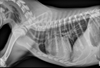
A cat cardiac silhouette radiograph is _____ at the crainal and caudal (compaired to a dog) and ends and appears to have a “_____” shape. The position of the apex is more _____ than a dogs, usually located on or left of midline but sometimes right of midline. In older cats, the cardiac silhouette is more ______ in position.
thinner
lemon
vairable
horizontal (more parallel with the sternum)
(picture A)

In cats, the width of the cardiac silhouette on a lateral radiograph is ___ intercoastal (b/t ribs) spaces. Width on lateral rad is ____ the width of the thoracic cavity.
2
1/2 - a dog is 2/3 the width of the thoracic cavity.

How does the caudal trachea differ in the cardiac silhouette of a cat vs. a dog?
Cat tracheas DO NOT bend ventrally at the heart base.
Dog traches DO curve slightly ventral tat the heart base
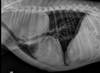
The aorta arises from the ____ of the heart base and becomes the aortic arch as it makes a U-turn _______ and to the left. The descending aorta continues caudally as a linear, soft tissue opacity structure, just ____ of the spine on properly positioned DV/VD radiographs.
middle
dorsocaudally (facing the vertebrae and moving back)
left
(picture B)

Older cats loose the elasticity of the aortic wall resulting in an altered appearance to cardiovascular structures. The heart moves more ____ to the sternum and the aortic arch b/c more prominent (described as “____” or “_____”) and may be mistaken for a cranial mediastinal or pulmonary mass on (_____ radiographs). On _____ radiographs, the desecending aorta appears more tortuous, wavy , or redundant
parallel
Kinked or knuckled
VD/DV- (B)
lateral - (A) BTW, hypertension can also result in a more tortuous descending aorta

Main pulmonary artery segments (pulmonary trunk) arises near the heart base, ___ of the aorta). Not often identiied on lateral rads (blends with adjacent cardiovascular structures). Right pulmonary artery commonly appears as a rounded soft tissue opacity ____ to the tracheal bifucation.
left
ventral (may mimic lymphadenopathy or pulmonary mass) (RPA)
The left pulmonary artery (LPA) may mimic a left atrial enlargement, lymphadenopathy or heart base mass

The caudal vena cava is the only mediastinal structure located in the _____ hemithorax (one side of thorax). It is visible b/t the right hemidiaphragm and the cardiac silhouette in the _____ _____. Phases of cardiac cycle ( includes the diastole, the systole, and the intervening pause) may affect the size of the vena cava, but it is rarely evident on survey rads.

right
caudal mediastinum

The caudal vena cava is normally less than ____ times the width of the aorta (measured at the same intercostal space). Less than the length of the ____ thoracic vertebra. And, wider at the ____ of inspiration, when the intrathoracic pressue is low. Cranial vena cava is NOT normally seen on survey rads.
1.5
6th
height

Cardiac division. The Long Axis line (cardiac length) extends from the ____ ____ to the ___ ___., dividing the heart into right chambers cranially and left chambers caudally (Generally in a 3:2 ratio). The Short Axis line (cardiac width) is perpendicular to the long axis at the level of the caudal vena cava and divides the heart into ____ dorsally and _____ ventrally.
tracheal bifucation to the cardiac apex
atria ventricles

Cardiac chambers are NOT distinguished on survey rads, however, their locations can be inferred using imaginary lines on a ______ rad.
Lateral

The Left caudal lobar artery extends from the pulmonary trunk past the left heart boarder approximately ___ o’clock.
The Left caudal lobar vein enters the left atirum approximately at ___ o’clock.
The Right caudal lobar artery extends from the pulmonary trunk past the right heart border approximately ___ o’clock.
The Right caudal lobar vein enters the left atrium approximately ___ o’clock.
4
5
8
7

The Right mainstem bronchus divides into 4 lobar (_____) bronchi. The right cranial bronchus is first to branch from the trachea (near the ___ intercostal space) and commonly appears as a radiolucent circle near the ____ in the lateral rads.
The right caudal bronchus arises at the ____.
The middle bronchus originates from the ______ aspect of the right caudal bronchus, a few mm caudal to the carina. (Ventral position of this bronchus makes it especially vulnerable to deposition of _____ material.
The accessory bronchus originates from the ____ caudal bronchus.
4
5th
carina
carina
ventrolateral (bottom outside)
aspirate
right

The left mainstem bronchus has ___ divisions.
The left crainal bronchus is ___ to branch from the trachea and quickly divides in to ____ and ____ segments to the left cranial lung lobe.
The left caudal bronchus arises at the ____.
2
second
cranial and caudal
carina


