Hemodynamics Flashcards

(184 cards)

1
Q

What is Hemodynamic Monitoring?

A

Hemodynamic monitoring is basically the assessment of several physiological parameters pertaining to the circulatory system.

It’s where we can measure blood pressure inside of the veins, arteries, and heart.

2
Q

What is the normal Mean Arterial Pressure (MAP)

A

93 mmHg

3
Q

What is the normal Central Venous Pressure (CVP)

A

2 – 6 mmHg

4
Q

What is the normal Pulmonary Artery Pressure (PAP)

A

25/8 mmHg

5
Q

What is the normal Pulmonary Capillary Wedge Pressure (PCWP)

A

4 – 12 mmHg

6
Q

What is the Normal Systemic Vascular Resistance (SVR)

A

900 – 1400 dynes/sec/cm

7
Q

What is the Normal Pulmonary Vascular Resistance (PVR)

A

150 – 300 dynes/sec/cm

8
Q

What is the normal Cardiac Output (CO)

A

4 – 8 L/min

9
Q

What is the normal Cardiac Index (CI

A

2 – 4 L/min/m2

10
Q

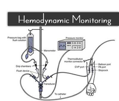
What is a Strain-gauge Transducer?

A

It’s a pressure-measuring device that records pressures by the expansion and contraction of a flexible metal diaphragm connected to electrical wires.

Basically, in healthcare, we can use it to continuously monitor blood pressure.

11
Q

What are the three values used to evaluate the forces influencing blood pressure?

A

(1) CVP (central venous pressure),
(2) PAP (pulmonary artery pressure), and
(3) PCWP (pulmonary capillary wedge pressure)

12
Q

What three factors affect blood pressure?

A

(1) The condition of the left ventricle (the pump),
(2) The volume of blood in the cardiovascular system (the volume), and
(3) The relative size of the intravascular space (the space).

13
Q

Which ventricle is composed of more muscle?

A

The left ventricle.

14
Q

Where is the majority of the systemic blood stored in the body?

A

In the veins.

15
Q

What happens during inspiration?

A

The drop in negative pressure in the thorax from -2 to -5 helps suck blood back toward the heart.

16
Q

What is the Swan-Ganz catheter?

A

An invasive method of measuring pressure within the heart and lungs

17
Q

What is another name for the Swan-Ganz catheter?

A

The triple lumen catheter.

18
Q

What is shock?

A

It is a lack of blood flow to any tissues/organs in the body.

19
Q

What is the distal lumen?

A

The fluid-filled line that transmits a wave of pressure from the tip of the catheter to the transducer.

20
Q

What is a transducer?

A

A device that converts one form of energy to another.

The transducer converts the pressure signal to an electrical signal then sends it on to the monitor.

21
Q

What does the monitor amplify?

A

It amplifies the signal and displays digital readings and/or a waveform.

22
Q

What does the distal port communicate with?

A

The pulmonary artery.

23
Q

Which chamber of the heart does the pulmonary artery come out of?

A

The right ventricle.

24
Q

If the catheter is properly inserted, where does it rest?

A

In a pulmonary arteriole.

25
What can the distal port help us measure?
Problems that originate in the lungs. For example, pulmonary edema
26
What is PCWP?
It stands for pulmonary capillary wedge pressure. It reflects what is going on with the left side of the heart.
27
What happens when the balloon is inflated?
It wedges in an arteriole and is able to obtain a pressure.
28
Where is the closest location that we can get to the left side of the heart?
The pulmonary arteriole.
29
The proximal port is also known as what?
The central venous pressure port.
30
Where is the CVP port located?
At the top of the right side of the heart, where the superior vena cava goes into the right atrium.
31
The lumen opens how many centimeters from the catheter’s port?
30 cm
32
How is cardiac output measured?
By injecting a bolus of saline at a known temperature that is less than the body’s, into the proximal lumen.
33
What does the SVO2 measurement tell us?
How much oxygen the body is using.
34
What is a normal SVO2 value?
70%
35
What measurement is the closest to the left side of the heart?
PCWP
36
What is the formula for cardiac output?
CO = HR x SV
37
Where can you obtain the PaO2?
In a blood gas sample.
38
Where can you find the PVO2?
Mixed venous blood obtained from the distal (PA) port of a Swan-Ganz catheter.
39
Where is the SVO2 obtained from?
The optical module connector in the top of the line Swan-Ganz catheter.
40
VO2 is the amount of what?
Oxygen that the body extracts from the blood every minute in order to feed the tissues.
41
What blood vessel comes in at the top of the right heart?
The superior vena cava.
42
What drains into the superior vena cava?
Internal jugular vein.
43
Where would the first choice be for the insertion of the Swan-Ganz catheter?
The internal jugular vein and subclavian vein because they are a direct shot into the right atrium (veins closest to the right side of the heart).
44
What pressure can be taken when the catheter is in the right atrium?
The central venous pressure (CVP).
45
What is a normal CVP?
The average = 0 – 5 mmHg. The normal range = -2 – +12 mmHg.
46
After the catheter is in the right atrium, where does it go next?
Through the tricuspid valve into the right ventricle.
47
Why does the waveform change as soon as the catheter moves thru the tricuspid valve?
Because the pressure recorded within right ventricle is dramatically higher than within the right atrium.
48
What is a normal right ventricle pressure for systolic?
15-28 mmHg
49
What is a normal right ventricle pressure for diastolic?
0-8 mmHg
50
What is the normal systolic pulmonary artery pressure?
15-30 mmHg
51
What is the normal diastolic pulmonary artery pressure?
4-12 mmHg
52
What is the normal mean pulmonary artery pressure?
6-18 mmHg
53
What does the dicrotic notch represent?
The closure of the aortic & pulmonic valves @ the end of systole
54
What does it indicate when there is no dicrotic notch?
You may have to reposition the catheter or the patient was possibly moving.
55
What happens each time the mitral valve opens?
There is a direct communication between the catheter tip and the left ventricle via pulmonary capillary bed.
56
What is a normal mean of PCWP?
6-12 mmHg
57
How do we know if someone is in shock?
They will demonstrate the following: decreased urinary output, decreased blood pressure, cyanotic, temperature changes of skin and cerebral perfusion compromise.
58
What does cardiogenic mean?
Beginning at the heart.
59
What is cardiogenic shock?
Left ventricular failing as a pump.
60
What pressure is closest to the left ventricle?
PCWP
61
Patients with CHF will have what?
An increased PCWP.
62
What is the most common cause of shock?
The inability of the left ventricle to produce adequate stroke volume.
63
What is stroke volume?
It is the amount of oxygenated blood pumped out of the heart during systole.
64
In an adult, what is the average volume for each heartbeat?
60 – 130 mL/beat.
65
What is an infarction?
Dead muscle.
66
What are some disorders that can cause cardiogenic shock?
(1) Infarction of more than 40% of the left ventricular muscle, (2) CHF, (3) Cardiac tamponade, (4) Chest trauma.
67
What is cardiac tamponade?
Air or fluid in the pericardial sac.
68
How do you know that a patient has CHF based on their chest x-ray?
Measure the CT ratio and if it is \> 50%, CHF is likely.
69
What happens when left ventricular failure gets worse?
Blood begins to back up into the left atrium.
70
What pressure is measured in pulmonary arteriole?
PCWP
71
Which factors are not affected by cardiogenic shock?
The skin’s color and temperature.
72
What does peripheral edema cause?
An increase in heart rate.
73
What can happen if you give a patient too much positive pressure on a ventilator?
It can cause cardiac tamponade (because it will squeeze the heart), among other things.
74
How is cardiac output affected in cardiogenic shock?
The left ventricle is failing so cardiac output is decreased.
75
What drugs would you give to increase cardiac output?
Positive Inotropes (digitalis, digoxin), which will increase the contractility of the heart.
76
What is the total amount of blood volume in the venous system is?
64%
77
Venous return is the amount of blood volume returning to the?
Right heart.
78
The patient cardiac index is the cardiac output based on what?
Actual body size.
79
What is end-systolic pressure?
The amount of blood in the ventricle after ejection.
80
What is end-diastolic pressure is?
The amount of blood in the ventricle after filling.
81
What does cardiac tamponade do to blood entering the heart?
It restricts the blood entering the heart.
82
What is the Fick equation?
CO= VO2/C(a-v)O2 x 10
83
What is the normal right atrial pressure (RAP)?
2 to 6 torr.
84
In general, PAP is concerning what?
The lungs.
85
In general, PCWP is concerning what?
The left heart.
86
In general, CVP is concerning what?
The right atrial pressure or fluid levels.
87
The systemic artery blood pressure (SABP) generally looks at what?
The blood pressure throughout the body.
88
What is preload?
The stretch on the ventricle muscle fibers before contraction.
89
What is afterload?
The resistance of external factors that oppose ventricular contraction.
90
Stroke volume is determined by what?
Preload, afterload, and contractility.
91
A decrease in potassium and sodium causes what?
Atrial fibrillation.
92
What is the procedure for placing an arterial line? | (9 steps)
1. Assemble all equipment, 2. perform the Allen’s test, 3. drape the patient, 4. inject 1% lidocaine, 5. insert the catheter at a 30-degree angle, 6. hold the needle and advance the catheter, 7. remove the needle and secure, 8. attach drip and 9. observe the waveform.
93
What is the procedure for placing a pulmonary catheter?
It is done by the physician, 1. check the balloon for patency, 2. it is inserted into a selected sight until it reaches the 3. right atrium, 4. then inflate the balloon.
94
What are the common sites for placing a pulmonary catheter?
Subclavian or internal jugular vein.
95
What are the common sites for placing an arterial catheter?
Radial, brachial, or femoral arteries.
96
What is Systemic Vascular Resistance?
It is the pressure on the vessel’s throughout the body (from the aorta).
97
What is Pulmonary Vascular Resistance?
It is the pressure on the pulmonary artery.
98
What factors affect the contractility of the heart?
Coronary blood flow, sympathetic nerve stimulation, Inotropic drugs, Physiologic depressants, damage to heart.
99
What are the left atrial filling pressures?
Preload and PCWP.
100
What are the right atrial filling pressures?
The amount of blood in the right atrium or CVP.
101
What is MAP and what is the normal range?
It stands for mean arterial blood pressure and the normal value is 90 mmhg (80-100).
102
What is CVP?
It stands for central venous pressure.
103
What is PCWP and what is the normal range?
It stands for pulmonary capillary wedge pressure that the normal ranges is 5 – 10mm Hg (\<18).
104
What is an increased CVP associated with?
Fluid overload, right ventricular failure, hypercapnia, valvular stenosis, PE, cardiac tamponade, pneumothorax, PPV, PEEP, left ventricular failure.
105
What is associated with an increased PAP?
Pulmonary hypercapnia, left ventricular failure, fluid overload.
106
What is associated with an increased PCWP?
Left ventricular failure, fluid overload, \> 20 interstitial edema, \>25 alveolar filling, \>30 pulmonary edema.
107
What does an increase or decrease in CVP represent?
An increase represents fluid overload, L-R shunt, cor pulmonale. A decrease represents hypovolemic shock.
108
Why should all blood pressure measurements be taken at the heart level?
To eliminate the effect of gravity on hydrostatic pressure.
109
What is the definition of stroke volume?
The volume of blood ejected from the heart with each beat.
110
How does heart rate affect diastolic pressure?
It changes the duration of diastole and the pressure continues to fall until the next systole.
111
Low pulse pressures can indicate what two things?
CHF and shock.
112
What is the average driving force in the arterial system throughout the cardiac cycle known as?
Mean arterial pressure
113
MAP can be used to calculate what?
SVR and PVR
114
What is the normal for MAP?
70-105
115
An MAP below 60 indicates what?
Circulation to vital organs may be compromised and poor tissue perfusion.
116
What are the indications for Arterial cannulation?
It’s indicated for hemodynamically unstable patients, patients on vasoactive drips, patients with IAB, and perioperative patients.
117
What are the hazards of arterial cannulation?
Hemorrhage, thrombus, air embolism, systemic infection, site infection, arterial spasm, vascular occlusion.
118
Why is invasive hemodynamic monitoring needed?
Because clinical assessment alone may not accurately predict hemodynamics.
119
What must be considered before a catheter is placed in a patient?
The risk-benefit ratio of invasive monitoring.
120
What is hemodynamic monitoring performed to do?
To evaluate the intravascular fluid volume, cardiac/vascular function; and to identify sudden changes in hemodynamics.
121
Why is invasive monitoring needed?
To obtain an accurate evaluation of hemodynamics.
122
What type of patient may a physician want to place an arterial catheter in?
Those with significant hemodynamic instability or the need for frequent arterial blood draws.
123
What conditions are likely candidates for arterial pressure monitoring?
Severe hypotension (shock) or HTN; respiratory failure,
124
What patient may benefit from arterial pressure monitoring?
Those in need of medication that affects blood pressure. Examples include vasodilators and inotropic agents.
125
Where is the arterial catheter usually placed?
Radial, ulnar, brachial, axillary, femoral.
126
Where is the arterial line most often placed and why?
The radial artery, because it is readily accessible/adequate collateral circulation.
127
What is low blood pressure a late sign of?
Deficits in blood volume or cardiac function.
128
What are the causes of hypotension?
Low blood volume (bleeding), cardiac failure/shock (heart attack), vasodilation (sepsis).
129
During the administration of what drugs should the diastolic pressure be watched carefully?
Vasodilators, such as sodium nitroprusside.
130
What are 6 causes of an increased central venous pressure?
Fluid overload, heart failure, pulmonary hypertension, tricuspid valve stenosis, pulmonary embolism, and increased venous return.
131
What are the 4 causes of a decreased central venous pressure?
Vasodilation, reduced circulating blood volume, leaks in pressure system/air bubbles, spontaneous inspiration.
132
When can pneumothorax occur during hemodynamic monitoring?
When the catheter punctures the pleural lining
133
What can the accidental opening of the central venous line stopcock allow and result in?
Air to enter the vein which can cause an air embolus.
134
What does the PAC allow the assessment of?
It allows the assessment of the filling pressures of the left side of the heart.
135
What are pulmonary artery catheters also called?
Swan-ganz catheters
136
What is the balloon at the tip of the catheter used for?
To float the catheter into position (into the right side of the heart and into the pulmonary artery) and obtain wedge pressure measurements.
137
What can resistance to pulmonary flow (increased PVR) be caused by?
Constriction, obstruction, or compression of the pulmonary vasculature or backpressure from the left heart.
138
What conditions cause an increased pulmonary vascular resistance?
Pulmonary emboli; acute/chronic lung disease; cardiac tamponade; left heart failure.
139
What should be immediately available at both the insertion and removal of a pulmonary artery catheter?
Lidocaine and emergency resuscitation equipment.
140
What should be optimized to decrease the risk of dysrhythmias?
Blood gases and serum electrolytes.
141
What is not normal and is an indication for obtaining a chest x-ray to assess the cause?
Catheter resistance
142
What are the factors that control blood pressure?
The heart, blood/fluid levels, and vessels.
143
The amount of blood pumped out of the left ventricle in a minute is known as what?
Cardiac Output
144
What is the cardiac Output normal range?
4 – 8 LPM
145
What is the formula for Cardiac Output?
CO= HR X SV
146
What is the volume or amount of blood ejected with each beat known as?
Stroke Volume
147
What is the stroke volume normal range?
60 – 130 mL/beat
148
What is the formula for stroke volume?
SV= CO / HR
149
The amount of blood that is ejected from the heart depends on what 3 things?
Preload, afterload, and contractility.
150
The right ventricle pumps against what?
Pulmonary vascular resistance.
151
The left ventricle pumps against what?
Systemic Vascular Resistance.
152
What happens when blood pressure is too low?
The tissues in the body won’t receive adequate oxygen.
153
What happens when blood pressure is too high?
It causes a strain on the heart and will eventually cause failure.
154
What is the pump that creates blood pressure?
The heart.
155
The distal port samples what type of blood?
Mixed venous blood.
156
What is the Respiratory Therapist’s job when assisting the physician with the insertion of a pulmonary artery catheter?
Set up the bag, zero, check the ports, inflate the balloon, and deflate the balloon.
157
The central venous pressure measures what 3 things?
Intravascular volume, venous return, and right ventricle preload.
158
What causes an increased central venous pressure?
Fluid overload, right ventricle failure, right sided valve disorders, cardiac tamponade, and obstructive RA tumor.
159
What causes a decreased central venous pressure?
Hypovolemia and shock
160
A PCWP that is greater than 18 may represent?
Pulmonary vascular congestion
161
A PCWP that is greater than 30 may represent?
Pulmonary edema
162
What causes an increased PCWP?
Left heart failure, intravascular volume overload, cardiac tamponade/effusion, Obstructive LA tumor.
163
What are the factors which can cause an increased PVR?
Hypoxemia, acidosis, PPV/PEEP & vasoconstrictors.
164
What are the factors which can cause a decreased PVR?
Oxygen, Nitrogen, Alkalosis & vasodilators
165
What are the factors which can cause an increased SVR?
Hypovolemia, certain shocks & vasoconstrictive drugs
166
What are the factors which can cause a decreased SVR?
Vasodilators, morphine, certain shocks
167
What are the normal values for CVP?
2 – 6 mmHg
168
What are the normal values for RAP?
2 – 6 mmHg
169
What are the normal values for RVP?
20-30 / 0-5 mmHg
170
What are the normal values for PAP?
15-25 / 8-15 mmHg
171
What are the normal values for MPAP?
10-20 mmHg
172
What are the normal values for PCWP?
5-12 mmHg
173
What are the normal values for MAP?
85-115 mmHg
174
What are the normal values for PVR?
155 – 255 dynes\*sec/cm5
175
What are the normal values for SVR?
950 – 1300 dynes\*sec/cm5
176
How many lumens do PAC catheters have?
4-6
177
Which part of the PAC rests in the right atrium for the CVP measurement?
The Proximal Lumen.
178
Where does the distal lumen rest?
In the pulmonary artery.
179
Which lumen measures the PAP, PCWP, and obtains mixed venous samples?
The distal lumen because it is in the pulmonary artery.
180
When inserting a PAC, how do you know if it’s in the right atrium?
The pressure should read 2-6 mmHg.
181
How does negative pressure affect the heart?
It increases venous return and preload.
182
What can cause an increased PCWP?
Left ventricular failure, hypervolemia, mitral valve stenosis, and technical causes.
183
What would be seen on a chest x-ray with a PCWP \> 18 mmHg?
The onset of pulmonary vascular congestion.
184
What would be seen on a chest x-ray with a PCWP \> 25 mmHg?
Obvious pulmonary edema.