Heritable Bleeding Disorders Flashcards

1
Q

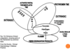
Describe the formation of a haemostatic plug

A
  1. Normal platelets in flowing blood
  2. Damaged endothelium leads to platelets adhering to the wall (bind to collagen in sub endothelium) and becoming activated
  3. Activated platelets aggregate to form a thrombus (platelet plug)
  4. Receptors are expressed and fibrinogen binds to platelets
  5. Granules are released, stimulating further platelet activation
  6. Coagulation cascade follows – clotting factors are soluble proteins and contribute to fibrin formation.
2
Q

Which enzyme converts fibrinogen to fibrin?

A

thrombin (serine protease)

3
Q

How is thrombin formed?

A

Prothrombin (coagulation factor II) is cleaved to form thrombin

4
Q

What is Von Willebrand factor?

A

Von Willebrand factor helps platelets aggregate and adhere to (collagen in) walls of blood vessels.

5
Q

What is the most common heritable bleeding disorder?

A

Von Willebrand disease

6
Q

What is Von Willebrand disease?

A

low or dysfunctional Von Willebrand factor –> bleeding

7
Q

What acts as receptors on platelet surfaces?

A

Membrane glycoproteins

8
Q

The membrane glycoproteins of platelets act as receptors that mediate 2 important functions. What are these?

A
  1. adhesion to the subendothelial matrix
  2. platelet aggregation
9
Q

What are the 2 important membrane glycoproteins found on platelets? What is each one a receptor for?

A
  1. 2b3a –> receptor for fibrinogen
  2. 1b9 –> receptor for vWf
10
Q

Inside the platelets are granules. What are the 3 different types of granules found?

A
  1. alpha granules
  2. dense granules
  3. lysosomal granules
11
Q

What do alpha granules contain?

A

contain adhesion proteins and calcium (role in coagulation cascade)

12
Q

What do dense granules contain?

A

contain ATP and ADP which are released when platelets become activated (ADP forms positive feedback loops – activates more platelets)

13
Q

Metabolic processes are going on inside cytoplasm of platelets when they become activated. What 2 major metabolic processes are going on?

A
  1. Formation of prostaglandins
  2. Formation of thromboxane
14
Q

How are prostaglandins formed in the cytoplasm of platelets?

A
  • Mobilisation of fatty acids from platelet membrane
  • Conversion of the fatty acid arachidonic acid to intermediate prostaglandins
15
Q

How is thromboxane formed in the cytoplasm of platelets?

A
  • Synthesised by activated platelets from arachidonic acid (via the enzyme cyclooxygenase)
  • Released from phospholipid membrane upon platelet activation
  • Secreted into surrounding and stimulates activation of new platelets as well as platelet aggregation
16
Q

What enzyme converts arachidonic acid to thromboxane?

A

cyclooxygenase

17
Q

how does aspirin act as an anti-thrombotic agent?

A

Aspirin inhibits cyclooxygenase (COX) –> prevents production of thromboxane A2

18
Q

Name 4 anti-platelet drugs

A
  1. Aspirin
  2. Clopidogrek
  3. Dipyridamole
  4. IIb and IIa anatagonists
19
Q

How does Clopidogrel act as an anti-platelet drug?

A
  • blocks ADP receptor (P2Y12 inhibitor)
    • P2Y12 is a chemoreceptor for ADP
20
Q

How does Dipyridamole act as an anti-platelet drug?

A

Stimulated prostacyclin to inhibit binding site for collagen, thrombin, thromboxane A2

21
Q

How do IIb and IIa antagonist act as an anti-platelet drugs?

A

blocks GPIIb/IIIa

22
Q

Overview of coagulation cascade

A
23
Q

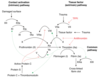
Describe the steps of the extrinsic pathway

A
  1. Coagulation is triggered by tissue factor
  2. Factor VII binds to tissue factor and becomes activated –> VIIa
  3. VIIa can directly activated X –> Xa
24
Q

What is tissue factor? Where is it found?

A

a cellular protein found on surface of cells – damaged endothelium, inflamed blood vessels, WBCs, brain cells

25
Describe the steps of the intrinsic pathway
Doesn’t rely on molecules external from the blood but instead becomes activated when blood is exposed to a foreign surface: 1. Factor 12 is activated when exposed to a foreign surface --\> XIIa 2. XIIa then activates XI --\> XIa 3. XIa then activates IX --\> IXa 4. IXa (with cofactor VIII) activates X --\> Xa
26
Describe the steps of the common pathway of the coagulation cascade
1. Xa (with cofactor Va) will convert prothrombin --\> thrombin 2. Thrombin converts fibrinogen to fibrin (crosslinked by Factor 13)
27
What is the 'prothrombin time'?
* Used to measure extrinsic pathway: * Adding a tissue factor substitute (thromboplastin) to plasma in a test tube * Time how long it takes blood to clot
28
What is the Activated Partial Thromboplastin Time (APTT) used to measure?
Intrinsic pathway
29
What is the thrombin clotting time?
* Used to measure final step * Thrombin is added to plasma * Time how long it takes to clot
30
What is the thrombin clotting time dependent on?
* Very dependent on level of plasma fibrinogen * Dependent on there being nothing in blood inhibiting formation of fibrin from fibrinogen * E.g. heparin
31
What is often the first line of investigation of someone with abnormal bleeding?
*Prothrombin Time (PT)*
32
What does a coagulation screen comprise?
1. Prothrombin time 2. Activated Partial Thromboplastin Time (APTT) 3. Thrombin Clotting Time
33
Why are the top parts of the intrinsic pathway is less physiologically important?
* Extrinsic pathway is thought to start it off. * Intrinsic pathway acts as amplification system instead of initiator. * Thrombin has positive feedback effect Factor 11 can therefore be activated through extrinsic pathway activating thrombin and then the thrombin activating Factor 11 through a feedback system.
34
Does a patient with Factor VII deficiency have a bleeding disorder? What will the APTT be like?
No - this factor is less important But will have a long APTT (instrinsic pathway time)
35
Will a patient with a factor XI deficiency have a bleeding disorder?
Yes but only mild
36
Protein C and Protein S also play a role in the coagulation cascade. What is their function?
Both have a role in **anticoagulation**: * Protein C plays an important role in regulating **_anticoagulation_** by **inhibiting Factor V** * Protein S has a role in the **_anticoagulation_** pathway, where it functions as a **cofactor** to **Protein** **C** in the inactivation of Factors Va and VIIIa
37
Antithrombin also has a role in the coagulation cascade. What is its function?
Anticoagulant effects; It inactivates several enzymes of the coagulation cascade, in particular thrombin and factor Xa
38
The fibrinolytic system also has a role in the coagulation cascade. What is its function?
Anticoagulant: The fibrinolytic system comprises an inactive proenzyme, **plasminogen**, which can be converted to the active enzyme, **plasmin**, which in turn **degrades fibrin into soluble fibrin** **degradation** products.
39
When conducting an assessment of a suspected bleeding disorder, clinical history is key. What questions should you ask?
* Date of onset * Previous history (longer is congenital, shorter if acquired) * Clinical pattern (more severe presents in childhood) * Response to challenges: surgery, dental extraction * Young children: bleeding from umbilical stump, vaccinations, circumcision * Requirement for medical/surgical intervention to ascertain extent of bleeding * Systemic illness, **drug** **history**, **family** **history** – helps to identify if congenital
40
When conducting a clinical examination during an assessment of a suspected bleeding disorder, waht should you look for?
* Pattern of any bruising * Signs of underlying disease: Joints, muscles, skin
41
Exampls of blood tests done during an assessment of a suspected bleeding disorder
* FBC and blood film * Coagulation screen * Clauss fibrinogen (D-dimer if suspicion of acquired disorder) * Mixing studies: if clotting tests are longer than expected, they can be performed with the patient’s plasma and normal plasma. Test will correct significantly if there is a lack of factors. It will not correct significantly if the patient’s blood contains an inhibitor of clotting. * Von Willebrand profile * Coagulation factor assays * Inhibitor assays (antiphospholipid type) * Platelet function tests * If all normal and suspicion remains: factor 13 and alpha2antiplasmin assays.
42
During mixing studies, if clotting tests are longer than expected, they can be performed with the patient’s plasma and normal plasma. * If the clotting test corrects significantly, what does this indicate? * If the clotting test does not correct significantly, what does this indicate?
* Lack of factors in patients blood * Patients blood contains an inhibitor of clotting
43
If a patient has a normal coagulation screen (APTT, PT, TCT), what are other potential causes of a bleeding disorder?
* Thrombocytopenia (low platelets) * Disorder of platelet function * vWD * Factor 13 deficiency * Mild coagulation factor deficiency * Vascular disorder * Disorder of fibrinolysis (rare) * Platelet/vessel wall defects (1ary haemostasis) --\> give rise to prolonged bleeding time
44
How can a factor 13 deficiency lead to bleeding? How does this bleeding present?
* Factor 13 covalently crosslinks the fibrin polymer and stabilises it * Patients with this deficiency typically present with late bleeding (a few days after sometimes)
45
How can platelet/vessel wall defects give rise to bleeding disorders?
* Reduced number of platelets (thrombocytopenia) * Abnormal platelet function (could be iatrogenic, caused by aspirin) * Abnormal vessel wall * E.g. Ehlers-Danlos syndromes; a group of rare inherited conditions that affect connective tissue --\> bleeding due to defects in collagen * Abnormal interaction between platelets and vessel wall * E.g. vWD – reduction in platelet adhering molecules to the wall.
46
What is Ehlers-Danlos syndromes?
Ehlers-Danlos syndromes; a group of rare inherited conditions that affect connective tissue --\> bleeding due to defects in collagen
47
Difference in presentation between vascular/platelet defects and coagulation defects
48
What is this? What would it indicate?
*Petechiae* * Little red spots often around hair follicles * Don’t blanch with pressure * Not palpable (flush against skin) * Think about; thrombocytopenia, meningitis, vascular/platelet defects
49
There are 3 types of **Von Willebrand Disease**. How does each type differ?
* Type 1: * **Reduced** production of **normal** vWf, tend to have normal/slightly reduced Factor VIII * Common, **mild**; especially in blood group O * Variable penetrance in a family with all the same genetic defect. * Type 2: * Patient is producing enough vWf but often is **structurally abnormal** so is dysfunctional (low molecular weight polymers) * Slightly more severe than type 1 * Type 3: * vWf is **absent altogether** and Factor VIII tends to be greatly reduced too * **Severe** and **spontaneous;** * **​**bleeding into joints and vessels as well as mucosal surfaces
50
Why can vWd often present with reduced Factor VIII levels?
Vwf is a carrier protein for factor VIII (carries it around circulation)
51
Inheritance pattern of vWf (remember different types)?
* Mainly autosomal dominant inheritance. * Type 3 is autosomal recessive.
52
Which blood group is often associated with low vWf levels?
Blood group O (this is not vWd)
53
Symptoms of vWd?
* Postoperative/post-partum bleeding * Mucocutaneous bleeding * Menorrhagia
54
vWf has 2 functions. What are these?
1. binds to **protein 1b9 on the platelet surface** and helps it bind to the vessel wall (**collagen**) 2. forms a complex with factor 8 (**carrier** protein) to activate factor 10
55
What factors are deficient in vWd?
In vWD, there is less 8a, F10a and fibrin and so bleeding is more likely
56
Treatment options for vWd:
* **Anti**-**fibrinolytics** --\> tranexamic acid * DDAVP * **Factor concentrates** containing vWF (pool plasma derived) * Vaccination again hepatitis * Combined OCP for menorrhagia (or Mirena coil)
57
What is tranexamic acid? What is it useful in treating?
Anti-fibrinolytic - good for bleeding of the mouth, periods and nosebleeds.
58
What is DDAVP? What is it used to treat?
* DDAVP is an analogue of vasopressin * Analogue of **vasopressin** * Side effect of promoting secretion of vWf from where it is stored inside endothelial cells inside lining of blood vessels --\> short term solution
59
What is Factor I?
Fibrinogen
60
What is Factor II?
Prothrombin
61
Haemophilia A is a defect in what?
Factor VIII
62
Haemophilia B is a defect in what? What is this also known as?
Factor IX - Christmas disease
63
Summary of **Heritable Coagulation Factor Deficiencies**
64
Inheritance of haemophilias?
X-linked recessive
65
How does haemophilia affect men vs women? Why?
* Typically expressed in males and carried by females. * In females, the normal X chromosome will compensate for the faulty X chromosome so tend to be carriers only
66
Is haemophilia A or B more common?
**Haemophilia A**: 1 in 5000 males Haemophilia B: 1 in 30,000 males
67
Which part of the coagulation cascade is affected in haemophilia A/B?
8 and 9 are **intrinsic** **pathway** factors and contribute to the **common** **clotting** cascade.
68
Factor VIII or IX levels determine if haemophilia is mild, moderate or severe. What are the Factor VIII or IX levels for each of these?
* Normal: Factor VIII or IX level between 50-150% * Mild: Factor VIII or IX level between 6-50% * Moderate: Factor VIII or IX level between 1-5% * Severe: Factor VIII or IX level less than 1%
69
How does mild haemophilia present?
* Bleeding only with moderate trauma or surgery. * Diagnosis often due to family history or later in childhood
70
How does moderate haemophilia present?
Variable, occasional spontaneous bleeds, often proceeding trauma
71
How does severe haemophilia present?
Frequent bleeds into muscle and joints
72
Types of bleeding - reflect the severity of haemophilia
* Spontaneous/post traumatic * Can be diagnosed after surgery, trauma, dental procedure etc * Joint bleeding = haemarthrosis, sets of synovitis and causes abnormal, damaged joints * Muscle haemorrhage – hallmark of severe haemophilia, leading to contractures, compartment syndrome, shortening, MSK disability * Soft tissue – bleeds into tongue, pharynx, primary teeth eruption, can threaten the airways (life threatening). * Life threatening bleeding – intracranial bleeding in severe/moderate
73
Treatment of haemophilia;
* Replacement of missing clotting protein (daily): on demand or prophylaxis. * Using factor concentrates; recombinant are products of choice, no longer blood derived * DDAVP (only for mild/moderate haemophilia A) * Antifibrinolytic agents – for mucosal bleeding, GI bleeding * Vaccination against hepatitis A and B * Supportive measures: icing, immobilisation, rest
74
What are the supportive measures for haemophilia?
icing, immobilisation, rest
75
Which haemophilia does DDAVP treat?
A
76
One complication of treatment of haemophilia is the potential development of inhibitors. What are these?
Antibodies formed against Factor VIII or IX that stop the factor concentrates from working due to immune response: * More common in haemophilia A (25%) * Genetic predisposition
77
Presentation of development of inhibitors during haemophilia treatment?
* Poor recovery, bleeding difficult to manage, muscle/joint damage. * Poor clinical response to treatment, factor ineffective or less effective * Occurring early in course of condition, around 10 exposure days
78
Emicizumab is a treatment used for Haemophilia. Mechanism?
Used for **haemophilia** **A** only, with and without inhibitors * Is a “**monoclonal** **antibody**” * Monoclonal antibodies are proteins that are designed to bind to a specific target in the body * They can be used to treat diseases or conditions * Emicizumab: * **Mimics the action of the missing FVIII** in the clotting pathway * The cue for its action is FIXa, which appears after an injury * Emicizumab binds to FIXa and FX, forming a link between these two clotting factors * FIXa can then activate FX, allowing the pathway to continue and the blood to clot
79
Factor VIII Prophylaxis can also be used in haemophilia A treatment. How does this work?
* FVIII concentrate replaces missing or low levels of FVIII * Prophylaxis is the regular administration of FVIII concentrate, to try and prevent bleeds * FVIII rises quickly after infusion and then decreases over a number of hours