Histo: Bone Pathology Flashcards

1
Q

What are the main functions of bone?

A
  • Mechanical - support and site for muscle attachment
  • Protective - vital organs and bone marrow
  • Metabolic - store of calcium
2
Q

Outline the composition of bone.

A
  • Inorganic (65%) - calcium hydroxyapatite, storehouse of 99% of body calcium (and 85% of phosphorus, 65% of sodium and magnesium)
  • Organic (35%) - bone cells and protein matrix
3
Q

In which part of a bone is the growth plate found?

A

Metaphysis

4
Q

What are the main features of cortical bones?

A
  • Long bones
  • 80% of skeleton
  • Appendicular
  • 80-90% calcified
  • Mainly mechanical and protective
5
Q

What are the main features of cancellous bones?

A
  • Vertebrae and pelvis
  • 20% of skeleton
  • Mainly axial
  • 15-25% calcified
  • Mainly metabolic
  • Large surface
6
Q

Describe the microanatomy of bone.

A
  • Made up of several micro-columns
  • Haversian canals are found at the middle of the micro-columns
  • Volkmann canals are canals that connect the Haversian canals
  • Around the Haversian canals are concentric lamellae and in between these units will be interstitial lamellae
  • Around the entire bone you will find circuferential lamellae
  • In the middle the bone will be trabecular lamellae
7
Q

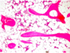
Describe the histological appearance of cancellous bone.

A

Trabeculae are dark pink areas and in between you will find adipose tissue with haemopoietic cells

8
Q

What are the three main types of bone cell and what are their roles?

A
  • Osteoblasts - build bone by laying down osteoid
  • Osteoclasts - multinucleate cells of the macrophage family that resorb bone
  • Osteocytes - osteoblasts-like cells that sit in the lacunae
9
Q

Which protein is important in regulating the action of osteoclasts?

A

Osteoblasts produce osteoprotegrin which blocks the RANK-RANKL interaction which prevents the differentiation of an osteoclast precursor into a fully functioning osteoclast

10
Q

Define metabolic bone disease.

A

A group of diseases characterised by disordered bone turnover due to imbalance of various body chemicals.

NOTE: overall effect tends to be osteopaenia

11
Q

What are the three main categories of metabolic bone disease?

A
  • Non-endocrine (e.g. age-related)
  • Endocrine (e.g. vitamin D, PTH)
  • Disuse osteopaenia
12
Q

Where is bone usually sampled from for histological analysis of bone in metabolic bone disease?

A

Iliac crest

NOTE: the sample be processed and un-decalcified for histomorphometry

13
Q

Which static parameters are measured in the histological analysis of bone in metabolic bone disease?

A
  • Cortical thickness
  • Trabecular bone volume
  • Thickness, number and separation of trabeculae
14
Q

Which technique is used to measure histodynamic parameters when investigating metabolic bone disease?

A

Tetracycline labelling

15
Q

What are the most common causes of osteoporosis?

A

90% are due to insufficient calcium intake or post-menopausal oestrogen deficiency

16
Q

List some primary and secondary causes of osteoporosis.

A
  • Primary - age, post-menopause
  • Secondary - steroids, systemic disease
17
Q

Broadly speaking, what are the two rutes by which disordered bone turnover can cause osteoporosis?

A
  • High turnover results in high bone resorption
  • Low turnover results in low bone formation
  • Both resulted in reduced bone mass
18
Q

What are the effects of long-term steroid use on bone?

A
  • Affects all three bone cell types
  • Leads to decrease in bone quality
  • Ultimately results in osteonecrosis and fracture
19
Q

Describe the ways in which osteoporosis can present.

A
  • Back pain
  • Pathological fractures
    • Wrist (Colles’)
    • Hip (NOF, intertrochanteric)
    • Pelvic
    • Vertebra (60% asymptomatic)
20
Q

Which investigations may be used in a patient with suspected osteoporosis?

A
  • Serum calcium, phosphate and ALP (should be NORMAL)
  • Urinary calcium
  • Collagen breakdown products
  • Imaging
  • DEXA
21
Q

Which four organs are affected by PTH and have a role in calcium homeostasis?

A
  • Kidney
  • Bone
  • Proximal small intestine
  • Parathyroid gland
22
Q

Define osteomalacia.

A

Defective bone mineralisation

23
Q

What two underlying abnormalities can cause osteomalacia?

A
  • Deficiency of vitamin D
  • Deficiency of phosphate
24
Q

What is the main histological feature of osteomalacia?

A

Reduced amount of mineralised bone compared to the amount of osteoid

25
Q

List some consequences of osteomalacia.

A
  • Bone pain/tenderness
  • Fracture
  • Proximal weakness
  • Bone deformity
26
Q

Name some radiological features of osteomalacia.

A
  • Bowing of the legs in Rickets
  • Horizontal pseudofractures in Looser’s zones
27
Q

Describe how the urine excretion of calcium and phosphate changes in primary hyperparathyroidism.

A
  • Both increase

NOTE: PTH causes a minor increase in reabsorption of calcium from the urine, however, as it causes a large increase in serum calcium concentration (and hence an increase in the calcium concentration of the glomerular filtrate) it causes an overall increase in urine calcium excretion

28
Q

What skeletal change is seen in primary hyperparathyroidism?

A

Osteitis fibrosa cystica

29
Q

List the main primary and secondary causes of hyperparathyroidism.

A

Primary

  • Parathyroid adenoma (90%)
  • Chief cell hyperplasia

Secondary

  • Chronic renal deficiency
  • Vitamin D deficiency
  • Malabsorption
30
Q

What histological feature is typically seen in hyperparathyroidism?

A

Brown cell tumour - several multinucleated giant cells on a background of fibrous stroma with haemorrhage

31
Q

Define renal osteodystrophy.

A

Term used to describe all the skeletal changes of chronic renal disease:

  • Increased bone resorption (osteitis fibrosa cystica)
  • Osteomalacia
  • Osteosclerosis
  • Osteoporosis
  • Growth retardation
32
Q

What is Paget’s disease and what are its three phases?

A

Disorder of bone turnover

Three phases:

  1. Osteolytic
  2. Osteolytic-osteosclerotic
  3. Quiescent osteosclerotic

NOTE: the combination of osteoblast and osteoclast activity results in new bone formation

NOTE: 85% are polyostotic

33
Q

Describe the histological appearance of Paget’s disease.

A

Lines will be seen between areas of new bone formation so it looks like a mosaic/jigsaw puzzle.

34
Q

Which virus is associated with Paget’s disease.

A

Parvomyxovirus

35
Q

Describe the presentatino of Paget’s disease.

A
  • Most lesions affect the skull and lumbar spine
  • Pain
  • Microfractures
  • Nerve compression
  • Skull changes (can put the medulla at risk leading to haemodynamic changes)
  • Sarcoma
  • Paget’s disease of the tibia can cause bowing
36
Q

What are the four stages of fracture repair?

A
  • Organisation of a haematoma at the site of the fracture (pro-callus)
  • Formation of a fibrocartilaginous callus
  • Mineralisation of the fibrocartilaginous callus
  • Remodelling of the bone along weight-bearing lines
37
Q

List some factors that influence fracture healing.

A
  • Type of fracture
  • Presence of infection
  • Pre-existing systemic conditions (e.g. chronic conditions)
38
Q

Which sites are most commonly affected by osteomyelitis?

A
  • Vertebra
  • Jaw (secondary to dental caries)
  • Toe
  • Long bones (usually metaphysis)
39
Q

Describe the typical presentation of osteomyelitis.

A

General - FLAWS

Local - pain, swelling, redness

40
Q

Which investigations may be used in suspected osteomyelitis?

A
  • Blood cultures (positive in 60%)
  • X-ray (will eventually show lytic areas)

NOTE: osteomyelitis is almost always bacterial

41
Q

What are the routes of infection in osteomyelitis?

A
  • Haematogenous
  • Direct extension
  • Traumatic (penetrating injury)
42
Q

Which organisms can cause osteomyelitis?

A
  • Staphylococcus aureas (90%)
  • E. coli
  • Klebsiella
  • Pseudomonas (IVDU)
43
Q

Which bacterium is associated with osteomyelitic in patients with sickle cell disease?

A

Salmonella

44
Q

Describe the X-ray changes seen in osteomyelitis.

A
  • Usually appear about 10 days after onset
  • Mottled rarefaction and lifting of periosteum
  • First week changes - irregular sub-periosteal new bone formation (involucrum - layer of new bone that forms around dead bone)
  • Later changes - irregular lytic destruction
  • Some areas of the necrotic cortex may become detached (sequestra). This takes 3-6 weeks
45
Q

What are the potential consequences of TB osteomyelitis of the vertebrae?

A
  • aka Pott’s disease
  • May result in psoas abscess or skeletal deformity

NOTE: TB osteomyelitis tends to only occur in immunocompromised patients

NOTE: systemic amyloidosis may occur in some cases

46
Q

Describe the histological appearance of TB osteomyelitis.

A
  • Lots of inflammatory cells can be seen in between trabeculae
  • Langerhans-type giant cells (with a horse-shoe nucleus) will be seen
47
Q

What congenital skeletal lesions are associated with syphilis?

A
  • Osteochondritis
  • Osteoperiositis
  • Diaphyseal osteomyelitis
48
Q

List some acquired skeletal lesions that are associated with syphilis.

A
  • Non-gummatous periostitis
  • Gummatous inflammation of joints and bones
  • Neuropathic joints (tabes dorsalis)
  • Neuropathic shaft fractures
49
Q

What is lyme disease?

A
  • Inflammatory arthropathy that occurs as part of a complex multisystem illness from a tick bite
50
Q

What is the organism and vector in lyme disease?

A

Organism: Borrelia burgdorferi

Vector: Ixodus dammini

51
Q

What is the skin rash that is classically associated with lyme disease?

A

Erythema chronicum migrans

52
Q

Describe the clinical features of lyme disease.

A
  • Early localised - rash (90%) often on thigh, groin or axilla, 1-50 cm in diameter
  • Early disseminated - affects many organs (musculoskeletal, heart, nervous system)
  • Late, persistent - dominated by arthritis
53
Q

Outline the treatment of lyme disease.

A
  • Aim for prevention
  • Antibiotics
  • No effective prophylaxis
  • Diagnosis is clinical
54
Q

What is the difference between primary and secondary osteoarthritis?

A
  • Primary - age-related
  • Secondary - can occur at any age, occurs in a previously damaged or congenitally abnormal joint
55
Q

What are the main sites affected by osteoarthritis?

A
  • Vertebrae
  • Hips
  • Knees
  • DIP and PIP of the hand

NOTE: the synovium becomes very inflamed with lots of inflammatory cells (lymphocytes)

56
Q

What are the HLA associations of rheumatoid arthritis?

A

HLA DR4 and HLA DR1 (Chr6p21)

Other alleles: TFNA1P3, STAT4

57
Q

What is rheumatoid factor and what proportion of patient’s with rheumatoid arthritis are rheumatoid factor positive?

A
  • IgM antibody against IgG
  • 80% positive
  • May be responsible for extra-articular disease
58
Q

Which sites tend to be affected by rheumatoid arthritis?

A
  • Small joints of the hand and feet (except DIP)
  • Wrists, elbows, ankles and knees
59
Q

List some characteristic deformities associated with rheumatoid arthiritis.

A
  • Radial deviation of the wrist
  • Ulnar deviation of the fingers
  • Swan neck and Boutonnier deformity
  • Z-shaped thumb
60
Q

Describe the histological features of rheumatoid arthritis.

A
  • Proliferative synovitis with thickening of synovial membranes, hyperplasia of surface synoviocytes, intense inflammatory cell infiltrate and fibrin deposition and necrosis
  • A pannus can form (exuberant inflamed synovium)
61
Q

Which type of multinucleate giant cell may be seen in rheumatoid arthritis?

A

Grimley-Sokoloff cell (like a Langerhans cell but does not have horseshoe nuclei)

62
Q

What type of crystals cause gout?

A

Needle-shaped negatively birefringent urate crystals

NOTE: big toe is involved in 90% of cases

NOTE: a tophus is pathognomonic of gout

63
Q

Which crystals cause pseudogout?

A
  • Calcium crystals
  • Calcium pyrophosphate - mainly knees
  • Calcium phosphate (hydroxyapatite) - knees and shouldrs
  • Rhomboid-shaped positively birefringent crystals
64
Q

Which tumours most commonly metastasis to the bone in:

  1. Adults
  2. Children
A

Adults:

  • Breast
  • Prostate
  • Lung
  • Kidney
  • Thyroid

Children:

  • Neuroblastoma
  • Wilm’s tumour
  • Osteosarcoma
  • Ewing’s
  • Rhabdomyosarcoma
65
Q

Name three types of primary malignant bone tumours.

A
  • Osteosarcoma
  • Chondrosarcoma
  • Ewing’s sarcoma/PPNET
66
Q

What are the key clinical and radiological features of osteosarcoma?

A
  • Peak in adolescence
  • 60% around the knee
  • X-ray: usually metaphysial, lytic, permeative, elevated periosteum (Codman’s triangle)
67
Q

Describe the histological appearanc of osteosarcoma.

A

Malignant mesenchymal cells with or without bone and cartilage formation

68
Q

What are the main clinical, radiological and histological features of chondrosarcoma?

A
  • Malignant cartilage producing tumour
  • Affects axial skeleton, proximal femur and proximal tibia
  • X-ray: lytic with fluffy calcification
  • Histology: malignant chondrocytes with or without chondroid matrix
69
Q

What are the main clinical, radiological and histology features of Ewing’s sarcoma?

A
  • Highly malignant small round cell tumour
  • Occurs in <20 years
  • Mainly affect diaphysis and metaphysis of long bones
  • X-ray: onion skinning of the periosteum, lytic with or withour sclerosis
  • Histology: sheets of small round cells
70
Q

Which chromosomal translocation is associated with Ewing’s sarcoma?

A

11;22 (ESWR1:Fli1)