HST 3 Flashcards
(42 cards)
What are the three main digestive glands, and their function?
- Major salivary glands - associated with the oral cavity thru independent excretory ducts 2. Exocrine pancreas - secrete alkaline aqueous and enzymatic products into the duodenum 3. Liver - exocrine and endocrine gland with extensive access to blood circulation
What is the acinus? duct?
- blind-sac of secretory cells that synthesize and release product into the duct 2. conducting passageway for product to be released into lumen
What comprises the salivon?
acinus, intercalated duct, and excretory duct
What is the role of saliva? what produces? how is its production controlled?
- lubricates/cleanses oral mucosa, contains immunoglobulins/minerals/electrolytes/buffers/enzymes/metabolic wastes, aids in digestion of food via enzymes, and tooth maintenance 2. Secretory cells 3. ANS
What is the role of myoepithelial cells?
assist in moving secretory products towards the excretory duct
What is the pathway of salvia? What epithelium is associated with the layers)
acinus –> interlaced duct (low cuboidal epi) –> striated duct (simple cuibodal to columnar epi) –> excretory duct (simple cuboidal to pseudo stratified columnar/stratified cuboidal)
When comparing parotid, submandibular, and sublingual glands, which has the longest intercalated duct? striated duct? Excretory duct?
- parotid > sublingual > submandibular 2. submandibular > parotid > sublingual 3. sublingual > submandibular > parotid
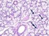
Parotid Gland
-Serous only -Largest salivary gland with adipocytes **serous only, large amounts of adipose tissue, CN VII passes through
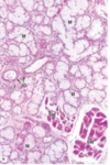
Sublingual Gland
-Mixed, but predominantly mucous -Lacks defined capsule, but CT divides it into small lobules
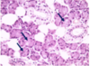
Submandibular Gland
-MIxed, but predominantly serous -Mucus cells containing acini are capped by serous demilunes
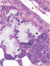
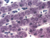
Where do we find centroacinar cells? what is their role?
- duct cells inside the acinus of the pancreas 2. secrete HCO3-, Na+, and H2O to alkalinize secretions *stain lightly inside dark staining acinar cells
What do pancreatic acinar cells contain to aid in digestion?
20 different proenzymes (ex. trypsinogen)
Pancreatitis 1. Cause 2. Result - acute vs. chronic
- premature activation of pancreatic enzymes results in the autodigetsion of the pancreatic gland 2. a. acute - usually follows trauma, heavy meals, or excessive alcohol ingestion, or biliary tract disease; causes severe abdominal pain, nausea, and vomiting b. chronic - fibrosis, and partial or total destruction of pancreatic islet; alcoholism
When does the serous covering NOT line the liver?
where it directly adheres to the diaphragm/other organs
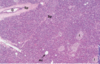
What is the role of hepatocytes?
metabolism, storage, and bile* production *drains into bile canaliculus between adjacent cells; eventually join to contribute to the biliary tree
Where does blood of the portal vein and hepatic artery mix?
sinusoids of the lobules central venue –> sub lobular veins
Terminal hepatic venule (central vein)
collects blood from the sinusoids
Bile and lymph flow in ____ directions.
opposite
Perisinusoidal space of Disse
separates hepatocytes plate from the blood sinusoidal space; site of material exchange b/t blood and liver cells
Periportal space (space of Mall)
excessive fluid in the space of Disse is collected here and drained by lymphatic vessels
What cell can be used to distinguish liver sinusoids?
Kupffer cells - macrophages that phagocytose RBCs
Portal lobule
triangle between 3 central veins that outlines bile drainage from adjacent lobules into the same bile duct
Liver Acinus
most clinically relevant with 3 zones *ischemia affects zone 3 the most **toxins affect zone 1 the most
Hereditary hemochromatosis
-characterized by increased iron absorption & accumulation in lysosomal hepatocytes -complications include cirrhosis & cancer of the liver