L3 Spinal cord to the diencephalon Flashcards
(111 cards)
•Central nervous system is formed from _____derm
•Central nervous system is formed from ectoderm
List the 3 steps in neuro
- Neuroectoderm cells receive inductive signals from notochord
- Cells thicken to form neural plate
- Lateral neural plate margins fold inwards to form neural tube
What is neurulation?
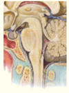
Development of the nervous system
At embryonic day 20, the cells at edges of neural plate are called _________ cells
Neural crest cells
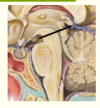
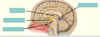
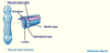
Label the diagram of neurulation


At embryonic day 24, ________ cells migrate into _______ and differentiate
At embryonic day 24, neural crest cells migrate into periphery and differentiate
Neural crest cells migrate into periphery and differentiate into…
(1) Autonomic and sensory neurons and glia
(2) Cells of the adrenal gland
(3) Melanocytes
(4) Skeletal/connective tissue of the head
What does the mantle layer become?

- Becomes brain parenchyma
What does the Ependymal layer become?

Lines the ventricles
What happens at embryonic day 24 after neural crest cells migrate into periphery and differentiate?
•Neural tube thickens
What does the lumen become?

- Becomes ventricles + central canal
Complete the diagram on embryonic day 24 of neurulation

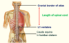
•Neural tube defects occur in ~1/______ established pregnancies
1000
What is anencephaly?
Failure of anterior neuropore to close
= Anencephaly (fatal)
What is spina bifada?
Failure of posterior neural tube to close
= Spina bifida (divided by a cleft)
Name the types of neural tube defect circled on the diagram
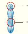

What type of spina bifida does this show?

Spina bifida occulta (hidden, vertebral arch defect only)
What type of spina bifida does this show?

Spina bifida cystica (e.g.; meningocele = meninges projects out)
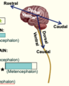
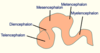
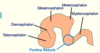
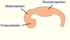
How are primary brain vesicles formed?
•Expansion of cranial end to form main brain regions (primary vesicles)
In what direction does the development of the cervical and cephalic flexures occur?
Sagittal
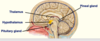
Complete the diagram of the primary brain vesicles


Label where the cervical and cephalic flexures are on the diagram
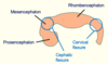
What does the telencephalon form?
(Cerebral hemispheres)
What do the optic vesicles form?
Eyes