Lameness Flashcards

(82 cards)

1
Q

What can you add to a ruminants diet if high grain diets to rebalance high P: CaCO3?

A

Calcium carbonate (limestone)– immediately for growing ruminants, if > 8 weeks full feeding for adults

2
Q

High serum phosphate from a diet high in cereal grains in growing cattle and sheep does what?

A

Antagonises serum Ca2+–> Low Ca2+ stimulates PTH–> Ca resorbed from the bone–> tends to cause osteoporosis in sheep and cattle

3
Q

How can we assess secondary nutritional hyperparathyroidism?

A

* Urinary P excretion… compare urein: serum % of phosphorous and creatinine (baseline metabolite that the kidney doesn’t reabsorb)

4
Q

What can happen with low dietary vitamin D?

A

Vitamin D needed for active Ca & P absorption from gut–> low Ca &/0r P means bone can’t be mineralised–> Rickets in growing animals or Osteomalacia in adults (remodelled bone is soft)

5
Q

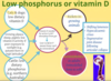
How do you ensure enough vitamin D in a ruminants diet?

A

Avoid rachitogenic factors (anti vit D) and high NH4 (antagonises dietary Ca absorption) e.g. in oat crops

** can give oral or injectable supplements in young sheep if signs are developing to help in situations of Ca deficiency

* Vit D toxicity causes abnormal tissue mineralization

6
Q

Keys in a ruminants diet?

A

* Enough energy, protein, Ca, sunlight, Cu

* Parasitism can have a big effect

* Ca: P balance especially with cereal supplements

7
Q

How much calcium do you need to add to a diet to fix Ca:P ratio?

A
8
Q

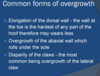
How can nutrition affect lameness?

A
9
Q

90-90-90 rule?

A

90% of a herd’s lameness from the feet

90% of those lamenesses are in the back feet

90% lateral claw

10
Q

Most cattle lameness from what lesions?

A
11
Q

Economic losses due to?

A
12
Q

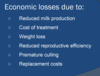
Approximate cost of lameness in an individual treatment

A

$200 per lame cow (1998) in AUS

13
Q

Average loss of production in a lame cow?

A
14
Q

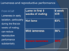
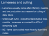
Lameness and reproductive performance?

A
15
Q

What percentage of cows that are culled are culled for lameness?

A
16
Q

Multifactorial aetiology of lameness in cattle

A
17
Q

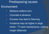
Predisposing causes of lameness

A

* Infectious agents- Fusobacterium necrophorum

* Nutrition- rumen acidosis and laminitis

* Hereditary- e.g. cork screw claw, sore ulcer

* most lameness due to over-worn, bruised soles or stones in interdigital cleft

* Increased when farm track had steep slope, course gravel or broken sections

* Bail feeding associated lower incidence lameness

* Cows should not be hurried on farm tracks

** Risk factors on track

  • below avg track maintenance, bad cow flow on track (number of congestion points), patience of farmer- motor bikes, biting dogs

**Risk factors in shed

  • biting dog, contentment of cows, cow density in yard, aggressive use of backing gate

** Risk factors in animals

  • higher Friesians, white feet
18
Q

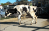
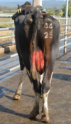
Lameness prevalence picture

A
19
Q

Examination of the lame cow

A

* Observe standing free in yard

* History- how long lame, treatment?

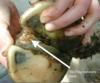
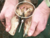
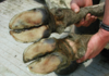
* Clean with scrubbing brush, water… hoof knife, rasp or angle grinder

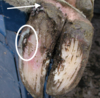
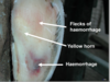
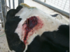
* Look for swelling at the coronary band

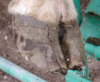
* look for defects or penetrations of the sole

* Use hoof testers to check for pain

* Examine for axial, abaxial wall cracks

* Thoroughly explore any defects detected

8 point exam

  1. Which leg is the cow lame in?
  2. Is the leg swollen and obviously painful above the hoof?
  3. Are cracks present in external surface of the hoof wall of lame leg?
  4. Is there soft tissue swelling with foul swelling discharge and dead and damaged skin in the interdigital space?
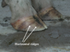
  5. Is there abnormal discolouration in the sole horn? Do not confuse with normal pigment (usually black)
  6. White line- expanded and filled with mud and gravel especially towards heel area?
  7. Are there any sensitive areas which cause withdrawal reflex?
  8. Seek vet advice OR withdrawal response detected– bruising- rest; toe/foot abscess or under run sole- trim, pare, grind until loose sole horn removed back to normal horn; pus detected- establish drainage
20
Q

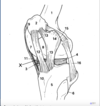
How do you lift the hind foot? What is a good idea if there is hindfoot lameness?

A

* possibly low dose Xylazine with hindfoot

21
Q

How do you lift the front foot?

A

* Loop around leg at level of dewclaws

* over rail and back under axilla

* then back over rail and lift

22
Q

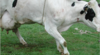
What’s the difference between Holstein and Friesian?

A

Holstein Friesians are a mixture

Holstein are bigger girls– genes from North America

Friesian gene is from Europe

** our herds are a mixture of the two so a middle size

23
Q
A
24
Q
A
25
Why are angle grinders potentially dangerous?
\* Electricity and water- use circuit breaker \* Only use on well restrained animal \* Must wear safety goggles \* Can generate excessive heat- proceed slowly \* Not recommended for use on front feet of standing animal unless very well restrained
26
Examination of a lame herd
\*\* A lameness problem on a farm needs to examine farm management factors in the herd rather than treating a long series of individual lame cows \* Define the problem-- how many cases, what are the lesions \* Ascertain likely aetiological factors-- farm tracks, animal handling, general farm environment, nutritional-- SARA and laminitis
27
Principles of treatment of lame cows
\* Adequate restraint +/- sedation \* Appropriate anaesthesia \* Suitabe equipment \* Avoid walking lame cows too far
28
Anaesthesia of the bovine digit?
\* Xylazine an excellent sedative \* Interdigital nerve block- 20 cc lignocaine injected between the digits
29
Regional intravenous anaesthesia?
\* Regional intravenous anaesthesia: - apply tourniquet - 20 cc lignocaine IV in digital vein
30
31
32
White line disease (25%)?
\* Disintegration junction between sole, wall \* Penetration dirt, stones at this site \* may track up to coronary band \* Usually lateral claw hind digit \* Variable lameness
33
Sole injury (26%)?
Sole penetration, abscess, under-run sole \* Infection through FB or horn defect \* Acutely lame \* Characteristic "sliding" lameness \* Swelling above coronary band (may be very slight) \* Careful search for hoof defect
34
Axial wall cracks (19%)?
\* Occur adjacent to axial groove of medial claw of hoof \* Often no lameness \* Lameness often follows heavy rain- fine mud forced into crack
35
Footrot (infectious necrobacillosis 10%)?
\* Footrot- Fusobacterium necrophorum \* Necrotising infection interdigital dermis, cellulitis of digital area \* Signs - sudden onset lameness VERY PAINFUL - swelling affected area - responds antibiotics
36
Bruised sole?
\* Commonly affects hind feet \* Sole worn flat \* Sole may be palpably thin \* Bruising, haemorrhage evident
37
Excess hoof wear?
\* Feet kept moist due to environmental conditions \* On concrete at time of milking \* Plus walking laneways \* May lead to excess hoof wear
38
Sole ulcer?
\* More common in housed cows \* Specific lesion sole/bulb junction, usually nearer abaxial margin \* Lateral hind claws \* If exposed dermis, granulation tissue commonly develops at defect \* Treatment- expose diseased tissue, remove necrotic horn
39
Inter-digital dermatitis?
\* Cause- bacteroides nodosus \* Can produce benign footrot sheep \* Inter-digital dermatitis, but little swelling \* Often affects more than one foot, calves particularly susceptible
40
What is the actual correct term for cattle laminitis?
Coriosis
41
Digital dermatitis (Hairy Heelwart 1%)? Clinical Signs?
\* very painful, contagious disease causing wart-like areas on the back of the hind feet, on the bulb of the heel or near the interdigital cleft \* Common in dairy herds in the US, uncommon in AUS at the moment \* precise cause is unknown, Bacteroides species and an invasive Treponema spiroacheate have both been implicated \* Clinical features- lesions of digital dermatitis can be of at least two distinct types: erosive/ reactive (strawberry like) and the proliferative (wart-like) forms-- US, EU, Chile now seeing non-healing lesion; cleansing exposes reddish granulation tissue (strawberry) with concave profile); lesion is very sensitive and easily bleeds but the soft tissue is not swollen; animal is often lame; proliferative form may give rise to the papillomatous type, which is characterised by a mass of hard, fine tendrils which can be several cm in length and cover a considerable area
42
Diagnosis Digital dermatitis (Hairy Heelwart 1%)? Treatment?
\* Based on history of an epidemic onset of discomfort and lameness in the herd- commonly seen as shifting lameness \* Treatment: - Lesions affecting individual animals should be treated topically - after cleaning and drying, the lesion should be sprayed with Oxytetracycline - herd outbreak with a footbath containing antibiotic - parenteral antibiotics have not shown any affect on either the erosive or proliferative form of digital dermatitis
43
44
Interdigital skin hyperplasia?
\* Excess epidermal, hypodermal tissue \* More common hind feet \* Appear as protuberance of skin \* Degree of lameness varies \* Treatment surgical
45
Aseptic Laminitis?
\* Diffuse aseptic inflammation of the corium \* Laminitis is inflammation of the soft tissue between the hoof wall and the sole and the underlying pedal bone within the claw \* Inflammation results in a reduction in the quantity and quality of hoof material produced
46
Acute laminitis?
\* Acute laminitis is not common in cattle in our environment- may follow acute illness \* Clinical signs similar to horse, but more variable \* Affected animals - move slowly, arched back - stand with hind legs extended forward, fore legs forward
47
48
Subclinical laminitis
\* Occurs much more commonly than acute laminitis \* It's an important cause of other hoof diseases, for example, white line disease, sole abscesses, sole ulcer and claw deformities \* Produces no immediate signs but is recognized by the changes in the hooves which occur several months after episodes of sub clinical laminitis-- claw changes shape, becomes longer and the front wall of the hoof is concave when viewed from the side... sole becomes wider and flatter, wall develops horizontal ridges or ripples due to partial interruptions in its production \* Changes are a deterioration in hoof horn quality and haemorrhages in the sole of the hoof \* The white line develops a yellow colour and has small red dots of haemorrhage along it
49
Subclinical laminits/ chronic laminitis
50
Sand Crack- vertical split in the hoof wall extending from coronet to a variable distance along the direction of growth \* Large incomplete sandcracks often not associated lameness \* two distinct vertical fissures-- fissures of coronet, fissures of wall
51
\* Horizontal fissures of hoof wall-- discontinuity of wall of hoof in a plane parallel to coronet - often occurs in all hooves - related to severe upset of metabolism-- mastitis, metritis, other acute diseases
52
Abnormalities of hoof growth
53
54
Indications of amputation of a digit? Considerations?
55
Could be?
56
Principles of treatment of the bovine hoof
\* Adequate restraint \* Suitable local anaesthesia \* under-run areas opened to allow drainage \* avoid exposing too much corium when trimming claw cracks- otherwise granulation tissue \* in painful conditions, raised affected claw
57
Applying blocks or cowslips
\* Clean (defat) area with alcohol \* Mix glue and use this to attach block or cowslip \* Keep foot restrained until glue dry \* must be sure the claw is sound \* Use angle grinder to clean sole, walls. Place shallow grooves on sole
58
Footblocks
\* Wood or rubber blocks attached to the sound claw opposite the affected claw of the lame foot \* Block attached to sound claw with Bovibond \* Very effective, well accepted by farmers
59
Assessing for upper limb lamess
\* Vast majority of bovine lameness is in foot \* if suspect upper limb-- sedate the cow and then walk cow around the yard, palpating joints of upper limb-- abnormal clunks, crepitus
60
Diagnosis? Treatment?
Coxofemoral luxation \* Dianosis: stance of animal, crepitus is usually evident, palpation head of femur, affected limb abducted excessively, palpate greater trochanter-- upwards and forwards, upwards and back, downwards and forward- rectal, downwards and back- rectal \* Treatment- sedate heavily with Xylazine... lateral recumbency, affected limb uppermost, restrain the animal with rope through inguinal region, traction- block and tackle or calving jack.... rotate limb while applying traction, can change direction of traction if needed, feel clunk-- immediately release traction \*\* Better prognosis if cow able to stand before reduction, cow less than 3 years, less than 400 kg, duration dislocation less than 12 hours
61
Pain relief for cows?
Metacam or Ketoprofen for example
62
Stifle instability
Stifle instability-- second most common form of high limb lameness \* Shortened stride affected leg \* Weight bearing on toe, heel raised \* Restricted movement stifle joint
63
Pelvis fractures-- body of ilium, ischium or pubis \* rest \* beware of risk bony callus- dystocia
64
Hip dysplasia
\* Seen young rapidly growing, heavy beef bulls \* Swaying gait of hind limbs or drag hind limbs slightly \* More severe cases click as pushed from side to side or crepitus \* INherited defect
65
Partial or complete separation joint surfaces sacrum, ilium \* Mild cases- slight ataxia, knuckling fetlock
66
Patellar luxation \* Patellar temporarily fixed on upper part of femoral trochlea \* Clinically the animal progresses with jerky action, leg remains extended back until patellar frees itself \* Treatment- medial patellar desmotomy (incision or divison of a ligament) Patellar luxation- correction: \* Identify middle and medial patellar ligaments \* Prepare area anterior to and over medial patellar ligament for surgery \* local anaesthesia \* 1.5 cm incision just anterior to medial patellar ligament, insert tenotome beneath medial patellar ligament, incise ligament completely, clunk, suture skin
67
11? 12? 13?
68
Degenerative arthritis
\* Primary degeneration of articular cartilage, generally accompanied by secondary hypertrophy cartilage, bone-- coxitis, gonitis (inflammation of the stifle (femorotibial) joint.) \* Usually affects older animals, major weight bearing joints-- hip, stifle, hock \* Usually slow onset \* Weight loss and muscle atrophy as condition progresses \* Hoof dragging, rolling gait \* In advanced cases, crepitus obvious
69
Rupture cranial cruciate ligament
\* Cranial ligament is shorter of the two, more prone to injury \* palpate and hear audible dislocation of distal femoral, promixal tibial articular surfaces-- sedation often aids in diagnosis
70
Fractured limbs
\* Below carpus, tarsus respond well to casting provided not compound \* High fractures- conservative treatment
71
Infectious arthritis
\* joint penetration or septicaemia \* calves- streptococcal and coliform polyarthritis frequently follows calf scour syndrome \* Adult cattle- may follow mastitis
72
73
74
75
Peroneal paralysis- susceptible where it runs down over the lateral femoral condyle, head of fibula \* knuckling of fetlock, over-extension of hock- in severe cases dorsal aspect fetlock joint contacts the ground
76
77
Prevention of digital disease
\* Environment- farm track maintenance, gate ways, troughs, selection of concrete for yards \* management- don't hurry, force cows along track, try and avoid turning on concrete... design of laneways-- minimal abrasive qualitie, avoid sharp stones, adequate drainage, eliminate congestion points.... minimize time on concrete \* Concrete in yard - too smooth- slipping, injury - too rough- very abrasive \* Reduce twisting, turning on concrete \* Do not force, hurry cattle \* Foot bathing-- needs to be changed very frequently \* Mate heifers 3 weeks ahead of herd \* Site dairy at centre of farm \* Claw trimming
78
\*very little effect on the middle group of cows in the mob \* Overall speed of herd will hardly change- this is set by the dominant cow group in the herd THEREFORE \* impatience and attempting to hurry cows will save little time \* but may lead to a marked increase in a number of lame cows in the herd
79
Building an appropriate track
1. Track drainage- prevent water seeping into the track base from the surrounding ground-- must have somewhere for water to flow and fenced off drains so the cattle won't walk on them 2. Remove grass and topsoil- topsoil containing grass has no strength and can pug rapidly.. needs to be removed before constrcution of the track commences 3. Track base-- well compacted, crowned surface which is above the surrounding ground and has drainage provided on either side 4. Crowning- suffiently crowned to shed water, but not to make walking difficult... avg. cross between 3 and 6% with a suggested maximum of 10% 5. A suitable compacting device such as a vibrating roller is used. The base is built in layers not exceeding 150 mm and each layer is thoroughly compacted\ 6. Track bearing surface-- suitable surface for cows to walk on-- must prevent seepage of water into the underlying track base-- gravel, sand and clay-- finer particles will fill the pores between the larger particles, binding the material and forming a hard wearing and relatively smooth surface
80
If a cow goes down with paralysis who is BAR and the farmer is unwilling to move her from the paddock, how should you proceed with treatment?
Euthanasia
81
What else can present a problem?
\* Exits from the dairy can also present a problem
82
Stage one-- trim the medial toe to the correct length- commonly the dorsal wall at the toe measures 75 to 80 mm... the lateral toe is then trimmed to a similar length Stage two-- remove excess sole horn- most importantly at the toe.. the weight bearing services across both toes should be flat and balanced Stage three-- equalize weight bearing on the medial and lateral claws Stage four-- remove the axial ledge- the central sole area beneath the flexor tuberosity over the pedal bone should be non-weight bearing Stage five-- if necessary remove excess overgrowth of horn heel horn, commonly on the outside the heel of the hind limb