Lecture 12 (Exam II) Flashcards
(24 cards)
Which cell types can recognize a specific epitope on an intact, unprocessed antigen
B cell and y delta T cell
Which Th2 cytokine has a predominant role in inhibiting a Th1 response
IL10
List the three professional antigen presenting cells
- Denditric cells
- B cells
- Macrophages

give brief explanation of how/when dendritic cells present antigen
- Special MHCII antigen presenting cells that are especially important in activating a naïve T cell and triggering a primary response
- Presents on MHC II. If antigen is opsonized it has been done w/ IgG to bind to Fc portion (could also be done w/acute phase proteins or C3b).
- It would perform phagocytosis by exogenous pathway.
- The lysosomes break it up and in the endoplasmic reticulum the phagolysosome and clip is then removed.
- Then loads membrane and puts it on the membrane.

give brief explanation of how/when B cells and macrophages present antigen
- Both present antigen on MHCII to memory TH cells (ones that have already met its antigen before)
- Presents on MHC II. If antigen is opsonized it has been done w/ IgG to bind to Fc portion (could also be done w/acute phase proteins or C3b).
- It would perform phagocytosis by exogenous pathway.
- The lysosomes break it up and in the endoplasmic reticulum the phagolysosome and clip is then removed.
- Then loads membrane and puts it on the membrane.

Lecture objective
- List the three professional antigen presenting cells and give brief explanation of how/when they present antigen
- Dendritic cells
- Special MHCII antigen presenting cells that are especially important in activating a naïve T cell and triggering a primary response
- B cells
- Present antigen on MHCII to memory TH cells
- Macrophage
- Present antigen on MHCII to memory TH cells
Regarding Immature dendritic cells
- Where are immature dendritic cells found?
- What are they efficient at doing?
- Do they have a low or high surface MHCII expression?
- Do they have low or high intracellular MHCII ready for expression?
- Found in the tissues
- are efficient at antigen uptake and processing
- Low surface MHCII expression
- High intracellular MHCII ready for expression

Regarding Immature dendritic cells
- Do they have low or high Fc receptors and what will those receptors do?
- Low or high costimulatory molecules?
- Low or high cytokine production?
- High Fc receptors bind antibody opsonized antigen
- Low costimulatory molecules
- Low cytokine production

Once an immature dendritic cell takes up an antigen and recieve danger signals what generally happens to them?
- Once immature dendritic cells take up antigen and receive danger signals, they rapidly mature and migrate through the lymphatics to the T cell area of lymphoid tissue.
- They secrete cytokines that attract T cells to them
Regarding mature dendritic cells
- Where are they found?
- What are they efficient at doing?
- Do they have a low or high surface MHCII expression?
- Do they have low or high intracellular MHCII ready for expression?
- Found in T cell area of lymphoid tissue,
- secrete cytokines that attract T cells, and are efficient at antigen processing and presentation
- High surface MHC II- presenting a lot of peptides
- It is not specifically stated in the notes but I would imagine low intracellular MHC II since they are mostly on the surface now

Regarding mature dendritic cells
- Do they have low or high Fc receptors and what will those receptors do?
- Low or high costimulatory molecules?
- Low or high cytokine production?
- Low Fc receptors
- High costimulatory molecules
- High cytokine production

Lecture objective
- Explain the difference between immature and mature dendritic cells.
- Immature dendritic cells (when in tissues)
- Found in the tissues and are efficient at antigen uptake and processing
- Low surface MHCII expression
- High intracellular MHCII ready for expression
- High Fc receptors bind antibody opsonized antigen
- Low costimulatory molecules
- Low cytokine production
- Once immature dendritic cells take up antigen and receive danger signals, they rapidly mature and migrate to the T cell area of lymphoid tissue.
- Mature dendritic cells
- Found in T cell area of lymphoid tissue, secrete cytokines that attract T cells, and are efficient at antigen processing and presentation
- High surface MHC II- presenting a lot of peptides
- Low Fc receptors
- High costimulatory molecules
- High cytokine production

Lecture objective
- Define Langerhans cell, also explain what they do
They are immature dendritic cells present in the skin which pick up antigen then go through the lymph to a regional lymph node in the T cell rich area of it, mature and present antigen to the T cell.

Describe how follicular dendritic cells and dendritic cells are different
- location
- Origin
- Follicular Dendritic cell
- Location- B Cell Rich area of the lymph node Follicular area
- Origin- stromal cell derived. (Not derived from a bone marrow precursor)
- Dendritic Cell
- Location- Different tissues, then travels to the lymph node (T cell rich areas)
- Origin- Made in Bone Marrow
Describe how follicular dendritic cells and dendritic cells are different
- receptors
- Antigen presentation
- Follicular Dendritic cell
- Receptors- Fc and C3b
- Antigen presentation-
- Trap and hold antigen for weeks (does not internalize antigen).
- The follicular dendritic cell forms beads of membrane with trapped antigen on the surface; these are called iccosomes. B cells can bind and internalize the antigen and then present it on MHCII
- Dendritic Cell
- Receptors- Fc, mature do not have C3b receptors
- Antigen presentation- Process antigen (Exogenous Pathway)

Lecture objective
- Describe how follicular dendritic cells and dendritic cells are different, i.e. origin, antigen presentation, locations.

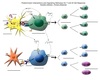
What are the 6 Different cell interactions that stimulate adaptive immunity
- 3 primary responses
- Parasitic infections
- Intravesicular/Extracellular Pathogens
- Cytoplasmic Pathogens
- 3 secondary responses
- Parasitic infections
- Intravesicular/Extracellular Pathogens
- Cytoplasmic Pathogens
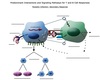
Lecture objective
- Label this diagram for a primary response to a parasitic pathogen (you may need to do this own your own)
- The blank images are on blackboard
The diagram and labels should include cell surface molecules, important binding between cells, signals to nucleus, cytokines involved, and response/outcome for a parasitic infection, intravesicular or extracellular bacterial infection, and viral infection.
- Dendritic cell:
- Binds parasite PAMPS via its TLR and becomes activated
- Phagocytoses the parasite
- Processes the antigen via exogenous pathway and presents parasite peptide on MHCII
- Secretes IL4 and IL1
- Puts costimulatory molecules on its surface
- TH0 (naïve T cell):
- Binds parasite peptide and MHC II via its TCR and CD4 molecule which results in CD3 signaling to the nucleus
- Binds IL4 via its IL4 receptor and that sends a signal to nucleus
- Binds co-stimulatory molecules on dendritic cell which results in another signal to nucleus
- Differentiates in response to the signals
- Upregulates IL2 receptors on its membrane and secretes IL2
- Undergoes mitosis/clonal expansion; this continues for 10 days to two weeks
- Becomes differentiated effector and memory TH2 cells
IL2 acts in an autocrine fashion and binds to IL2 receptors (CD25). IL2 stimulates mitosis of lymphocytes
- Meanwhile…..
- Follicular dendritic cell and B cell interaction
- Follicular dendritic cell binds C3b that has opsonized the parasite or parasite parts
- BCR (IgM with transmembrane portion) recognizes an epitope on the pathogen
- Three signals to B cell nucleus result in clonal expansion:
- BCR binding to antigen
- IL1 from the dendritic cell binds to the B cell’s IL1 receptor
- IL2 receptors are upregulated and IL2 from the TH cell binds to the B cell’s IL2 receptor (IL2 receptor= IL2R = CD25)
- B cell clone expands - some memory cells and some plasma cells secreting IgM result

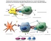
Lecture objective
- Label this diagram for a primary response to a bacterial pathogen (you may need to do this own your own)
- The blank images are on blackboard
The diagram and labels should include cell surface molecules, important binding between cells, signals to nucleus, cytokines involved, and response/outcome for a parasitic infection, intravesicular or extracellular bacterial infection, and viral infection

- Main difference from the parasitic response and this one is IL12 is being secreted by the dendritic cell instead of IL4
- Dendritic cell:
- Binds bacterial PAMPs via TLRs and becomes activated
- Phagocytoses pathogen
- Processes the bacterial pathogen via exogenous pathway and presents antigen on MHCII
- Secretes predominately IL12, instead of IL4 which was secreted in response to the parasitic pathogen, and it also secretes IL1
- Puts costimulatory molecules on its surface
- TH0:
- Binds bacterial peptide and MHCII via its TCR and CD4 molecule which results in CD3 signaling to nucleus
- Binds IL12 via IL12 receptor and sends signal to nucleus
- Binds costimulatory molecules on dendritic cells which results in another signal to the nucleus
- Differentiates in response to the signals:
- Upregulates IL2 receptors on membrane and secretes IL2 (IL2 receptor= IL2R = CD25)
- Undergoes mitosis/clonal expansion
- Becomes differentiated effector and memory TH1 cells

Lecture objective
- Label this diagram for a primary response to a cytoplasmic pathogen (you may need to do this own your own)
- The blank images are on blackboard
The diagram and labels should include cell surface molecules, important binding between cells, signals to nucleus, cytokines involved, and response/outcome for a parasitic infection, intravesicular or extracellular bacterial infection, and viral infection

Virus infects a dendritic cell and /or enters via endocytosis or phagocytosis
- Dendritic cell:
- Is infected by the virus and cytosolic viral proteins are processed via the endogenous pathway and presented on MHC I or takes up the virus by endocytosis/phagocytosis and processes the virus via the exogenous pathway and presents viral peptides on MHC II and some virus escapes to the cytoplasm from the endocytic vesicle. The viral proteins in the cytosol are processed via the endogenous pathway and presented on MHC I. The dendritic cell presents viral peptides on both MHCI and MHC II, this is called cross presentation.
- Secretes IL12 and IL1
- Puts costimulatory molecules on its surface
- NaiveTC cell:
- Binds the viral peptide and MHCI via its TCR and CD8 molecules and this causes CD3 to signal the nucleus
- Binds IL1 from dendritic cell via its IL1R and this signals the nucleus
- Binds costimulatory molecules on the dendritic cell which also signals the nucleus
- Differentiates in response to the signals:
- Upregulates IL2R and binds IL2 from TH cell
- Undergoes mitosis/clonal expansion
- Becomes differentiated effector and memory TC cell
- TH0 cell:
- Binds the viral peptide and MHC II via its TCR and CD4 molecules and this causes CD3 to send a signal to nucleus
- Binds IL12 from dendritic cell via its IL12R this sends asignal to nucleus
- Binds costimulatory molecules on dendritic cell which results in another signal to the nucleus
- Differentiates in response to the signals:
- Upregulates IL2 receptors and secretes IL2
- Binds IL2 via the IL2R and undergoes mitosis/clonal expansion
- Becomes differentiated effector and memory TH1cells

Lecture objective
- Label this diagram for a secondary response to a parasitic pathogen (you may need to do this own your own)
- The blank images are on blackboard
The diagram and labels should include cell surface molecules, important binding between cells, signals to nucleus, cytokines involved, and response/outcome for a parasitic infection, intravesicular or extracellular bacterial infection, and viral infection
Memory B cell can serve as an antigen presenting cell in the secondary response.
- Memory B cell:
- Binds parasitic epitope via its BCR this signals the nucleus
- Endocytosis the parasite and processes antigen through the exogenous pathway to present the parasitic peptide on MHC II
- Upregulates costimulatory molecules and cytokine receptors
- Binds cytokines from TH2 cell, e.g. IL4, and this signals the B cell nucleus
- B cell undergoes mitosis/clonal expansion
- Rearranges DNA and makes IgE as its BCR
- Some in the clone further differentiate into IgE secreting plasma cells
- The combinations and concentrations (milieu) of cytokines secreted by T helper cells determine which class of antibody the B cell will make.

Lecture objective
- Label this diagram for a secondary response to a bacterial pathogen (you may need to do this own your own)
- The blank images are on blackboard
The diagram and labels should include cell surface molecules, important binding between cells, signals to nucleus, cytokines involved, and response/outcome for a parasitic infection, intravesicular or extracellular bacterial infection, and viral infection
- Macrophage phagocytoses the pathogen and has difficulty killing it
- The antigen is processed through the exogenous pathway and presented on MHCII
- The macrophage also upregulates costimulatory molecules and cytokine receptors
- Memory TH1:
- Recognizes peptide and MHCII via the TCR and CD4 molecules and this causes the CD3 to signal the nucleus
- Binds costimulatory molecules which signal nucleus
- Secretes TH1cytokines in response to the signals from the macrophage
- IL3 and GM-CSF - stimulate bone marrow
- IL2, IFNγ, TNF
- TH1 cytokines activate macrophages to become M1 macrophages which are better able to kill bacteria. These cytokines can also enhance the function of other cells like neutrophils, NK cells, and CTLs
Meanwhile…..
- The antigen can be opsonized and caught by a follicular dendritic cell in the lymph node.
- Memory B cell:
- Binds the epitope on the bacteria being held by the follicular dendritic cell and this sends a signal to the nuclus:
- The B cell internalizes the antigen and process it through the exogenous pathway and presents antigen on MHCII
- Costimulatory and cytokine receptors are upregulated in the B cell
- Differentiates and undergoes mitosis in response to the TH1 cell cytokines
- B cell undergoes mitosis/clonal expansion
- Rearranges DNA and makes IgG as its BCR; these are IgG memory B cells
- Some in the clone further differentiate into IgG secreting plasma cells

Lecture objective
- Label this diagram for a secondary response to a bacterial pathogen (you may need to do this own your own)
- The blank images are on blackboard
The diagram and labels should include cell surface molecules, important binding between cells, signals to nucleus, cytokines involved, and response/outcome for a parasitic infection, intravesicular or extracellular bacterial infection, and viral infection

- Virus infects a tissue cell (example respiratory epithelium) and replicates in the cytoplasm. The viral peptides are processed through the endogenous pathway and the peptide is presented on MHCI.
- The infected cell upregulates costimulatory molecules
- The memory CTL:
- Binds to the viral peptide and MHCI with its TCR and CD8 molecule causing CD3 to signal the nucleus.
- Binds the costimulatory molecules and this sends a signal to the nucleus.
- Becomes activated and kills the infected cell by inducing apoptosis via its killing mechanisms (will be discussed in detail in a later lecture):
- Perforin/granzyme
- TNF/IFNγ
- Fas/FasL binding

