Lecture 2 - Lower Respiratory System Flashcards
What parts of the resp. system are in the Lower respiratory system?
- Trachea
- Bronchi (R & L)
- Bronchioles
- Alveolar units
What does the air-conducting tract in the LR system consist of?
- Trachea
- Bronchi
- Primary (main)
- Secondary (Lobar)
- Tertiary (segmental)
- Bronchioles
- Terminal bronchioles (No alveoli present)
What does the respiratory tract in the lower respiratory tract consist of?
- Respiratory bronchioles (has alveoli unlike terminal bronchioles)
- Alveolar units
Describe the trachea
- It’s structure
- The Cartilage arrangement
- What muscle lies posteriorly in between the C-shaped cartilage
- What kind of tissue lies between the adjacent cartilage bars
The lumen of the trachea is D shaped in transverse section
It’s made of 15-20 C-shaped cartilages which maintain the patency of the airway.
Posteriorly in the gaps between the C shaped cartilage lie bundles of smooth muscle called Trachealis muscle
Between the adjacent bars of cartialge the narrow gaps are filled by fibrous connective tissue called annular ligaments, which also contain numberous elastic fibres to give elasticity to the wall
Where is the thyroid gland located?
In front of the upper cartilage rings and at the sides of the trachea
Describe where the trachea extends from, and how it terminates
The trachea extends from C6 to T4 level inferiorly and is about 10cm in expiration (& postmortum), and on inspiration extends to T5-6 level inferiorly (with 15cm in length).
The trachea terminates by dividing (tracheal bifurcation) into left and right main bronchus at the level of the imaginary plane between the sternal angle & T4
The carina is a ridge of cartilage in the trachea that occurs in the lumen at the tracheal bifurcation.

Describe the bronchi divisions
- There’s 2 (main) bronchi (left and right) divide into 3 secondary (lobar) bronchi on right and 2 secondary (lobar) bronchi on the left (One for each lobe of the lung).
- The secondary (Lobar) bronchi divide into tertiary (Segmental) bronchi - one for each bronchopulmonary segment in the lobe.
- The tertiary (Segmental) bronchi divide to give progressively smaller and smaller tubes - the bronchioles
Which bronchi are intrapulmonar and extrapulmonar?
Right main bronchus, left main bronchus and right superior lobar bronchus are extrapulmonary; all other bronchi are intrapulmonary
Compare the extrapulmonary bronchi structure to that of the trachea
Extrapulmonary bronchi structure closely resember that of the trachea - and only differ from it by having a smaller diameter with cartilage rings still incomplete, posterior deficiency is still occupied by smooth muscle and still has D-shaped lumen.
Compare the structure of the intrapulmonary bronchi to the trachea
Intrapulmonary bronchi differ from the trachea alot more than extrapulmonary bronchi.
They are spherical in outine, and don’t show posterior flattening which is seen in trachea or C-shaped cartilage rings.
The cartilage is arragned into irregular plates and the smooth muslce fibres are arranged in spirals around the bronchus, together with elastic fibres.

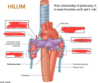
Label this

Label this my g

A conducting tube of less than 1mm diameter is regarded as a _______
Bronchiole
Describe the changes seen in cilia, goblet cells and submucosal glands when moving further down towards the bottom of the respiratory tree
Cilia extend further down the respiratory tree than do goblet cells and submucosal glands, thus preventing the respiratory tissue from becoming waterlogged or occluded by mucus.
In the smallest bronchioles where cilia are absent, macrophages take over the function of internal drainage
Describe the terminal bronchioles
They are narrowest part of the air-conducting system, which has cuboidal epithelium with no goblet cells, and has only patches of ciliated cells. It is surrounded by respiratory tissue.
Describe the respiratory bronchioles
Contains alveoli on their wall, so therefore they are not apart of the air-conducting system, but are the first component of the respiratory system (=gaseous exchange) where blood in capillaries is seperated from air with a very thin mass of material so that they can exchange CO2 & O2. This arrangement occurs in the respiratory bronchioles to alveoli.


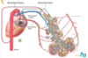
Describe how respiratory bronchioles eventually form the alveoli
Respiratory bronchioles branch into alveolar ducts.
(Alveolar ducts are thin walled tubes which connect the respiratory bronchioles to alveolar sacs)
The alveolar duct opens to an alveolar sac, each of which contains a collection of alveoli (small pouches made of flattened epithelial cells that allow gas exchange)
What is an acinus?
A pulmonary acinus comprises the distal unit that contains gas-exchange surfaces of the lung in the form of respiratory bronchioles, alveolar ducts and alveoli.
An acinus contains these three structures
What occurs at the air-blood barrier? (resp. membrane)
Gas exchange occurs when CO2 in blood (carried to the lungs in pulmonary arteries) diffuses across the blood capillary wall into the alveoli, and O2 diffuses from the air in the alveoli acorss the capillary wall into the pulmonary veins. The oxygenated blood returns via the pulmonary veins to the left atrium of the heart.
The layers of tissue that constitute the gas exchange is called the air-blood barrier (Respiratory membrane).

How do the lungs stay in their position in the thoracic cavity?
They lie free in the thoracic cavity except where they are attached by their roots to the heart and trachea.
Describe the shape of the lungs
They are conical in shape with apex, base and 3 surfaces
Apex = upper tapered part (lies in the plane of thoracic inlet)
Base = Concave, lower part (overlies the dome of the diaphragm)
Surfaces are costal, diaphragmatic, medial
What are the surfaces of the lungs?
- Costal - is convex and fits the thoracic wall formed by the sternum, ribs and costal cartilages
- Diaphragmatic - is concave and fits the domeof diaphragm
-
Medial - is divided into
- Mediastinal part which shows a concavity caused by the heart and pericardium (Cardiac impression)
- Vertebral part in contact with the sides of thoracic vertebrae
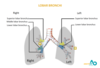
Label this