Lecture 4 - Lung histology Flashcards
(29 cards)
What does conduction portion of the respiratory system do?
- Delivers air to and from the respiratory portion
- Prevents food entering (epiglottis stops)
- Ensures air entering respiratory portion is
- Clean
- Warm
- Humid
In the conducting portion of epithelium, what basal cells are present?
Regenerative cells, and there are some endocrine cells
What type of epithelium is found on the conducting portion of epithelium?
Pseudostratified columnar epithelium
What kind of neurons are olfactory neurons?
They are bipolar neurons - they have an axon leaving at the base to combine with other axons to form the olfactory nerve - they are an interface between the external environment and the brain
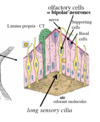
What kind of basal cells are found in the olfactory epitethelium?
There are globose, horizontal basal cells and mesenchymal stem cells - they are quite different due to the proteins found in their cells.
These are progenitor cells which can replace cells which may be damaged by inhaled substances.
We may be able to take this tissue out and grow it into other tissues.

What are the basal cells of the epithelium?
Globose basal cells or horizontal basal cells - these help regenerate the olfactory epithelium.
So when injury/neuronal cell death occurs -> these help regeneration. And then maintain the sensory system.
The GBC can either form sustaining cells and neurons - these mature in a period of 50 days.

Is the mucosa folded in the esophagus?
No
Trachea
What epithelium is found
What is in the mucosal tissue
What is found in submucosa? And what role does this have?
and what role does the cartilage in trachea have?
Epithelium: Pseudostratified, ciliated with mucous cells.
Mucosal tissue: Elastin +/- some smooth muscle cells. It’s flexible, resilient, moblie and elastic.
In the submucosa serous and mucous glands are found - they produce fluid and mucus to moisten airways and trap particulate matter.
The cartilage makes the trachea resilient, flexible and compressible - thereby keeping the airway patent.
What happens to the epithelium, cartilage and smooth muscle as you move down the bronchi?
The epithelium decreases in height, becomes columnar as bronchi become narrower.
The cartilage is in seperate blocks throughout bronchus wall.
The smooth muscle is arranged into a circular layer, and decreases in size as bronchi become narrower
What is the function of ciliated cells?
Movement of fluid towards pharynx
What is the main function of goblet cells?
Production and secretion of mucus - similar to sub-mucosal glands
What happens if mucous isn’t moved towards the pharynx by the mucociliary escalator?
Mucus can deposit in resp. bronchioles and can block the alveoli.
We need to maintain the balance between mucous cells and ciliated cells - in smoking you get more mucous cells, so you end up coughing it up to prevent blockage
What happens when we see changes in respiratory epithelium due to things such as: chronic coughing etc.
The chronic coughing causes forceful airflow in epithelium, this is sensed by epitehlium - they have a protective change.
The basal cells start producing alternative type of epithelial cell, where there is a change from columnar to squamous cells.
The squamous is effet against the sensed stress, but is less effective for moving mucous, since we need beating cilia.
This is what is seen in smoking.
Describe changes seen in epithelium when you move from trachea to alveolar sacs
The epithelium becomes flatter and flatter, and eventually loses it’s goblet cells. Once these are gone resident macrophages take over to clean debri.

What are bronchioles?
branching tubular structures that are less than 1mm in diameter, and they terminate in the alveolar ducts.
What kind of epithelium, protein secreting cells are found in the bronchioles?
And describe the smooth muscle, and is there cartilage in the bronchioles ?
Epithelium: Columnar and ciliated. Goblet cells are only located in larget bronchioles.
Clara cells present: They secrete lipoproteins (CC16) to prevent luminal adhesion - keeps bronchioles patent. In smaler bronchi you have clara cells instead of goblet cells.
Smooth muscle: It’s a distinct, thin layer
Cartilage is never located in bronchioles

Describe the terminal bronchioles, and what kidns of cells are found in the epithelium
They are the smallest conducting bronchioles, they have simple cuboidal epithelium.
In the epithelium of the terminal bronchioles there are no goblet cells, and clara cells are present - which secrete lipoprotein (CC16), which is a surface active agent. This prevents luminal surfaces adhering on bronchiole collapse (expiration).
What can be used as a marker of bronchoalveolar damage in serum?
CC16
What allows air to pass between alveolar sacs?
Every sac doesn’t need to be associated with a bronchiole - there are pores for air to move into other sacs so that there can be an equal pressure, and so that air can link up with other units if theres a blockage somewhere.
What are the types of cells found in alveoli?
- Type I pneumocytes - they are the main interface cells (cover 90-95% of lung surface)
- Type II pneumocytes - these produce surfactant to reduce surface tension (so that the lung doesn’t collapse on expiration)
- Macrophages (known as dust cells)
The pneumocytes are supported by connective tissue
The thin flattened type I pneumocytes allow for a narrow interface between air and blood.
What maintains the shape of alvolar sacs?
Connective tissue


What makes up the blood air barrier? (respiratory membrane)
- Endothelim (capillary wall)
- Shared basement membrane
- Epithelium (type I pneumocyte)
as O2 demand increases this barrier gets thinner
What do type II pneumocytes produce? (50-60% of cells)
And what is found in this?
Type II pneumocyte produce a surfactant layer, which decreases surface tension to allow us to expand the lungs.
- DPPC (phospholipid is found here)
- also has hydrophobic+hydrophilic proteins


