Lecture 5 Lab Flashcards
(45 cards)
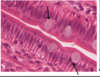
What is depicted by the areas highlighted below

Microvilli – brush border
Terminal web – only seen with EM. The web is a mat of filaments found below the apical surface of the cell. The microfilaments of the microvilli are anchored into the terminal web.

What is depicted by the boxed portion of the image

brush or striated border.
What is depicted by the boxed portion of the image

brush or striated border.
What is depicted in the image

Stereocilia
›What is depicted in the image

Stereocilia
Stereocilia
are actually long, branching microvilli. With LM, stereocilia look like wispy, spiky projections off the apical surface of the cells. Similar to microvilli , they contain a core of actin microfilaments. They are seen in certain parts of the male reproductive system.
What is depicted in the images

Cilia
C – cilium
Mv – microvillus
BB – basal body
What is this a cross section of and what is indicated by the arrows

What structures are depicted by the arrows


Basal infoldings
are found on the basal surface of a cell, adjacent to the underlying connective tissue. The folds are projections of the plasmalemma that invaginate into the cell cytoplasm, increasing surface area. Mitochondria are frequently found in the cytoplasm between adjacent folds. Basal infoldings are easiest to see at EM, although they are visible with LM as faint striations on the basal surface of the cell that run perpendicular to the base of the cell. In salivary glands, they are present in the striated ducts.
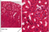
what is the pic depicting and what is shown by the arrows
Depicts: Basal infoldings
Red: Mitochondria
Blue: Plasmalemma
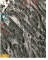
What is depicted in the image as well as what are the arrows pointing to

Basal infoldings
are visible with LM as faint striations on the basal surface of the cell that run perpendicular to the base of the cell. In salivary glands, they are present in the striated ducts.
Arrows= Serous acini
What is depicted by the image


What is shown in the image

Hemidesmosome
Half of a desmosome, found on the basal surface of the cell
what type of epithelium is depicted

Simple squamous epithelium
consists of a single layer of very flat cells. The cells are so flat that their nucleus may bulge above the level of their cytoplasm. The cytoplasm is extremely thin and attenuated.
what type of epithelium is depicted

Simple cuboidal epithelium
single layer of cube-shaped cells. Cells are as wide as they are tall. The nucleus is usually round and centrally located. Secretory units will also be formed by simple cuboidal epithelium. In the secretory units, however, the cells are not actually cubeshaped. They are usually shaped like a pyramid, with the base of the cell resting on the basement membrane at the periphery of the secretory unit and the apex of the cell abutting the lumen.
What type of epithelium is depicted

Simple columnar epithelium
single layer of columnar cells. The cells are taller than they are wide. The large nuclei are usually oval in shape and located near the base of the cell. A negative Golgi is frequently seen in the cytoplasm between the nucleus and the cell apex. A simple columnar epithelium may have a brush or striated border (microvillus border). Goblet cells are also sometimes present.
What type of epithelium is depicted

Pseudostratified columnar epithelium
single layer of cells of differing shapes. Their nuclei appear at different levels, so the epithelium appears layered when it is not (hence the term ‘pseudostratified’). The epithelium usually can be distinguished by the fact that the nuclei usually do not line up in distinct rows, as they do in other simple and stratified epithelia, so that the pseudostratified epithelium has a random or disorganized appearance. This type of epithelium is frequently associated with a ciliated border, and may have goblet cells.
What type of epithelium is depicted

Transitional epithelium (uroepithelium)
Transitional epithelium (uroepithelium)
The distinguishing characteristic of this epithelium is the presence of the layer of domeshaped large umbrella cells on the luminal surface. The surface cells have a rounded apical surface. They often look as if they are bulging above the rest of the epithelium. The cells are also frequently binucleate. Basal cells are small and form a single layer that is in contact with underlying connective tissue. It contains stem cells. Several layers of intermediate cells are located between the basal and umbrella cells.
What type of epithelium is depicted

Stratified squamous epithelium
This type of epithelium may be nonkeratinized (nuclei are found in all cell layers) or keratinized, which means it has a layer of dead cells on its luminal surface.
What type of epithelium is depicted

Stratified squamous keratinized epithelium
What type of epithelium is depicted
What type of epithelium is depicted

Stratified cuboidal
epithelium is usually made up of only two layers of cells. Cells in both layers are cuboidal in shape.