Lines Flashcards
(43 cards)
Sella Turcica Size normal values

-AP diameter: 5-16mm average of 11mm -Vertical Diameter: 4-12 mm average of 8mm - enlargement = neoplasm, empty sella syndrome, pituitary adenoma or aneurysm
when taking a lateral xray of the cervical spine, an enlarged sella turcica is seen, what is the next best step?
a) CT scan
b) MRI
c) xray of lateral skull
d) repeat lateral cervical spine xray
c) xray lateral skull
central beam aimed at EAM, we do this because beam can get distorted in the lateral cervical view causing the illusion of an enarged sella turcica
Acromegaly is seen with?

an enlarged sella turcica
an enlarged frontal sinus
(Martin’s)Basilar angle
aka Welcker’s

-Projection: Lateral skull
- Landmarks and methods: Draw a line from the
nasion (frontal-nasal junction) to the center of
the sella turcica and one from the center of the
sella turcica to the anterior margin of the
foramen magnum and measure the angle
between them.
-Normal values: Should vary anywhere from 123 ° to 152° with an average of 137°
-Significance: An increased angle is indicative of platybasia, which may or may not be associated with basilar impression.
platybasia
skull is long and shallow
McGregor’s line (AKA Basal line)
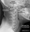
- Most accurate and reproducible method for basilar impression
- Normal values: The odontoid should not lie above this line more than 8mm in males, and 10mm in females. In children younger than 18 years, these values diminish with decreasing age.
- Significance: If odontoid protrudes further than normal, there is suspicion of basilar impression that can be caused by Paget’s, osteomalacia,or fibrous dysplasia.
Chamberlain’s line (aka palato-occipital line)

- Normal values: The tip of the odontoid should not project above the line, however there might be a normal variation of 3mm. A projection of 7mm or more is definitely abnormal
- Significance: If odontoid protrudes further than normal, there is suspicion of basilar impression that can be caused by Paget’s, osteomalacia,or fibrous dysplasia.
-Landmarks and methods:Draw a line from the
posterior margin of the hard palate to the
posterior margin of the foramen magnum.
Assess the relationship to the odontoid.
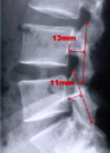
Digastric Line (AKA Biventer Line)

- Normal values: The distance from the line to the dens value varies from 1 to 21mm with an average of 11mm. The distance from the line to the Co-C1 joint varies from 4 to 20mm with an average of 4mm.
- Significance: Both measurements decrease with platybasia caused by any of the bone softening disease listed previously.
-Landmarks and methods: Locate the digastric
grooves medial to the base of the mastoid
processes and draw a line between them.
Then measure the vertical distance to the
dens, as well as to the Co-C1 joint.
-dens shouldnt cross the line
Atlantodental interspace (ADI) (AKA Atlas- odontoid space, predental space, atlas-dens interval)
- Normal values: The space should be a minimum of 1mm in both children and adults and a maximum of 5 in children and 3 in adults.
- Significance: A decrease in the space is often due to degenerative changes. An increased space can be caused by trauma, occipitalization, Down’s syndrome, pharyngeal infections, inflammatory arthropathies.
-Landmarks and methods: Measure the
distance between the posterior margin of
the anterior tubercle and the anterior
surface of the odontoid.
Sagital Dimension of the Cervical Spinal Canal
- Landmarks and methods: Measure the sagital
diameter from the posterior surface of the
midvertebral body to the nearest surface of
the same segmental spinolaminar junction
line.
-Normal measurements: Average
C1-22mm
C2-20mm
C3-18mm
C4-7-17mm
Torg ratio
-Landmarks and methods: Measure the sagital
diameter from the most posterior surface of
endplate to the nearest surface of the same
segmental spinolaminar junction line.
Compare that to the AP dimension of the
midbody.
-Normal measurements: Canal to body ratio
should not be less than 0.80
Cervical Gravity Line
-Landmarks and methods: Draw a vertical
line through the apex of the odontoid
process:
-Normal measurements: The line should pass through the body of C7.
-Significance: The line allows the assessment of the location of gravitational stresses at the cervicothoracic junction.
Stress line of the C spine (AKA Ruth Jackson’s lines)

-Projection: C spine, neutral, flexion and
extension.
- Landmarks and methods: Draw a line
along posterior body of C2and one along
posterior body of C7. Measure the angles
in both flexion and extension - Normal values: In flexion the lines should intersect at the level of C5-6 disc or facets. In extension they should intersect at the level of the C4-5 disc or facets.
- Significance: Muscle spasm, joint fixation and DDD may alter the stress point.
Prevertebral soft tissues
- Projection: Lateral C spine
- Landmarks and methods, and normal values:
Bony landmarks are the anterior arch of C1,
inferior corners of C2 and 3, superior corner of
C4 and inferior corners of C5-C7. - At C2-3 we have the retropharyngeal space,
which shouldn’t exceed 7mm. - At C4-5 we have the retrolaryngeal space, which
shouldn’t exceed 20mm. - At C5-7 we have the retrotracheal space that
shouldn’t exceed 22 mm.
-Significance: soft tissue masses may increase the measurements. Examples are posttraumatic hematomas, abscesses, neoplasms.
Cobb method for scoliosis (AKA Cobb-Lippman)

-Projection: AP T spine
-Landmarks and methods: Draw lines along the superior and inferior endplates of the two vertebraes that are at the extremes of the scoliosis. Then draw two perpendiculars and measure the angle in betweeen.
YR
-Significance: Curvatures less than 20 ° require no bracing or surgical intervention, however if they are present in a patient between the age of 10 and 15 they should be monitered for a progression of more than 5° in a 3 month period. Curves between 20° and 40° should be braced. Curvatures that excess 40° may require surgical intervention.
Risser-Fergusson method
- Projection:AP T spine
- Landmarks and methods: Choose the two end vertebrae like in Cobb’s method and then choose an apical vertebrae. For each of the vertebraes, draw two diagonals that cross each other at the center of the vertebrae. Finally connect the lines going through the centers of the vertebraes and measure the resulting angle at the apex.
- Significance: This method gives values about 25% less than those of Cobb, and its use is discouraged.
Thoracic Cage Dimension (AKA Straight Back Syndrome evaluation)
Projection:Lateral chest
- Landmarks and methods: Measure the distance between the posterior sternum and the anterior surface of the 8 thoracic vertebral bodies.
- Normal values: in males the distance should vary between 11 and 18 cm with an average of 14cm. In females it should be between 9 and 15cm with an average of 12.
- Significance: A distance below 13cm in males and below 11 in females may indicate the presence of straight back syndrome. If abnormality is detected, check the heart for murmurs.
Lumbar Lordosis
Projection: lateral L spine
- Landmarks and methods: Draw a line along the superior endplate of L1 and one along the superior endplate of S1. Then draw two perpendiculars and measure the angle between them.
- Normal values: There is a wide variation, however an average is between 50 ° and 60°.
- Significance: There is a wide spectrum of opinions. An increase in the angle seems to move the nucleus pulposis anterior, however that is of unclear significance.
Lumbosacral angle (AKA Sacral base angle, Ferguson’s angle)

Projection: lateral L or S spine
- Landmarks and methods: Draw a line along the superior endplate of S1 and intersect it with a true horizontal. Measure the angle between them.
- Normal values: there is a wide variation. The minimum angle is 26 °. The maximum 57°. The average is 41°. From recumbent to upright position there is a variation from 8 to 12°. One standard deviation is +/- 7°.
- Significance: There is no consensus on the significance of an decreased or increased angle. An increased angle my put more stress on the lumbosacral posterior joints.
Lumbosacral Disc angle
Projection: lateral L or L/S spine
- Landmarks and methods: Draw a line along the inferior endplate of L5 and one along the superior endplate of S1. Measure the angle between them.
- Normal values: the angle should be between 10 and 15°.
- Significance: There is a correlation between an increased angle and LBPn due to facet impaction.There may be a decrease in the value in the presence of acute disc herniation of the 5th lumbar disc.
Lumbar gravity line (AKA Ferguson’s weight bearing line, Ferguson’s gravity line)

Projection: Lateral lumbar.
- Landmarks and methods: Find the center of the body of L3 by drawing two diagonals. Then draw a true vertical from this center.
- Normal measurements: The vertical line should intersect S1.
- Significance: If the line is more than 10mm anterior to S1 there may be an increase in shearing stresses in an anterior direction between the lumbosacral apophyseal joints. A posterior shift may increase the stress on the same joints and produce LBPn.
Macnab’s Line

Projection: Lateral lumbar
- Landmarks and methods: Draw a line through and parallel to the inferior endplate of the lumbar vertebraes. Look at the relationship with the adjacent tip of the superior articular process (SAP) of the vertebrae above.
- Normal measurements: The line shouldn’t intersect the (SAP).
- Significance: An intersection may be an indication of facet imbrication. However, the reliability of this line has not been documented. Originally it was applied on recumbent radiographs, therefore its validity on upright ones is doubtful.
Hadley’s “S” curve

Projection: AP and oblique L spine
- Landmarks and methods: Draw a curvilinear line along the inferior margin of the TP and down along the inferior articular process to the apophyseal joint space. Then go across the joint and along the superior articular process of the vertebrae below.
- Normal measurements: It should look like an S.
- Significance: An interruption indicates facet imbrication. A wide facet joint was linked to disc problems.
Van Akkerveeken’s measurement of lumbar instability

- Projection: lateral neutral, flexion, extension of the L spine
- Landmarks and methods: draw two lines along opposing segmental endplates until they intersect posteriorly. Measure the distance from the posterior body margin to the point of intersection.
- Normal values: there should be less than 1.5mm difference in measurements from one posterior body margin to another.
- Significance: If the difference is greater than 1.5mm then there might be disc, or posterior ligament damage. Valuable in flexion and extension.