Liver & Biliary Overview Flashcards

1
Q
A

B - non-alcoholic steatohepatitis

  • she has type II diabetes, is obese and consumes alcohol regularly
    • these are all risk factors
  • simvastatin can be hepatotoxic, but she has only been taking this for 2 years so it is unlikely
2
Q
A

A - full recovery

  • HBsAg shows that they have a current infection
    • this could be acute or chronic
  • Anti-HBcAg IgM (IgM core antibody) shows that they must currently have an acute infection
  • someone with an acute hep b infection is most likely to make a full recovery
3
Q
A

D - IV cefotaxmine and oral lactulose

  • the patient has encephalopathy
  • they also have spontaneous bacterial peritonitis
    • fever
    • worsening abdominal tenderness / distenstion
    • high neutrophil count
  • IV cefotaxime is an antibiotic that will treat the SBP
  • lactulose reduces ammonia production in the gut to prevent encephalopathy from getting worse
4
Q

What is the underlying cause of jaundice?

A

it is caused by an increased concentration of bilirubin in the blood

(hyperbilirubinaemia)

5
Q

What are the steps involved in the bilirubin metabolism pathway?

A
  • old RBCs are broken down in the spleen
  • the haemoglobin from RBCs produces iron** and **unconjugated bilirubin
  • the unconjugated bilirubin travels in the blood, bound to albumin, to the liver
  • in the liver it is conjugated by UDPGT enzyme
  • conjugated bilirubin then enters the biliary system and forms part of the bile
  • conjugated bilirubin enters the duodenum in the bile via the common bile duct
  • here it is converted to urobilinogen and stercobilinogen
    • urobilinogen is excreted in the urine
    • stercobilinogen is excreted in the faeces and gives them a dark colour
6
Q

What causes pre-hepatic jaundice?

What type of hyperbilirubinaemia is present?

A
  • caused by excessive RBC breakdown
    • or impaired uptake of RBCs by the liver
  • this overwhelms the liver’s ability to conjugate bilirubin
  • there is an unconjugated hyperbilirubinaemia
  • any bilirubin that is conjugated will be excreted normally, but the excess unconjugated bilirubin will remain in the bloodstream to cause jaundice
7
Q

What are the 2 major causes of pre-hepatic jaundice?

Why do these lead to jaundice?

A

Haemolysis:

  • increased RBC breakdown leads to an increase in unconjugated bilirubin concentration
  • the liver cannot conjugate the bilirubin fast enough, leading to an increase in unconjugated bilirubin in the blood
    • there is nothing wrong with the liver, there is just a massive excess of bilirubin

Gilbert’s Syndrome:

  • this is a deficiency of the UDPGT enzyme
  • UDPGT enzyme is not working as well as it would in a healthy person, so when this individual becomes stressed / gets an infection they can appear jaundiced
8
Q

What causes hepatocellular (intrahepatic) jaundice?

What type of hyperbilirubinaemia is produced here?

A
  • caused by dysfunction of the hepatic cells
  • the liver loses some of its ability to conjugate bilirubin, however this is not the main problem
  • if the liver becomes cirrhotic, it compresses the intra-hepatic portions of the biliary tree to cause a degree of obstruction
    • the conjugated bilirubin cannot get into the biliary system
  • this produces a mixed conjugated and unconjugated hyperbilirubinaemia
    • ​it is mainly conjugated hyperbilirubinaemia
9
Q

What are the main causes of hepatocellular jaundice?

A

Caused by anything that damages the hepatocytes:

  • alcoholic liver disease / cirrhosis
  • hepatitis
    • viral, autoimmune
  • hepatocellular carcinoma / liver mass
  • haemochromatosis
  • iatrogenic e.g. medication
10
Q

What causes post-hepatic jaundice?

What type of hyperbilirubinaemia is produced?

A
  • this is jaundice caused by obstruction of biliary drainage
  • the liver is still functioning and conjugating bilirubin as normal
  • the conjugated bilirubin cannot get into the duodenum, so it enters the bloodstream instead
  • this produces a conjugated hyperbilirubinaemia
11
Q

How can the causes of post-hepatic jaundice be divided into 3 categories?

A

Intra-luminal causes:

  • gallstones

Mural causes:

  • strictures
  • cholangiocarcinoma
  • drug-induced cholestasis
  • PSC / PBS

Extra-mural causes:

  • pancreatic cancer
  • abdominal masses (e.g. lymphomas)
12
Q

How can looking at the urine determine what kind of hyperbilirubinaemia might be present?

A
  • conjugated bilirubin is water soluble and so can be excreted in the urine
  • unconjugated bilirubin cannot be excreted in the urine
  • dark (“coca-cola”) urine occurs in conjugated or mixed hyperbilirubinaemia
    • hepatocellular or post-hepatic jaundice
  • normal urine is seen in unconjugated hyperbilirubinaemia
    • ​pre-hepatic jaundice
13
Q

In what type of jaundice do the stools appear different?

Why does this occur?

A
  • in post-hepatic jaundice the stools will appear paler
  • this occurs when there is an obstructive picture as there are reduced levels of stercobilin entering the GI tract
    • this normally colours the stool
  • there will also be dark urine and pruritis
    • ​itching is caused by bile salts, as the blockage affects the drainage of bile salts into the duodenum
14
Q

What blood tests should anyone presenting with jaundice have?

A
  • liver function tests
  • coagulation studies
    • prothrombin time can be used as a marker of liver synthetic function
  • FBC
    • ​anaemia, raised MCV and thrombocytopenia can all be seen in liver disease
  • U&Es
15
Q

What tests are included in a liver screen?

What does each of these things measure?

A

Bilirubin:

  • quantifies the degree of suspected jaundice

Albumin:

  • marker of liver synthetic function

Transaminases - ALT & AST:

  • markers of hepatocellular injury
    • ​AST : ALT ratio > 2 means likely alcoholic liver disease
    • AST : ALT ratio = 1 means viral hepatitis more likely

Alkaline phosphatase (ALP):

  • raised in biliary obstruction
    • ​as well as during pregnancy, bone disease and certain malignancies

Gamma-GT:

  • more specific for biliary obstruction than ALP
16
Q

What results would you expect to see for each type of jaundice on LFTs?

A

Pre-hepatic:

  • raised bilirubin only

Hepatocellular:

  • there is damage to hepatocytes so you would expect to see raised AST & ALT

Post-hepatic:

  • there is an obstruction / bile duct damage so you would expect to see raised ALP / GGT
17
Q

What is meant by hepatitis?

What are the possible causes of this?

A
  • hepatitis is inflammation of the liver
  • it presents with raised AST and ALT
  • causes can be acute or chronic and include:
    • alcoholic hepatitis
    • non-alcoholic steatohepatitis (NASH)
    • viruses
    • drugs
    • autoimmune
18
Q

How does hepatitis tend to present?

A
  • all types of hepatitis tend to present with similar symptoms
  • RUQ pain
  • jaundice (hepatocellular)
  • hepatomegaly
  • joint pain
  • nausea
  • fatigue
  • dark urine
19
Q

How long does hepatitis have to persist for to become chronic?

What are the possible outcomes of acute and chronic hepatitis?

A
  • acute hepatitis resolves within 6 months
    • it becomes chronic if it lasts for longer than 6 months
  • acute hepatitis can resolve on its own, progress to chronic hepatitis or (rarely) result in acute liver failure
  • chronic hepatitis may progress to cirrhosis, liver failure and hepatocellular carcinoma
20
Q

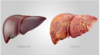
What are the 3 conditions that make up the spectrum of alcoholic liver disease?

A
  • steatosis occurs after a few days of heavy drinking
    • this is completely reversible
  • alcoholic hepatitis (inflammation) occurs after long term alcohol use (not a binge)
    • ​this is reversible, especially if mild
  • if alcohol consumption is continued this can cause cirrhosis
    • ​this is IRREVERSIBLE as it involves scarring of the liver
21
Q

What are the symptoms of mild and severe alcoholic hepatitis?

A

Mild:

  • nausea
  • anorexia
  • weight loss
  • hepatomegaly

Severe:

  • fever
  • jaundice
  • tachycardia
  • tender hepatomegaly
  • bruising
  • encephalopathy
  • ascites
22
Q

What causes the inflammation associated with alcoholic hepatitis?

A
  • alcohol metabolism requires NAD+
  • when there is excessive alcohol consumption, there is not enough NAD+ available for glycolysis
  • this promotes fatty infiltration into the liver, leading to inflammation
23
Q

What would a full blood count show in someone with chronic high alcohol intake?

A

macrocytic anaemia

  • this presents as low haemoglobin and high mean cell volume (MCV)
24
Q

What would liver function tests show in someone with chronic high alcohol consumption?

A
  • AST : ALT ratio > 2
    • remember “alcohol then toAST”
  • increased bilirubin
  • decreased albumin
  • ALP may be normal or raised
  • GGT is raised in someone who drinks a lot of alcohol over long periods of time
    • raised GGT indicates biliary damage, but also chronic alcohol consumption
25
What would a clotting screen show in someone with chronic alcohol consumption?
* clotting screen is a functional assessment that tells you how well the liver is working * the liver makes **clotting factors**, so ***clotting time increases*** when the ***liver is damaged*** * **_increased prothrombin time_** is a sensitive marker of **significant liver damage**
26
What imaging is performed in alcoholic liver disease? What would a liver biopsy show?
* **_hepatic ultrasound scan_** is performed * liver biopsy is diagnostic, but rarely needed the presence of **_Mallory bodies_** indicates **hepatitis** * these are damaged intermediate filaments within the hepatocytes there may also be **_ballooning degeneration_** of hepatocytes * this is a form of liver parenchymal cell death
27
What is involved in the management of alcoholic liver disease?
* **alcohol abstinence** * **_diazepam_** may be given in hospital to manage alcohol withdrawal * this prevents **withdrawal symptoms** and **seizures** * **nutrition** * ​enteral preferred - calories and vitamins - alcoholics are often malnourished * weight loss / smoking cessation * **_steroids_** are given in **_severe alcoholic hepatitis_**
28
What is meant by non-alcoholic fatty liver disease (NAFLD) and how can this progress?
* the presence of a **fatty liver** in those who **_do not consume alcohol_** in amounts generally considered harmful to the liver * **steatosis** is completely reversible * this progresses to **steatohepatitis (NASH)**, which involves **inflammation** * this is reversible, especially if it is mild * this progresses to **cirrhosis**, which is irreversible
29
What are the risk factors associated with NAFLD?
* **obesity (truncal)** * **insulin resistance / type II diabetes** * hyperlipidaemia * hypertension * metabolic syndrome * short bowel syndrome * total parenteral nutrition (TPN)
30
What additional symptoms may someone with NAFLD present with?
* they may have signs of **insulin resistance** * polyuria * polydipsia * acanthosis nigricans
31
What are the investigations involved in NAFLD?
* LFTs * **AST : ALT ratio \< 1** * GGT / ALP may also be raised * **blood glucose** to check for diabetes / how well controlled it is
32
33
What is involved in the management of NAFLD?
* diet and exercise * controlling risk factors * statins for hypercholesterolaemia * good blood sugar control in diabetes * metformin for diabetics
34
What is the difference between steatosis and non-alcoholic steatohepatitis (NASH)?
* **steatosis** involves **_fatty accumulation_** in the liver * a liver can remain fatty without disturbing liver function​ * by various mechanisms and insults to the liver, steatosis can progress to **non-alcoholic steatohepatitis (NASH)** * NASH involves steatosis combined with **_inflammation_** and sometimes **_fibrosis_**
35
How does someone with NAFLD usually present?
* usually **no noticeable symptoms** and is only detected during routine blood tests or unrelated abdominal imaging * may have some symptoms related to liver dysfunction * fatigue * malaise * dull RUQ abdominal discomfort
36
What type of infection is usually caused by viral hepatitis A & E? What are the exceptions to this?
* hep A & E usually cause **_acute hepatitis_**, which very rarely progresses to chronic hepatitis * *when you think of A&E - think of ACUTE problems* * hepatitis A&E can become **chronic** in **_immunosuppressed individuals_** * **hepatitis E** can cause **_severe liver failure_** in **_pregnant women_**
37
How are hepatitis A & E spread? What is the management for this infection?
* they are spread via the **_fAEco-oral route_** * this is via contaminated water or sex * management is **supportive** as this infection is acute and will regress on its own * **_​paracetamol_** and **_alcohol_** should be avoided
38
What does the presence of IgM and IgG in Hep A / E serology tell you?
* **IgM** tells you that the **_infection is acute_** * they currently have the infection * **IgG** tells you that they **have had the infection**, but have **_now cleared it_** * IgM = now* * IgG = gone*
39
Do hepatitis B and C tend to be acute or chronic? What is the risk associated if an infection can become chronic?
* Hep B & C can be **acute or chronic** * **Hep _C**_ most commonly becomes _****C**_hronic** * **Hep B** usually stays **acute** (80-90% cases) * as they can become chronic, there is a risk of **cirrhosis / hepatocellular carcinoma** * this risk is greater with Hep C infection as it is more likely to become chronic
40
Under what circumstances can Hep D infection occur? What is the associated risk?
* Hep D is a **_coinfection / superinfection of Hep B_** * it can only occur if someone is already infected with Hep B * Hep D infection **increases the risk of _liver failure_** in someone with Hep B infection
41
What is the treatment for acute Hep B infection?
* it **_does not require treatment_** as the infection is cleared naturally in 90% cases * if Hep B persists for **\> 6 months** then **antiviral treatment** is required
42
What is the treatment for chronic Hep B infection? What is the problem with this treatment?
* antiviral treatment ***suppresses HBV DNA replication*** * it is **_NOT curative_**, so drug withdrawal leads to **relapse** * if the liver is working well (i.e. no decompensated cirrhosis) then **_peginterferon-a-2a_** is given * if the liver is functioining poorly (i.e. decompensated cirrhosis), then give **_tenofovir_** or **_entecavir_**
43
What is the treatment for hep B when there is a co-existing hep D infection?
peginterferon-a-2a **_AND_** tenofovir/entecavir
44
What is the treatment for hepatitis C infection?
* this needs to be treated with antivirals as infection is likely to become chronic * treat with **_sofosbuvir_** or **_ledipasvir_**
45
What are the 2 antigens associated with the hepatitis B virus? Which one can be useful in detecting Hep B infection?
***_Surface antigen:_*** * HBsAg is on the **outside** of the molecule * if HBsAg is present, there is a **_current active Hep B infection_** ***_Core antigen:_*** * HBcAg is in the **middle** of the virus and is **not detectable in the blood** * this is not useful in telling whether there is a current active infection
46
What are the 2 antibodies that are associated with Hep B infection? What does their presence tell you about current infection?
***_Surface antibody:_*** * HBsAb tells you that the person has **_cleared_ a Hep B infection** * they had the infection in the past, but do not have it currently * OR they have been **_vaccinated_** against Hep B ***_Core antibody:_*** * this is only derived from **natural infection** * if **_IgM_** is present, there is an **_acute infection_** * if **_IgG_** is present, this person may have a **_chronic infection_**
47
Complete the table
48
What does hepatitis E antigen represent?
HBeAg represents **degree of replication**, and thus **_infectivity_**
49
How is hepatitis B spread? What are children more at risk of if they get this infection?
* most adults clear this infection * 10% become carriers and 10% develop chronic infection * children are more likely to become **carriers / chronic infection** * this can be transmitted: * **​sexually** * **IVDU / blood products** * **vertical transmission**
50
How is hepatitis C spread? What is the major risk associated with this infection?
* **_blood product spread_** * IVDU / transfusion * 80% of adults develop chronic hepatitis, which increases risk of hepatocellular carcinoma * infection is usually asymptomatic
51
What are the key features to distinguish between the different hepatitis viruses in a history?
***_Hep A & E:_*** * travel to endemic area / faeco-oral (contaminated water) ***_Hep B:_*** * unprotected sex / MSM * vertical transmission ***_Hep C_******_:_*** * blood products - IVDU sharing needles * **_FEVER_** in all of these infections **_AST / ALT will be in 1000s_**
52
What is the main drug cause of hepatitis? What is investigation and management?
* **paracetamol overdose** leading to **_acute liver failure_** * AST / ALT will be in 1000s * need to measure **serum paracetamol concentration** * treatment is **N-acetyl-cysteine**
53
What are other common causes of drug induced hepatitis? By which mechanisms can drugs cause this?
* some drugs will affect the **hepatocytes** themselves * paracetamol * NSAIDs * rifampicin * erythromycin * some drugs **induce cholestasis** by paralysing the bile duct * ​oral contraceptive pill * co-amoxiclav * nitrofurantoin
54
55
What are key features to pick up in the history for autoimmune hepatitis? Why is it important to identify?
* prevalence is highest in **_young females_** and people with **_other autoimmune diseases_** * e.g. type 1 diabetes * hashimoto's thyroiditis * coeliac disease * ​it is a chronic disease that can lead to **cirrhosis** and **liver failure** if left untreated
56
What tests are used to detect autoimmune hepatitis?
autoimmune liver screen * this is an autoantibody test that identifies some important antibodies responsible for autoimmune liver disease * ANA (antinuclear antibody) * ASMA (anti-smooth muscle antibody) * AMA (antimitochondrial antibody)
57
What is the definition of haemochromatosis?
condition leading to **_abnormal iron deposition_** in certain organs * skin * **liver** * **pancreas** * heart * pituitary gland * joints * the levels of iron build-up in the organs over many years, leading to unpleasant symptoms
58
What is the difference between primary and secondary haemochromatosis?
***_Primary haemochromatosis:_*** * hereditary - it is autosomal recessive * there is an **_inability to stop iron being absorbed_** from the GI tract, leading to its accumulation in affected organs ***_Secondary haemochromatosis:_*** * this is due to **_iron overload_** * e.g. having multiple transfusions
59
What is the pathophysiology involved in why primary (hereditary) haemochromatosis occurs?
* a defect in the **_HFE gene_** means that **transferrin binds poorly to its receptor** * this causes the liver to **_reduce hepcidin production_** * hepcidin usually **inhibits ferroportin** (enterocyte iron transporter) * with reduced hepcidin production, **_ferroportin activity_** in the enterocytes in **_unregulated_** * **more iron is transported across the enterocytes**, leading to **_iron accumulation_** and damage to affected organs
60
What are the main symptoms associated with haemochromatosis? What can this progress to?
* 75% of cases are asymptomatic * hepatomegaly * **diabetes mellitus onset** * **bronze skin** * arthralgia * male impotence / testicular atrophy
61
Why do males experience an earlier onset of haemochromatosis than females?
* females have an additional method of **excreting iron** through **_menstruation_** * this is not possible in males, so **iron accumulates more quickly**
62
What are the investigations involved in haemochromatosis?
* first line investigation is **_transferrin saturation_** and **_serum ferritin_** * transferrin saturation will be raised * serum ferritin will be raised (this is non-specific as it is an acute phase protein) * **gene typing of HFE** * gold standard is **_liver biopsy_** to see iron deposits
63
What is Wilson's disease? Who is affected by this?
* **_impaired copper excretion_** leads to **copper accumulation** in the **_liver_** and **_brain_** * it is an autosomal recessive genetic disorder * age of onset is **5 - 35 years**
64
What are the clinical features of Wilson's disease?
* characterised by **hepatitis + dementia + parkinsonism** in a **young person** * hepatosplenomegaly * abdominal pain * jaundice * ascites * portal hypertension * **Kayser-Fleischer rings**
65
What are the investigations conducted to diagnose Wilson's disease?
* 1st line investigation is LFTs + bloods + 24-hour urinary copper * raised transaminases & bilirubin * **reduced serum ceruloplasmin (copper transport protein)** * **increased serum free copper** * **increased urinary copper excretion** * slit lamp examination * gold standard is genetic testing / liver biopsy and measurement of copper content
66
What is meant by cirrhosis? Does the liver still function if it is cirrhotic?
* normal liver is replaced by **_fibrosis_** and **_nodules_** of **regenerating hepatocytes** * it can be **stable** or **_decompensated_** (liver failure) * the liver still functions when it is cirrhotic, but cirrhosis can eventually lead to liver failure, which can be life-threatening * there is also increased risk of HCC
67
What are the most common causes of cirrhosis? What type of cirrhosis is seen in viral causes and alcoholic causes?
* alcohol misuse * viral hepatitis (B / C) * autoimmune hepatitis * haemochromatosis * NASH * chronic biliary disease * ***viral hepatitis*** leads to **_macronodular cirrhosis_** (nodules \> 3mm) * ***chronic alcohol abuse*** leads to a **_micronodular cirrhosis_** (nodules \< 3mm)
68
What are the most common stigmata of chronic liver disease?
* clubbing * spider naevi * Dupuytren's contracture * palmar erythema * gynaecomastia * increased bruising * a patient can be clincally well with hepatitis or cirrhosis, with signs of chronic stable liver disease
69
What is the normal portal circulation? What is the purpose of this?
* blood from the spleen (**_splenic vein_**) and bowels (**_inferior_** and **_superior mesenteric veins_**) travels through the liver via the **portal vein** * this blood is full of **ingested toxins / pathogens** * the blood passes through the liver to be **detoxified** and then becomes part of the **systemic circulation** via the **inferior vena cava**
70
What is meant by portal hypertension? Why does it occur?
* an **_increased pressure in the portal vein_** due to cirrhosis (of any cause) * it occurs due to a **blockage in the liver** (e.g. due to cirrhosis) leads to a **_backlog of blood_** into the portal system * this raises pressure within the portal vein
71
How does blood flow from the portal to systemic circulation in portal hypertension?
* blood in the portal vein **has not yet been processed** by the liver * it transfers into the systemic circulation at sites of **_porto-systemic anastomoses_** * lower oesophagus * anal canal * umbilicus * splenorenal * this **increases the pressure** in the **systemic circulation** at the porto-systemic anastomosis points
72
What are clinical signs of portal hypertension?
* distended veins (varices) - **_caput medusa_** * due to increased pressure as a result of porto-systemic anastomoses * **_ascites_** * blood is backing up into the veins and leaking out into the abdomen * **_splenomegaly_** * the spleen isn't being drained properly via the splenic vein, so it becomes engorged in blood * **_oesophageal varices_**
73
What is involved in the management of cirrhosis?
* treat cause + **_avoid hepatotoxic drugs_** * alcohol * NSAIDs * sedatives * opiates * monitor the **risk of complications** * ​MELD score * 6-monthly USS * endoscopy upon diagnosis and every 3 years following
74
Why does decompensated cirrhosis / liver failure lead to encephalopathy?
* the liver functions to **remove gut products** from the blood * these arrive via the **portal vein**, and are removed before the blood returns to systemic circulation * in decompensated cirrhosis, the **hepatocytes are damaged** and **_less functional_** and there is **scar tissue** in the liver * the liver is **less effective at removing gut products** from the blood, so they make their way into the **systemic circulation** (**_intrahepatic shunting_**) * some blood from the gut **bypasses the liver** due to raised portal pressure at the sites of **_porto-systemic anastomoses_** * gut products in systemic circulation then impact on **_neurotransmission in the brain_**, causing encephalopathy * ​in this case, it is caused by **ammonia**
75
What is the major risk factor for encephalopathy in the presence of cirrhosis? What are other risk factors?
***_GI bleeding:_*** * bleeding into the gut acts as a **"protein meal"** * blood is broken down into **ammonia**, which causes encephalopathy * the same effect would be seen in a **_high protein diet_** ***_Other causes:_*** * infection * constipation * zinc deficiency * benzodiazepines
76
What is the treatment for encephalopathy?
* treat precipitating event + **_short term protein restriction_** * managing GI bleed, infection, etc. * **_oral lactulose_**, phosphate enema * ​this reduces ammonia production by gut bacteria * **avoid sedatives**
77
What is the treatment for ascites as a complication of cirrhosis?
* need to avoid fluid stasis in the abdomen as it could become infected * **sodium restriction** * diuretics - **furosemide + spironolactone** * **large volume paracentesis** * this is different to "ascitic tap" which is diagnostic paracentesis * this involves a needle into the abdomen to remove fluid
78
What is spontaneous bacterial perionitis? How is it diagnosed?
* this is the development of a **bacterial infection** in the peritoneum * it occurs when the **_ascitic fluid_** (i.e. an increased volume of peritoneal fluid) **becomes infected** **_without an apparent source_** * it is diagnosed using **paracentesis** (needle aspiration of ascitic fluid) * SBP is diagnosed when **neutrophil count** of the aspirate is **_\> 250 cells per mm3_** in the absence of another reason for this
79
What is the treatment for spontaneous bacterial peritonitis?
* antibiotic treatment with **_cefurotaxime + metronidazole_**
80
What is the primary prophylaxis for varices? Why is it needed?
* varices are **_abnormally dilated vessels_** (usually veins) that are at **risk of rupture** * primary prophylaxis is in place to **_prevent rupture_** of varices * if ***small varices*** then give **_non-selective beta-blocker_** * if ***large varices*** then **_EVL_** (esophageal variceal ligation)
81
What is the management for ruptured varices when there is haematemesis?
* start with ABCDE approach then **_IV fluids and/or blood_** * need to replace blood when **_Hb \< 7 g/dL_** or patient could bleed to death * fluids will replace volume, but not clotting factors * then give **_terlipressin + antibiotics_** * ​terlipressin reduces blood flow to the gut, which reduces pressure on the portal system * **_EVL_** is performed after resuscitation when the patient is **haemodynamically stable** * ​this involves placing bands over the varices to stop bleeding
82
What is involved in secondary prophylaxis of oesophageal varices?
* give **_non-selective beta-blocker_** to replace terlipressin after 2-5 days * if EVL + beta-blocker fails to prevent rupture, then TIPS procedure is performed * this involves connecting the portal vein to the hepatic vein in the liver to reduce pressure in the portal system
83
What is the definition of liver failure? What is the difference between acute and subacute liver failure?
severe liver dysfunction leading to **_jaundice_**, **_encephalopathy_** and **_coagulopathy_** ***_Acute:_*** * encephalopathy occurs **1-4 weeks** after onset of jaundice * it is hyperacute if \< 7 days after onset of jaundice * seen in **_paracetamol overdose_** and **_viral hepatitis_** * associated with transaminitis (very high AST / ALT) ***_Subacute:_*** * encephalopathy develops **4-12 weeks** after onset of jaundice
84
What is meant by acute-on-chronic liver failure?
* this is **acute decompensation** in a patient who already has chronic liver disease
85
86
What is cholelithiasis? What is biliary colic and why does it occur?
* ***cholelithiasis*** refers to a **_gallstone in the gallbladder_** * ***biliary colic*** refers to presence of **_cholelithiasis + pain_** * the stone in the gallbladder moves into the **cystic duct** and **_obstructs the flow of bile_** moving into the common bile duct * the **gallbladder contracts vigorously against the stone**, which causes pain * this is worse after a **fatty meal** as this stimulates the gallbladder to contract (release of bile helps emulsification of fats)
87
What is meant by acute cholecystitis? What type of jaundice is present?
* acute cholecystitis refers to **_cholelithiasis + inflammation_ with or without infection** * there is **_no jaundice_** in acute cholecystis * the stone is within the gallbladder or cystic duct and is **not obstructing the flow of bile**
88
What investigation is performed to diagnose gallstones?
USS of the liver and biliary tree
89
What are the treatments for cholelithiasis, biliary colic and acute cholecystitis?
* no treatment for cholelithiasis as it is an incidental finding * biliary colic is treated with **analgesia + elective lap chole** * acute cholecystitis is initially treated: * clear fluids only (to prevent gallbaldder from contracting) * analgesics * fluid resuscitation * broad spectrum IV abx if infection then treated with **lap chole within 1 week** if uncomplicated * if fever and high WCC persists, they could have an abscess * perform delayed cholecystectomy after few weeks/months
90
What is meant by choledocholithiasis? What if there is also pain present?
* choledocholelithiasis refers to a **gallstone in the _common bile duct_** * if there is **choledocholelithiasis + pain** then this is **_biliary colic_** * this term means presence of pain due to gallstones
91
What happens if there is choledocholelithiasis + infection? Would the patient be jaundiced?
* an infection can develop **behind the blocked common bile duct** where there is **stagnant bile** * this is **_ascending cholangitis_** (infection of bile duct) * this is usually due to E. coli * the patient may be **_jaundiced_** with **_raised ALP / GGT_** as there is an obstruction to biliary flow * if the stone comes down the CBD to block the **pancreatic duct** then this can cause **_pancreatitis_**
92
What is involved in the investigations of choledochoelithiasis and ascending cholangitis?
* ultrasound of liver and biliary tree * ERCP if ascending cholangitis is suspected
93
What is the management for choledocholelithiasis and biliary colic?
* for both **_analgesia + ERCP + lap chole_** * in cholelithiasis, you do not remove the gallbladder if there is no pain * in choledocholelithiasis, remove the gallbladder even if there is no pain * the stone in the CBD will go on to cause complications, including pancreatitis and ascending cholangitis
94
What triad is associated with cholangitis?
**_Charcot's triad_** * pain * fever * jaundice
95
What is involved in the management of ascending cholangitis?
* initial treatment involves: * clear fluids only * analgesics * fluid resuscitation * broad IV antibiotics * ERCP to drain pus from common bile duct, then remove stones * delayed lap chole will be performed at a later date
96
97
What are the 5 Fs that describe the risk factors for gallstones?
* Fair * Fat * Female * Forty * Fertile (1 or more children)
98
What sign is often present in acute cholecystitis?
**Murphy's sign** * press hand into the RUQ when patient is breathing in * they will stop breathing as it hurts so much when the gallbladder presses into the hand
99
What type of conditions are PBC and PSC?
* ***primary biliary cirrhosis*** affects **_intrahepatic ducts_** only * remember "b**_i_**liary" - **_i_**ntrahepatic * ***primary sclerosing cholangitis*** affects both **_intrahepatic and extrahepatic ducts_** * remember "scl**_e_**ros**_i_**ng" - **_e_**xtra- and **_i_**ntra- hepatic * they are both **inflammatory conditions of the bile ducts** that cause **_cholestasis_**
100
What happens in primary biliary cirrhosis? What bile ducts are affected and who tends to be affected more?
* only affects **_intrahepatic ducts_** * more common in **females** * autoimmune condition in which the intrahepatic bile ducts are **slowly destroyed** * this leads to a **build-up of bile** and **other toxins in the liver** (**_cholestasis_**)
101
What other conditions is PBC associated with? How is it diagnosed?
* it is associated with other **autoimmune conditions** including **_Sjorgen's syndrome_** and **_rheumatoid arthritis_** * raised **_antimitochondrial antibodies_** are usually diagnostic * a **_"florid duct lesion"_** will be present on histology * this is a granulomatous destruction of bile ducts
102
What are the complications of PBC?
* cirrhosis * hepatocellular carcinoma * **_granuloma_** * aggregation of macrophages that occurs due to chronic inflammation
103
What is primary sclerosing cholangitis? Which ducts are affected and which gender is affected more often?
a long-term progressive disease of the liver and bile ducts characterised by **_inflammation and scarring of the bile ducts_** that usually allow bile to drain from the liver * affects both **_intra- and extra- hepatic ducts_** * affects **_males_** more than females
104
What condition is associated with PSC? What are potential complications?
* associated with **_ulcerative colitis_** * complications include cirrhosis, hepatocellular carcinoma and **cholangiocarcinoma**
105
How is PSC diagnosed? What would it look like on histology?
* it is diagnosed with **_MRCP_** * this shows a **_beaded appearance_** - ***segmental fibrosis with saccular dilation*** * histology will show **_"concentric onion skin fibrosis"_** * many antibodies, including **_pANCA_**, may be raised
106
What are the 2 main symptoms of pancreatic cancer? What is the main form of pancreatic cancer?
* **_p_**ancreatic cancer presents with ****_p_**ainless jaundice** and a ****_p_**alpable gallbladder** * pancreatic cancers are mostly **adenocarcinomas** from exocrine tissue * 75% are found in the **head of the pancreas**
107
What are the risk factors for pancreatic cancer?
* smoking * obesity * type 2 diabetes mellitus * chronic pancreatitis
108
What are the signs and symptoms of pancreatic cancer?
* it commonly has a **_delayed presentation_** due to **non-specific signs** * malaise * weight loss * abdominal pain * **_post-hepatic jaundice_** is a later sign * if the tumour in the head of the pancreas **obstructs the bile duct**, this leads to **painless jaundice** and a **palpable gallbladder** * **metastasis** can lead to **_hepatomegaly_**
109
What are the investigations for pancreatic cancer? What tumour marker will be raised?
* 1st line can be **USS** but **_pancreatic protocol CT_** is ***more sensitive*** and shows the extent of ***local or distant spread*** * gold standard is **_biopsy_** via ERCP / EUS * **_CA19-9 tumour marker_** will be raised
110
What are the 2 different categories of malignant liver tumours?
***_Primary tumours:_*** * these originate in the liver * 90% are **hepatocellular carcinomas** * these occur in people who have had cirrhosis for a long time * **cholangiocarcinomas** are cancer of the bile ducts ***_Secondary tumours (most common):_*** * these are liver tumours that have **_metastasised from elsewhere_** * common sites are: * **bowel** * **breast** * **oesophagus** * **stomach** * **pancreas**
111
What are the risk factors for hepatocellular carcinoma?
* this is a **primary malignancy** of hepatocytes that usually occurs in a **_cirrhotic liver_** * hepatitis B and C infection * alcoholic liver disease * autoimmune hepatitis * haemochromatosis * NAFLD * aflatoxin * PBC / PSC * smoking * obesity
112
What are the signs and symptoms of hepatocellular carcinoma?
* malaise * anorexia * weight loss * RUQ pain * jaundice * ascites (due to chronic liver disease) * cachexia * hepatomegaly
113
What are the investigations for hepatocellular carcinoma? Which tumour marker will be raised?
* **LFTs** and **viral serology** are first line * tumour marker is **_AFP_** * those at high risk of HCC have a **_6-monthly USS_** * if a liver mass is found, they are referred via 2ww pathway * **_liver CT_** to confirm the mass * gold standard is **_liver biopsy_**
114
What is cholangiocarcinoma? Where does it arise from?
* they are usually **adenomas of the bile duct epithelium** * can arise from **_intra-_** or **_extra-hepatic ducts_** or the **_gallbladder_**
115
What are the risk factors for cholangiocarcinoma?
* PSC * worm infections * cirrhosis
116
What does it present similarly to? How can they be distinguished\>
* signs and symptoms are the same as pancreatic cancer * pancreatic cancer is more common * they are distinguished by imaging / histology
117
What are the investigations for cholangiocarcinoma?
* first line is abdominal USS * this shows dilated intrahepatic ducts and mass lesion * gold standard is biopsy using ERCP
118