Acute Abdomen Flashcards

(144 cards)

1
Q

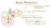
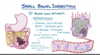
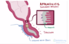
What type of pain would someone with appendicitis complain of?

A

central abdominal pain that moves into the right iliac fossa

2
Q

Typically, how does someone with appendicitis present?

A
  • tends to be a young person (5 - 40 years old)
  • acute onset within 12 - 24 hours
  • present with umbilical pain that moves to the right iliac fossa
  • nausea and/or vomiting
  • diarrhoea or constipation
  • fever
3
Q

What might be present on general inspection and palpation of someone with appendicitis?

A
  • in the early stages there is general pain and peri-umbilical pain on palpation
  • in the later stages, the person will often stay very still due to peritonitis
    • this occurs after the appendix has ruptured, and the peritoneum has become inflamed

on palpation, there will be right iliac fossa pain

4
Q

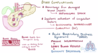
What signs might be present in appendicitis?

A
  • Rovsing’s sign
  • Cope’s sign
  • Psoas sign
  • rebound tenderness
5
Q

What is Rovsing’s sign?

A
  • pain is greater in the RIF than the LIF when the LIF is pressed
  • this is specific to appendicitis
6
Q

What is Cope’s sign?

A
  • there is pain on passive flexion and internal rotation of the hip
  • it indicates irritation to the obturator internus muscle
  • the appendix becomes inflamed and enlarged and may come into contact with the obturator internus muscle when this move is performed
7
Q

What is Psoas sign?

A
  • there is pain on extending the hip
  • pain indicates an inflamed appendix overlying the iliopsoas muscles
  • this only occurs with retrocaecal appendix
    • (as the iliopsoas muscle is retroperioneal)
    • this indicates that the inflamed appendix sits behind the caecum
8
Q

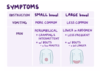
When might rebound tenderness be evident in appendicitis?

What is this?

A
  • this indicates that the infection is involving the peritoneum
  • there is pain upon removal of pressure from the abdomen rather than application of pressure to the abdomen
  • this is indicative of peritonitis
  • there may also be abdominal guarding - the abdominal muscles tense up to avoid pain
9
Q

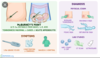
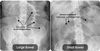
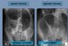
What are the investigations involved in appendicitis?

A
  • first line investigation is CT abdomen
    • USS can be done if CT is not available
  • this will show increased appendix diameter and increased wall enhancement
  • bloods - which will show leucocytosis and elevated CRP
10
Q

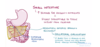
What is the most common cause of appendicitis in adults?

A
  • appendicitis results from obstruction of the appendix lumen
  • this may be due to a fecalith (hardened lump of faecal matter) that wedges itself within the lumen
    • it can also be due to undigested seeds or pinworm infections
11
Q

What is a common cause of appendicitis in adolescents?

A

lymphoid hyperplasia

  • this involves growth of the lymphoid follicles, which are dense collections of lymphocytes
  • these reach their maximum size in adolescence and can obstruct the lumen of the appendix
  • when exposed to viral infections or immunisations, the follicles can increase in size
12
Q

How does obstruction of the lumen of the appendix lead to pain?

A
  • the intestinal mucosa secretes mucus and fluids to keep pathogens from entering the bloodstream and to keep the tissue moist
  • even when obstructed, the appendix keeps secreting
  • there is a build up of fluid and mucus in the appendix, which increases the pressure
  • the appendix gets bigger and physically pushes on afferent visceral nerve fibres nearby, causing pain
13
Q

Why is there is an increase in serum WBC count in acute appendicitis?

What processes have to occur prior to this for it to occur?

A
  • as there is an obstruction, flora and bacteria in the gut are trapped
    • E. coli and bacteroides fragilis
  • these bacteria are now free to multiply
  • this causes the immune system to produce WBCs, which leads to the build up of pus in the appendix
14
Q

What happens if the obstruction in the appendix persists past the build-up of pus in the appendix?

A
  • the pressure in the appendix increases even further
  • it expands and begins to compress small blood vessels that supply it with blood and oxygen
  • without oxygen, the cells in the wall of the appendix become ischaemic and die
  • these cells were responsible for secreting mucus and keeping bacteria out, so now the growing colony of bacteria can invade the wall of the appendix
15
Q

What leads to rupture of the appendix?

What happens if the appendix ruptures?

A
  • as more cells in the wall of the appendix die, it becomes weaker and weaker
  • in a small proportion of patients, the appendix wall becomes so weak that it ruptures
  • this leads to bacteria entering into the peritoneum and causing peritonitis
    • this leads to abdominal guarding and rebound tenderness at McBurney’s point
16
Q

What is the most common complication of a ruptured appendix?

A
  • formation of a periappendiceal abscess
    • this is a collection of fluid and pus around the ruptured appendix
  • sometimes smaller subphrenic abscesses can form
    • ​these are below the diaphragm, but above the liver/spleen
17
Q

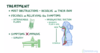
What is the treatment for appendicitis?

A

appendicetomy

  • this is surgical removal of the appendix, followed by antibiotics
  • if there is an abscess, this must be drained first
18
Q

What scoring system is used to determine the severity of appendicitis?

A

Alvarado score

  • score of 1 to 4 is discharged
  • score of 5 to 6 is observed
  • score of 7 to 10 needs surgery
19
Q

Which antibiotics are given following appendicetomy?

A
  • cefotaxime
  • metronidazole
    • this is an anti-anaerobe antibiotic for the gut
20
Q

What are the 3 possible complications of appendicitis?

A
  • perforation
  • appendix abscess
  • appendix mass
    • the inflamed appendix becomes covered in omentum and forms a mass
    • this tends to occur in older men who avoid coming to the doctors when they get pain
21
Q
A

B-hCG test

  • the first line investigation in any woman with an abdominal pathology should ALWAYS be a pregnancy test
22
Q

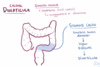
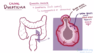
What is meant by diverticulosis?

A
  • the presence of diverticulae
  • these are outpouchings of the colonic mucosa and submucosa throughout the large bowel
  • high pressure in the bowel causes these outpouchings to form
    • e.g. chronic constipation
23
Q
A
24
Q

What is meant by diverticulitis?

Which part of the bowel is more commonly affected?

A
  • acute inflammation and infection of the diverticulae
  • most commonly affects the sigmoid colon
25
What is the structure of the large intestine wall like? What is the difference between a true diverticula and a pseudo-diverticula (false)?
* the wall of the large intestine is made up of **4 layers** * mucosa * submucosa * muscle layer * serosa * a ***true diverticula*** involves **_all 4 layers_** of the intestine * a ***false (pseudo) diverticula*** includes only the **_mucosa and submucosa_** * ​these 2 layers are covered by serosa only, and the muscle layer is not involved * these are more common
26
27
Why do diverticula form?
* they are formed by **_high pressure_** within the lumen of the large intestine * **smooth muscle** in the intestinal wall contracts to push food along the bowel * when it contracts, higher pressures are generated inside the lumen as it "squeezes" air inside * **contractions** in someone with diverticula are **_abnormal_** * instead of pressure being ***distributed evenly*** throughout the lumen, there are **_areas of very high pressure_** during abnormal smooth muscle contraction, which leads to diverticula formation
28
What is the most common location for diverticula to form and why?
**_sigmoid colon_** * this has the **smallest lumen diameter**, and so it subject to the highest intraluminal pressures
29
Does rectal bleeding (haematochezia) occur in diverticulosis and/or diverticulitis? Why?
* PR bleeding **_can_** occur in **_diverticulosis_** * a diverticulum can form where blood vessels traverse the muscle layer, as this point of the wall is weaker * the blood vessel becomes separated from the intestinal lumen only by mucosa * it is predisposed to rupture, meaning blood enters the large intestine * PR bleeding does **_NOT_** occur in **_diverticulitis_** * ​this is because the blood vessels become **scarred from inflammation**
30
What genetic conditions and lifestyle factors are associated with an increased risk of diverticular disease?
* anything that **increases the stress** on the intestinal walls or **decreases their strength** predisposes to diverticula * **_Marfan syndrome_** & **_Ehlers-Danlos_** are genetic conditions that affect **connective tissue** * diverticula can form in the absence of strong connective tissue supporting the intestinal wall * diets **_low in fibre_** and **_high in fatty foods_** and **_red meat_** increases risk of symptomatic diverticular disease
31
What symptoms may be present in diverticulosis?
* this is the presence of diverticula and usually has **_no symptoms_** * sometimes there will be **vague stomach pain** and the diverticula can **bleed** * they are often found incidentally on **colonoscopy** or **CT scan**
32
What are the 2 reasons why diverticula may become inflamed and cause diverticulitis?
* if a faecalith becomes lodged in the diverticula (less common) * due to erosion of the walls of the diverticula from higher luminal pressures
33
What is the most severe complication of diverticulitis?
* if the diverticula become **distended enough**, they can **rupture** and form a **_fistula_** * this is a connection with an adjacent organ or structure * a **_colovesicular fistula_** between the **large intestine** and the **_bladder_** might form, leading to **air or stool** in the **urine**
34
How does someone with acute diverticulitis typically present?
* **_left iliac fossa pain +/- bloating_** * anorexia * this refers to not eating much * fever * nausea & vomiting * may have bloody stools * may have urinary symptoms if colovesicular fistula has formed * e.g. brown urine due to the presence of faeces in the urine
35
What are the risk factors associated with diverticulitis?
* tends to present in **_50 - 70 year olds_** who have previously been asymptomatic * **_low dietary fibre_** * **smoking** * **chronic _NSAID_ use**
36
What would someone look like on general inspection and palpation in acute diverticulitis? What if they had peritonitis?
***_Acute diverticulitis:_*** * tachycardia * low-grade pyrexia * **_LIF tenderness_** on palpation ***_Peritonitis:_*** * this may occur if they have a **_perforated diverticulum_** * they will be **_lying very still_** to not aggravate the pain * on palpation, there will be **_guarding_** and **_rebound tenderness_**
37
What is the primary investigation for acute diverticulitis? What other investigation might be performed if perforation is suspected?
**_CT abdomen with contrast_** * an **erect CXR** may be performed if there are signs of perforation * the presence of **air under the diaphragm** confirms perforation * inflammation is shown as hyperdense tissue
38
What investigation might be performed once someone has recovered from diverticulitis?
**_barium enema_ +/- flexible sigmoidoscopy / colonoscopy** this is to confirm the presence of **chronic diverticulosis**
39
What is the treatment for mild and severe diverticulitis?
***_Mild / Uncomplicated:_*** * oral antibiotics (in more severe cases, IV antibiotics are given) * fluids * bowel rest (no food / water orally) ***_Severe:_*** * bowel resection is performed if there is recurrence / severe cases * this usually involves Hartmann's procedure
40
What is involved in Hartmann's procedure? Why is this performed to treat acute diverticulitis?
* this involves **emergency / acute removal** of a piece of bowel * this usually involves removal of the **sigmoid colon** * this results in formation of an **_end colostomy_** and **_anorectal stump_** * an immediate ***primary anastomosis*** is **_not possible_** due to ***inflammation*** and ***oedema*** * when the oedema starts to settle down, gaps will appear in the anastomosis and it will become leaky
41
What is the treatment for diverticulosis?
* encourage the patient to have a soluble, high-fibre diet * once the inflammation has settled down from diverticulitis, a primary anastomosis can be formed
42
What other operation may accompany formation of an anastomosis in distal large bowel cancers (e.g. rectal carcinoma)?
**_defunctioning loop ileostomy_** * this **diverts bowel contents** away from a distal anastomosis * this allows it **time to rest** prior to reversal of the loop ileostomy
43
What are the possible complications of diverticular disease?
* acute diverticulitis * faecal peritonitis * this tends to occur if there is rupture of a diverticulum * fistula formation * peri-colic abscess * colonic obstruction * perforation
44
C - diverticulitis * presence of bloody stools * she has had blood in the past - bleeding diverticula * fever * tenderness in left iliac fossa * ​this is the location of the sigmoid colon * low fibre diet
45
A - Hartmann's procedure * the presence of air under the diaphragm shows perforation * a primary anastomosis cannot be performed in the acute presentation
46
What is the definition of a hernia? What are the 2 major types?
a condition in which part of an organ is **displaced** and **protrudes** **through the wall** of the cavity containing it * this is often involving the intestine at a weak point in the abdominal wall * the 2 main types are **inguinal** and **femoral**
47
In what 2 different ways can abdominal hernias be classified?
* **_midline hernias_** * epigastric hernias * umbilical hernias * **_groin hernias_** * ​femoral hernias * inguinal hernias (much more common) * can also get **_incisional hernias_** when abdominal contents herniate through a scar from a previous abdominal surgery
48
What are the layers of the abdominal wall? What could cause something to protrude through these layers?
* deepest layer is the **visceral peritoneum** * this covers many abdominal organs and lines the peritoneal space * this layer wraps around to form the **parietal peritoneum** * extraperitoneal fat * **transversalis fascia** * muscle layer containing **external oblique**, **internal oblique** & **transversus abdominis aponeurosis** * fascia * anything that **_increases the pressure_ in the abdominal cavity** may result in a **sac** that forms in the abdominal wall through which **_organs might protrude_**
49
What are the 2 different types of midline hernias and when do they occur?
* a midline hernia occurs when organs protrude **_through the midline_** * an ***epigastric hernia*** occurs when organs protrude through the **_linea alba_**, between the xiphoid process and the umbilicus * an ***umbilical hernia*** occurs when organs protrude through the **_umbilicus_**
50
What is meant by an inguinal hernia? What are the 2 openings of the inguinal canal?
* an inguinal hernia occurs when abdominal contents protrude through the **_inguinal canal_** * the **_deep inguinal ring_** is an opening in the ***transversalis muscle fascia*** * the **_superficial inguinal ring_** is an opening in the ***external oblique muscle aponeurosis***
51
Why are inguinal hernias much more common in males?
* the **inguinal canal is much _larger_** and **more prominent** in males, which creates a **_site of weakness_** in the abdominal wall * this is due to the testes having to descend further than the ovaries during development, meaning that the **processus vaginalis** (now obliterated) may remain open
52
What is meant by an indirect inguinal hernia and why does it occur? Who tends to be affected by these?
* occurs when the **_processus vaginalis fails to close_** after the testes have passed through it * sometimes called a ***congenital hernia*** * when the processus vaginalis remains open, intestinal contents herniate through **_BOTH_** the **_deep_** and **_superficial inguinal rings_** and into the scrotum * ​think *"contents herniate INDIRECTLY through the inguinal canal"* * this is more common in **_infants and children_** but can be discovered in adulthood
53
What is meant by a direct inguinal hernia and why does it occur? Who tends to be affected by this?
* this results from **_weakness of the transversalis fascia_** * sometimes called an ***acquired hernia*** * the abdominal wall gets **weaker with age**, so these tend to occur in **_middle-aged_** and **_elderly people_** * the intestinal contents pass through the **_external inguinal ring_** ONLY * ​think *"contents are herniating DIRECTLY through the abdominal wall"*
54
In which location is a direct inguinal hernia most likely to occur? When does this happen?
* the transversalis fascia weakens most commonly in the p**osterior wall** of the inguinal canal * this region is called **_Hesselbach's triangle_** * hernia through Hesselbach's triangle occurs as a result of **increased abdominal pressure**, usually through **_coughing_** or **_heavy lifting_**
55
What happens in a femoral hernia?
* occurs when abdominal contents herniate beneath the inguinal ligament and into the femoral canal * these are less common than inguinal hernias
56
What is the difference between an uncomplicated hernia and an incarcerated hernia?
* in an ***uncomplicated hernia***, the hernia can be reduced back into the abdomen by **_pressing on the hernial sac_** * this is a pouch of peritoneum that covers the herniating organ * if the contents of the hernia **_cannot be pushed_ back inside the abdomen** then this is ***incarceration*** * ​there is reduced venous and lymphatic flow * this leads to **swelling and oedema** of the incarcerated tissue
57
What is meant by a strangulated hernia?
* if a hernia is **incarcerated**, eventually the tissue will **swell so much** that the **_arterial blood flow_** to the hernial contents is **_completely cut off_** * this is strangulation and it leads to **_ischaemia**_ and _**tissue necrosis_**
58
How would someone with a hernia typically present?
* small hernias are often **asymptomatic** * **_palpable lump_** in groin * **_pain_** in the groin * **scrotal swelling** (inguinal hernia) * **nausea & vomiting** / **fever** are signs of **_incarceration_** * incarceration may interrupt passage of contents through the intestines and lead to symptoms of **bowel obstruction** * if the hernia appears **red**, it is likely to be **_strangulated_** and there is blood trapped in the hernial sac * (groin pain and vomiting are red flags)*
59
What are key features to pick up in the history of someone who could have an inguinal or femoral hernia?
* **increasing age** * **obesity** * history of **chronic constipation** * history of **chronic cough** * recent **heavy lifting** (e.g. gym) * (obesity, constipation, cough and lifting all increase intra-abdominal pressure)*
60
Who tends to be more affected by femoral and inguinal hernias? Which is more likely to become strangulated and which is more likely to need surgery?
* inguinal hernias are the most common hernia in both sexes * but femoral hernias occur more often in women * ***femoral hernias*** are ***more commonly _strangulated***_ and _***surgery_ is recommended*** * **femoral hernias** affect **_older people_** and inguinal hernias affect younger people
61
What is the defintion of a strangulated hernia?
the compression around the hernia **prevents blood flow** into the hernial contents causing **_ischaemia to the tissues_** and **_pain_**
62
Where is a femoral hernia located and what is often contained within it?
* located **_lateral_** and **_inferior_** to the **pubic tubercle** * often contains **_omentum_**
63
What is the location of the inguinal hernia? What is often contained within it?
* located **_superior_** and **_medial_** to the **pubic tubercle** * often contains **_bowel_**
64
What are the features that are common to both femoral and inguinal hernias? What are the signs that they might have become strangulated?
* **swells / appears** **_on coughing_** and may **reduce on supination** * both may be **_reducible on pressure_** * signs of strangulation include: * **redness / tender** * **colicky abdominal pain** * **distension** * **vomiting**
65
What are the borders of Hesselbach's triangle? What type of hernia is this associated with?
* **rectus abdominus** * **inferior epigastric vessels** * **inguinal ligament** * associated with a **_direct inguinal hernia_** that will protrude through this weakness in the abdominal wall * this tends to occur in **older people, weight-lifters** and **manual labour workers**
66
How can you make a clinical diagnosis of an inguinal hernia and distinguish an indirect from a direct hernia?
* ***reduce** the hernia* * *place your finger over the **_deep inguinal ring_*** * just above the midpoint of the inguinal ligament * *ask the patient to **cough*** * *if the hernia **reappears**, it **_CANNOT_** be an **_indirect hernia_** (must be **direct**)* * ​your finger is blocking the deep inguinal ring so the indirect hernia could not pass through * if still unsure, an **USS** can be done to **locate the hernia** and decide whether it is composed of **bowel or omentum** * ​femoral = omentum * inguinal = bowel
67
Which type of hernia is more likely to need surgery?
* **femoral hernia** needs **_urgent surgery_** due to risk of strangulation * for inguinal hernia, surgery is less urgent * this tends to be mesh repair or more conservative measures like wearing a corset
68
What is meant by inflammation of the pancreas? What are the 2 different types?
* **_inflammation_** of the pancreas * leading to **_autodigestion_** of pancreatic tissue by the pancreatic enzymes, resulting in **_necrosis_** * this can occur in either an **_acute_** or **_chronic_** setting
69
What is the definition of acute pancreatitis?
sudden **_inflammation_** and **_haemorrhaging_** of the pancreas due to destruction by **its own digestive enzymes** (autodigestion)
70
What are the endocrine and exocrine roles of the pancreas?
***_Endocrine:_*** * alpha and beta cells release hormones like insulin and glucagon into the blood ***_Exocrine:_*** * acinar cells secrete digestive enzymes into the duodenum * these break down carbohydrates, proteins and lipids that are also found within the cells of the pancreas
71
How does the pancreas protect itself from autodigestion normally?
* the acinar cells manufacture **inactive forms** of digestive enzymes (**_zymogens_**) * zymogens are stored in vesicles (**_zymogen granules_**) with **_protease inhibitors_** * this allows for extra protection if the zymogens were to become prematurely active * the zymogens are released into the **pancreatic duct** and delivered to the **duodenum**, where they activated by **_trypsin_** * trypsin comes from the pancreatic zymogen **trypsinogen**, which is activated by **_enteropeptidases_** in the **duodenum**
72
What are the 2 main causes of acute pancreatitis? What is the mechanism behind this?
* **early activation** of trypsinogen or zymogens causes acute pancreatitis * this can occur due to **_injury to the acinar cells_** * or due to anything that **_impairs secretion of zymogens_** into the duodenum * the 2 main causes are **_alcohol_** and **_gallstones_**
73
What is the mechanism behind how alcohol can cause acute pancreatitis?
* alcohol **_increases zymogen secretion_** from acinar cells * it also **_decreases fluid and bicarbonate secretion_** from ductal epithelial cells * the pancreatic juices become very thick and **_viscous_** * this leads to a **_plug forming_**, which blocks the pancreatic ducts * plug formation leads to pancreatic juices **backing up**, **increasing the pressure** within and **_distending the pancreatic ducts_**
74
How can a blocked and distended pancreatic duct lead to acute pancreatitis?
* distended ducts make **membrane trafficking** become more **chaotic** * **zymogen granules may fuse with lysosomes**, bringing trypsinogen into contact with digestive enzymes * trypsinogen is converted to **active trypsin**, which starts the **cascade of enzyme activation** within the pancreas * this leads to **autodigestion** of pancreatic tissue
75
In what other ways can alcohol contribute to the development of acute pancreatitis?
* alcohol stimulates release of **_inflammatory cytokines_**, stimulating an immune response * **neutrophils** arriving at the scene release **proteases**, making the problem worse * high consumption of alcohol can produce enough **_reactive oxygen species_** to overwhelm cellular defences and damage the cells
76
How can gallstones lead to acute pancreatitis?
* gallstones can **_block the sphincter of Oddi_** * this blocks the release of pancreatic juices
77
What is liquefactive haemorrhagic necrosis? How does it occur?
* autodigestion / pancreatic tissue destruction can cause **tiny blood vessels** to become **_leaky_** and **_sometimes rupture_** * extra fluid causes **_swelling and oedema_** of the pancreas * this can **_activate lipases_** which **digest the _peripancreatic fat_** * this is the fat surrounding the pancreas * digestion and bleeding can **liquefy** pancreatic tissue * ​this is liquefactive haemorrhagic necrosis
78
What is a pancreatic pseudocyst and how does it form? What symptoms might be seen with this?
* **fibrous tissue** surrounds the **liquefactive necrotic tissue** of the pancreas * this forms a **cavity** that fills with **pancreatic juices** * typical symptoms include **_abdominal pain_**, **_loss of appetite_** and **_palpable abdominal mass_** following a bout of pancreatitis * **serum amylase, bilirubin** and **lipase** may be raised * the best way to image a cyst is with abdominal CT
79
What happens if the pancreatic pseudo-cyst becomes infected?
* it forms a **_pancreatic abscess_** * it is usually infected by ***E. coli*** * this presents with the same symptoms, with additional **fever** and **high WCC**
80
How does someone with acute pancreatitis typically present?
* intense **_epigastric pain_** that may **radiate to the back** * pain is **_worse on movement_** * pain is **relieved when** **_sitting forwards_**
81
What is the mnemonic to remember the causes of acute pancreatitis?
I GET SMASHED * I - idiopathic * G - gallstones * E - ethanol * T - trauma * S - steroids * M - mumps infection * A - autoimmune * S - scorpion venom * H - hypercalcaemia, hyperlipidaemia, hyperparathyroidism * E - ERCP * D - drugs (thiazides)
82
On examination, what signs might be present in acute pancreatitis?
* epigastric tenderness * fever * reduced bowel sounds * shock, tachycardia, tachypnoea * Cullen's sign & Grey-Turner's sign
83
What are Cullen's sign and Grey-Turner's sign and why do they occur?
* they occur due to **intra-abdominal bleeding** from pancreatic inflammation * ***Cullen's sign*** is bruising around the **_umbilicus_** * ***Grey-Turner's sign*** is bruising in the **_flanks_** * remember "you have to TURN over to see it"
84
What blood tests will be deranged in acute pancreatitis?
* increase in serum digestive enzymes, including **_amylase_** and **_lipase_** amylase is not very specific * it will be raised in any cause of acute abdomen, including pancreatitis lipase is more specific * **_hypocalcaemia_** occurs in acute pancreatitis * this occurs as **fat necrosis consumes calcium**
85
What other investigations may be performed in acute pancreatitis?
* **serum amylase** * should be \> 3x upper limit of normal * **serum lipase** * **USS** to look for the cause (i.e. gallstones) * erect CXR and **CT abdomen** * CT will show inflammation, necrosis and pseudocysts
86
Why can acute pancreatitis lead to hypovolaemic shock and bleeding? What is the most severe complication of acute pancreatitis?
* **_haemorrhage_** from a damaged blood vessel can lead to shock * there may be systemic activation of blood coagulation factors (**_DIC_**) this leads to **small blood clots** forming all over the body, **using up clotting factors** and making it easier to bleed * massive pancreatic inflammation can lead to **_acute respiratory distress syndrome (ARDS)_** * this makes it difficult to breath
87
How can the Modified Glasgow Score be used to determine the severity of pancreatitis?
* a score of \> 3 indicates severe pancreatitis * P - pO2 \< 7.9 kPa * A - age \> 55 years * N - neutrophils \> 15 x 109 / L * C - calcium \< 2 mmol / L * R - renal function * measured by urea \> 16 mmol / L * E - enzymes * ​LDH \> 600 U / L or AST \> 200 U / L * A - albumin \< 32 g / L * S - sugar \> 10 mmol
88
How do you expect serum calcium to change in acute pancreatitis? What would normal calcium suggest?
* **serum calcium _drops_** in acute pancreatitis due to sequestering of free Ca2+ by free fatty acids * **_very low_ Ca2+** has **_worst prognosis_** * **_normal_ Ca2+** supports an aetiology of **_hypercalcaemia_**
89
What is involved in the medical and surgical management of acute pancreatitis?
* supportive management with fluids and analgesia * enzyme supplementation * diabetes medications * if someone becomes insufficient (i.e. cannot produce sufficient insulin) * ERCP may be performed to remove a gallstone if present
90
What is meant by chronic pancreatitis? How is it different to acute pancreatitis?
***_Acute pancreatitis:_*** * involves **_sudden_ inflammation** of the pancreas caused by digestion by its own enzymes (**autodigestion**) * generally is **_reversible_** ***_Chronic pancreatitis:_*** * involves **_persistent_ inflammation** of the pancreas caused by **_irreversible_ changes** to pancreatic structure * this involves **fibrosis, atrophy** and **calcification** * usually caused by recurrent bouts of acute pancreatitis
91
What are the most common causes of chronic pancreatitis?
* it is caused by **_recurrent bouts of acute pancreatitis_** * this is most commonly due to **alcohol** * **trauma** to the pancreas * pancreatic **tumours** * **_cystic fibrosis_** * **_​_**this is the most common cause of chronic pancreatitis in children
92
How can cystic fibrosis cause pancreatitis?
* there is a mutation in the **_CFTR gene_**, which encodes an **ion transporter** * there is **disruption to ion transport** * this leads to **pancreatic secretions** becoming **_thick / sticky_** * viscous secretions will **_obstruct / block pancreatic ducts_**
93
How can repeated bouts of acute pancreatitis progress to chronic pancreatitis?
* with each bout, there is potential for **_duct dilatation_** and **_damage_ to pancreatic tissue** * as part of the subsequent healing process, pancreatic **_stellate cells**_ lay down _**fibrotic tissue_** * fibrotic tissue causes **_stenosis (narrowing)_** of the ducts * there is also **_acinar cell atrophy_** * **_calcium deposits_** of various sizes can accumulate on the protein plugs that have already formed within the ducts * this only happens in some causes - such as alcoholic acute pancreatitis
94
Why can early diagnosis of chronic pancreatitis be challenging? What is the most common presenting symptom?
* continuous or intense **_intermittent epigastric pain_** that may **radiate to the back** * this is usually related to **eating meals** * it tends to last for several hours * the pain is **relieved on sitting forwards** * in acute pancreatitis, there would be elevated serum amylase and lipase in chronic pancreatitis, there may not be enough healthy pancreatic tissue to make these enzymes ***_serum amylase / lipase is NORMAL_ in chronic states***
95
96
What imaging investigations are performed to diagnose chronic pancreatitis?
* **abdominal X-ray** may show **_calcifications_** within the pancreas * **ERCP** can be performed to **visualise the pancreatic ducts** * this will show a **_"chain of lakes" pattern_** due to alternating stenosis and dilatation of the ducts
97
What marker can be used to distinguish acute and chronic pancreatitis?
**_faecal elastase_** * faecal elastase will be **raised** in **_chronic_ pancreatitis** * it will be normal in acute pancreatitis * ***serum _amylase***_ will be raised in _***acute_ pancreatitis*** * ​it will be normal in chronic pancreatitis
98
As chronic pancreatitis progresses, what signs might someone with pancreatic insufficiency show? Why does this happen?
* **acinar cells** become impaired and produce **fewer digestive enzymes** * patient has **trouble absorbing food / dietary fats**, leading to **_weight loss**_ and _**deficiency of fat-soluble vitamins_** * the fat-soluble vitamins are A, D, E and K * fat passes through the intestines without being digested, leading to **greasy, foul-smelling stools** * ​this is **steatorrhoea**
99
What condition can develop as a long-term consequence of chronic pancreatitis?
**_diabetes mellitus_** * recurrent inflammation begins to damage the alpha and beta cells of the pancreas * this impairs insulin secretion
100
What is involved in the treatment of chronic pancreatitis?
* treatment mainly involves **controlling pain / risk factors** * drinking less alcohol * eating less meat * losing weight * pancreatic **enzyme replacement** / **nutritional supplements** are given in pancreatic insufficiency * **diabetes medications** are given if necessary
101
What is meant by bowel obstruction? What are the 2 categories of causes?
* when the normal flow of contents moving through the intestines is interrupted ***_​Mechanical Causes:_*** * there is an **_actual blockage_** in the large or small intestine * this blockage can be **partial** or **complete** ***_Functional Causes:_*** * this is causes by **_disruption to peristalsis_**
102
What is the difference between partial and complete bowel obstruction?
***_Partial obstruction:_*** * **gas** or **liquid stool** can pass through the point of narrowing ***_Complete obstruction:_*** * absolutely **nothing** can pass through the point of narrowing
103
What are the mechanical causes of small and large bowel obstruction?
***_Small bowel obstruction:_*** * post-operative adhesions * scar tissue that forms after surgery can form fibrous bands that cause organs to attach to the surgical site or other organs * the lumen of the bowel can become pinched tight in some locations * hernias ***_Large bowel obstruction:_*** * volvulus * a loop of intestine twists around on itself, kinking off the lumen * a volvulus can occur around a mass - e.g. colorectal cancer ***_Both:_*** * ingestion of a foreign body * intussusception * inflammatory bowel disease - which causes strictures & adhesions
104
What are the potential functional causes of bowel obstruction?
* anything that **_reduces smooth muscle contractility / peristalsis_** * post-operative ileus * transient paralysis of the smooth muscle following surgery * hypothyroidism * inflammation and infection * electrolyte abnormalitites * e.g. hypokalaemia, hypercalcaemia * some medications - e.g. opioids
105
How can oedema and ischaemia result from bowel obstruction?
* proximal to the obstruction, gas and stools start to **accumulate** * this leads to **_dilatation of the bowel_** and **_abdominal distension_** * as pressure within the bowel lumen increases, intenstinal contents will **compress blood and lymphatic vessels** within the intestinal wall * **veins and lymphatics** are compressed first as they have weaker walls * **increased pressure** within these vessels **pushes fluid** into the surrounding area causing **_mucosal oedema_** * if the pressure in the lumen continues to increase, the **_arteries can become compressed_** * this leads to **reduced blood flow to the intestinal wall** (ischaemia)
106
How can bowel ischaemia and hypoxia lead to damaged capillaries?
* hypoxia is accompanied by production of **_reactive oxygen species_** * these **damage mucosal cells**, leading to **_cell death / mucosal infarction_** * capillaries within the intestinal wall **rupture**, leading to blood entering the intestinal lumen
107
What happens once blood enters the intestinal lumen?
* the presence of **blood and stool** in the lumen acts as a **_nutritional supply for gut bacteria_** * gut bacteria **multiply rapidly** * gut bacteria enter the **intestinal wall** * **macrophages** rush into the intestinal wall and release **inflammatory cytokines** * cytokines **increase blood vessel permeability**, which increases **_mucosal oedema, inflammation and damage_**
108
What are the consequences in bowel obstruction when inflammatory cytokines have increased blood vessel permeability?
* there is increased mucosal oedema, inflammation and damage * the overall result is **_compromised ability_** of the mucosa to **_absorb food and water_** * this leads to **_dehydration_** and **_loss of electrolytes_**
109
How can perforation result from bowel obstruction?
* if pressure within the bowel lumen becomes high enough, it can lead to **compression of large arteries** * bowel ischaemia / infarction extends from just the mucosa to **all layers of the bowel wall** * this is **_transmural infarction_** * transmural infarction can lead to **perforation** * bacteria within the bowel lumen leak into the peritoneal cavity and cause **_peritonitis_**
110
How can peritonitis lead to sepsis?
* the layers of the peritoneum are highly vascularised * large numbers of bacteria **enter the bloodstream**, causing a **_massive inflammatory response_** * this is sepsis * blood vessels throughout the body become **"leaky",** allowing cells and fluid to leak into the interstitial space * this **reduces blood volume** and **_blood pressure_** * there is **less blood and oxygen** reaching vital organs, which is **_shock_**
111
What are the typical symptoms of bowel obstruction?
* **vomiting** * this is more common in small bowel obstruction * **abdominal distension** * **constipation** * **diffuse / cramping abdominal pain**
112
How can you tell the difference in symptoms of complete and partial bowel obstruction?
***_Partial obstruction:_*** * symptoms come on more **gradually** and are **milder** * e.g. abdominal pain after meals, constipation ***_Complete obstruction:_*** * symptoms tend to come on **very suddenly** * there is **_absolute constipation_** * absolutely nothing can pass - not even gas / liquid stool
113
114
How are the symptoms different in small and large bowel obstruction? How might you know if there has been a perforation?
***_Small bowel:_*** * **_vomiting_** is more common * pain tends to be **_periumbilical_** (around bellybutton) * bouts of **crampy intermittent** abdominal pain that last for a **_few minutes_** at a time ***_Large bowel:_*** * vomiting is less common * pain tends to be **_lower down_** in the abdomen * bouts of pain tend to be **_less frequent_**, but **_last for longer_** ***_Perforation:_*** * when there is progression from **vague / crampy pains** to more **_constant / focal pains_** * this indicates **peritoneal irritation** due to perforation
115
What are key things to pick up in the history of someone with a bowel obstruction?
* **malignancy** history * colorectal cancer can cause volvulus * **hernia** history * **surgical** history (risk of adhesions)​
116
How can bowel obstruction affect breathing?
* abdominal distension can **_push on the diaphragm_** and make it more difficult for the lungs to expand * this leads to **_respiratory distress_** * patient may present with **shortness of breath**, **tachypnoea** and **cyanosis**
117
What might you see on general inspection and auscultation in bowel obstruction?
* **_high-pitched, tinkling bowel sounds_** suggest ***mechanical obstruction*** * complete **_absence of bowel sounds_** suggests ***functional obstruction*** * **abdominal distension** * **pyrexia / sweating** - this is more prevalent if there is a perforation
118
119
What investigations are performed if bowel obstruction is suspected?
* **abdominal X-ray** * looking for signs of **volvulus** and/or **Rigler's sign** * **CT abdomen with contrast** * abdominal USS if CT is contraindicated * ​e.g. in pregnancy / contrast allergy * bloods * ​FBC and crossmatch - in case of perforation
120
How can you tell the difference between large bowel and small bowel on AXR?
* **_valvulae conniventes_** are seen in the ***small bowel*** * these are lines that **transect the entire bowel** * **_haustra_** are seen in the ***large bowel*** * ​these do not transect the entire bowel
121
How do you tell the difference between caecal and sigmoid volvulus on AXR?
* ***caecal volvulus*** is represented by the **_"embryo"**_ or _**"comma" sign_** * formed from the caecum folding up in RIF * **sigmoid volvulus** is represented by the **_coffee bean sign_**
122
What is Rigler's sign?
* if there is **air present on _both sides_ of the bowel wall** then this indicates **_perforation_** * also known as the ***double-wall sign*** * there is gas outlining both sides of the bowel wall * gas within the bowel lumen and gas within the peritoneum
123
What are the treatment options for bowel obstruction?
* generally, bowel obstructions resolve on their own so treatment focuses on **relieving symptoms** * IV fluids * nasogastric suction * **"drip and suck"** to remove gas / liquid from the stomach that has accumulated * if symptoms do not improve, or there is perforation, then surgery is performed
124
What is meant by intestinal ischaemia? What are the 2 different types?
* **_reduction in blood flow_** to the intestine, resulting in **ischaemia of the _bowel wall_** * if it affects the ***small bowel***, it is **_mesenteric ischaemia_** * if it affects the ***large bowel***, it is **_ischaemic colitis_** * this leads to **_infarction_** when part of the intestinal wall necroses/dies
125
126
What is the main source of blood to the small intestine? What type of blood supply exists to try and prevent small bowel ischaemia?
* blood supply mainly comes from the **_superior mesenteric artery_** * branches of this artery spread through the mesentery (**_mesenteric arteries_**) * mesenteric arteries penetrate the submucosa and branch into arterioles * small intestine has a **high demand for oxygen and nutrients** to sustain digestion, so it is **_susceptible to ischaemia_** * mesenteric arteries **branch and reconnect at points** forming a **_collateral circulation_** * if blood flow is reduced in one pathway, the tissue can still receive blood via another pathway
127
When does small bowel infarction occur?
* when there is **_significant reduction in blood flow_** to the small intestine * this **reduces the blood pressure** meaning that there is **_insufficient blood flow_ throughout the _collateral circulation_** * this initiates **ischaemic injury** in a **_wide region_** of tissue
128
What is meant by reperfusion injury?
* reperfusion injury occurs when **_blood flow returns to ischaemic tissue_** * influx of oxygen into an already damaged cell can **increase oxidative stress** and cause **further cell damage** * damaged /ischaemic cells release **_reactive oxygen species_**, which stimulate an **immune response** * immune cells remove damaged/dead cells and release **cytokines** * cytokines cause blood vessels to become more **permeable**, resulting in **_bowel oedema_**
129
What layers of the bowel are affected in mesenteric ischaemia? How do symptoms range in severity?
* mesenteric ischaemia becomes more severe as it extends from a **_mucosal infarct_** to a **_transmural infarct_** * it goes from only affecting the mucosa to affecting all the layers of the bowel wall * early-on, ischaemia causes **_ileus_** * this is where the bowel stops working * food lingers in the bowel and doesn't get pushed along * severe damage can cause a **_break in the epithelial lining_** of the bowel, allowing **bacteria in the lumen** to enter **blood vessels** within the bowel wall * bacteria can pass through the wall into the **peritoneal space**, causing **_bacterial peritonitis_** * they can also pass from the peritoneal space into lymphatics and blood vessels * if bacteria enter the bloodstream, this can lead to **_septic shock_** * ​this involves hypoperfusion of organs throughout the body​​​
130
What are the occlusive causes of mesenteric ischaemia? What type of infarct does this usually cause?
* this involves a **_physical blockage_** that prevents blood flow through the vasculature * usually causes a t**_ransmural infarct_** * **_thrombus formation_** in superior mesenteric artery or vein * this leads to thrombosis and occlusion of the vessel * **_thromboembolism_** occluding superior mesenteric artery * ​part of a blood clot has broken off, travelled through the bloodstream and lodged in SMA * **tumour, hernia, volvulus or intussusception** (telescoping of bowel) can physically **_compress the vascualture_** and occlude blood flow
131
What are the non-occlusive causes of mesenteric ischaemia? What type of infarct does this usually cause?
* non-occlusive causes are related to **_systemic decreases in blood flow_** * this usually leads to **_mucosal infarcts_** * **_hypovolaemia_** due to **severe haemorrhage** or **dehydration** * **_low cardiac output_** state * e.g. after an MI
132
What is the key triad of symptoms associated with mesenteric ischaemia?
* sudden, diffuse abdominal pain * shock * normal abdominal examination
133
What are the typical symptoms of mesenteric ischaemia?
* ischaemia causes **_sudden, severe abdominal pain_** * the abdomen is **normal on examination** and soft * infarction develops after 12 hours and causes **vomiting** and **bloody diarrhoea** (sometimes) * over time, the abdomen **becomes distended** and **bowel sounds stop** as the bowel stops moving
134
What are the signs that mesenteric ischaemia has lead to sepsis or peritoneal inflammation?
* signs of sepsis include **hypotension, tachycardia, tachypnoea** and **fever** * fluid accumulating in the abdomen leads to signs of peritoneal inflammation * these are **_rebound tenderness_** and **_guarding_**
135
What is involved in the investigations for mesenteric ischaemia?
* AXR and CT abdomen * to look for perforation, megacolon and dilation of intestine * **ABG** shows **_lactic acidosis_** * CT angiography * ECG to look for signs of AF * colonoscopy
136
Does mesenteric ischaemia tend to be more occlusive or non-occlusive? What are key risk factors to pick up from the history?
* tends to be more **_occlusive_** with **sudden, severe symptoms** * risk factors include **old age** and **cardiovascular disease** * risk factors for occlusive causes include * **_AF_** * **cocaine use** * **smoking** * risk factors for non-occlusive causes is **trauma**
137
What might be seen on abdominal CT and angiography in mesenteric ischaemia?
* abdominal CT shows **bowel dilatation** and **bowel wall thickening** from oedema/inflammation * it can also show **_intestinal pneumatosis_** (air in bowel wall) * CT angiography is used to **visualise blood flow** through the small intestine
138
What is involved in the treatment for occlusive and non-occlusive causes mesenteric ischaemia?
* occlusive causes require **thrombectomy** or **thrombolysis** * this involves dissolving a blood clot or re-establishing blood flow through surgery * non-occlusive causes are treated with **fluid resuscitation** * if there is **_gangrene_** (***ischaemic death of the bowel***) then a **_laparotomy_** is required * this requires surgical resection of the infarcted tissue
139
What are the common symptoms of ischaemic colitis?
* this presents with a much more **chronic picture** * **transient gut claudication** * **PR bleeding** * **post-prandial abdominal pain** (after eating) * **weight loss** * less calories are being absorbed from food
140
What is the blood supply to the large intestine? Which location is most commonly affected by ischaemic colitis?
* the **_superior mesenteric artery_** supplies from the ***2nd part of the duodenum*** up to the ***splenic flexure*** * the **_inferior mesenteric artery_** supplies the rest of the colon from the splenic flexure * if there is **hypovolaemia**, the **_splenic flexure_** will be affected first * this is due to crossover of blood supply from SMA to IMA * this is known as **Griffith's point**
141
Does ischaemic colitis tend to be more occlusive or non-occlusive in nature?
* it is more commonly **_non-occlusive_** * symptoms typically involve **transient claudication** * treatment is usually **conservative** with fluids and bowel rest
142
What are key factors to take from the history of someone presenting with ischaemic colitis?
* old age * cardiovascular disease ***_Occlusive causes:_*** * atrial fibrillation * smoking * cocaine use ***_Non-occlusive causes:_*** * history of trauma
143
What is involved in the management of ischaemic colitis?
* management is mainly supportive with **_IV fluids_** * **_drip and suck_** is performed if there is an **ileus** * **​**an ileus occurs when the bowel stops moving * this can cause an obstructive picture (**pseudo-obstruction**) * if it is more severe and there is gangrene (areas of infarction) then **_laparotomy_** is performed
144