Lower Extremities Flashcards

(87 cards)

1
Q

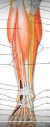
Name the components of the anterior compartment of the lower leg.

A

Extensor muscles: 1.) tibialis anterior 2.) extensor digitorum longus 3.) extensor hallucis longus 4.) fibularis (peroneus) tertius Anterior tibial artery and veins Deep fibular (peroneal) nerve [Plate 510]

2
Q

Name the components of the lateral compartment of the lower leg.

A

Fibularis (peroneus) longus muscle Fibularis (peroneus) brevis muscle Superficial fibular (peroneal) nerve [Plate 510]

3
Q

Name the components of the superficial posterior compartment of the lower leg.

A

Superficial flexor muscles: Soleus Gastrocnemius Plantaris (tendon) [Plate 510]

4
Q

Name the components of the deep posterior compartment of the lower leg.

A

Deep flexor muscles: Flexor digitorum longus Tibialis posterior Flexor hallucis longus Popliteus Posterior tibial artery and veins

5
Q

Name the 4 muscles which make up the quadriceps femoris (from lateral to medial)

A
  • Vastus lateralis
  • Rectus Femoris
  • Vastus intermedius (deep to rectus femoris)
  • Vastus medialis
6
Q

Name the muscles which comprise the anterior thigh muscle group.

A

From lateral to medial

  • Quadriceps:
    • vastus lateralis
    • rectus femoris
    • vastus intermedius
    • vastus medialis
  • Sartorius
  • IIiopsoas
7
Q

origin of the vastus medialis

A

intertrochanteric line, medial lip of linea aspera of femur [From Netter’s Anatomy, Table 7]

8
Q

insertion of the vastus medialis

A

base of patella and to tibial tuberosity via patellar ligament [From Netter’s Anatomy, Table 7]

9
Q

innervation of vastus medialis

A

femoral nerve [From Netter’s Anatomy, Table 7]

10
Q

main action of vastus medialis

A

extends leg at knee joint [From Netter’s Anatomy, Table 7]

11
Q

blood supply of vastus medialis

A

femoral and profunda femoris arteries

12
Q

origin of the vastus lateralis

A

greater trochanter, lateral lip of linea aspera of femur

13
Q

insertion of the vastus lateralis

A

base of patella and to tibial tuberosity via patellar ligament

14
Q

innervation of the vastus lateralis

A

femoral nerve

15
Q

main action of the vastus lateralis

A

extends leg at knee joint

16
Q

blood supply of the vastus lateralis

A

lateral circumflex femoral and profunda femoris arteries

17
Q

origin of the vastus intermedius

A

anterior and lateral surfaces of body of femur

18
Q

insertion of the vastus intermedius

A

base of patella and to tibial tuberosity via patellar ligament

19
Q

innervation of the vastus intermedius

A

femoral nerve

20
Q

main action of the vastus intermedius

A

extends leg at knee joint

21
Q

blood supply of the vastus intermedius

A

lateral circumflex femoral and profunda femoris arteries

22
Q

The ________ ________ is the only extensor of the knee joint.

A

quadriceps femoris [From https://www.kenhub.com/en/videos/vastus-medialis-3d-anatomy]

23
Q

What does musculus rectus femoris mean?

A

straight muscle of the thigh [From https://www.kenhub.com/en/videos/rectus-femoris-muscle-3d-anatomy]

24
Q

The femoral nerve originates from the lumbar plexus, specifically the ________ ____ of the ___ through ___ lumbar nerves.

A

anterior rami 2nd through 4th lumbar nerves [From https://www.kenhub.com/en/videos/rectus-femoris-muscle-3d-anatomy]

25
The rectus femoris has a double origin on the \_\_\_\_\_. The first is a straight tendon which arises from the ________ \_\_\_\_\_\_\_\_ _____ \_\_\_\_\_, while the other is a reflected tendon which arises just superior to the \_\_\_\_\_\_\_\_\_\_.
ilium anterior superior iliac spine acetabulum
26
The rectus femoris muscle is the only member of the quadriceps femoris group that crosses both the ___ and ____ joints.
hip and knee [From https://www.kenhub.com/en/library/anatomy/the-quadriceps-femoris-muscle]
27
Just like the femur, the proximal end of the tibia is formed by two condyles. On each of these condyles, you have an articular area which articulates with the corresponding condyle of the femur, and these are also collectively known as the ______ \_\_\_\_\_\_\_ especially in the clinical setting. The two areas are separated by a non-articular, irregular intercondylar area with the rugged raised area called the ________ \_\_\_\_\_\_\_\_.
tibial plateau intercondylar eminence [From Knee Joint video transcript, https://www.kenhub.com/en/start/anatomy-knee-joint]
28
The patellar ligament is strong flat band, which is actually just a continuation of the ________ \_\_\_\_\_\_\_ \_\_\_\_\_\_. It stretches from the apex of the patella to the tibial tuberosity. It’s joined medially by the medial patellar retinaculum and laterally by the lateral patellar retinaculum.
quadriceps femoris tendon [From Knee Joint video transcript, https://www.kenhub.com/en/start/anatomy-knee-joint]
29
A few bursae are actually continuous with the synovial cavity and that means that if there is an infection in one of the bursae, especially the large suprapatellar bursa, it can spread into the knee joint.
30
The sartorius muscle is a long, slim, superficially running muscle, formally belonging to the extensors of the thigh. The sartorius muscle moves both the hip and knee joint. Even though it is located in the anterior compartment of the thigh, it should not be confused as an _________ of the thigh.
extensor
31
Name the 4 muscle groups which comprise the gluteal muscles.
Gluteus maximus Gluteus medius Gluteus minimus Tensor fasciae latae
32
The posterior hip musculature can be found underneath the gluteal muscles. The four muscles which make up the posterior hip musculature are:
Piriformis Internal and external obturators Superior and inferior gemelli Quadratus femoris
33
The gluteus maximus is the ________ extensor of the hip joint, as well as the ________ rotator of the hip joint.
strongest external
34
The gluteus maximum originates from 3 places:
the sacrum, the ilium, and the sacrotuberous ligament.
35
The caudal fibers of the gluteus maximus insert at the....
....gluteal tuberosity of the femur.
36
The cranial fibers of the gluteus maximus insert on the....
....iliotibial fascia (the iliotibial tract on the fascia lata).
37
The gluteus maximum originates from 3 places:
the sacrum, the ilium, and the sacrotuberous ligament.
38
The caudal fibers of the gluteus maximus insert at the....
....gluteal tuberosity of the femur.
39
The cranial fibers of the gluteus maximus insert on the....
....iliotibial fascia (the iliotibial tract on the fascia lata).
40
What are the functions of the gluteus maximus? What do the cranial fibers vs what the caudal fibers do?
extension of the hip external rotation of the hip stabilization of the hip joint The cranial fibers abduct. The caudal fibers adduct.
41
The gluteus maximus is innervated by the...
...inferior gluteal nerve which comes from the sacral plexus.
42
what muscles insert onto the pes anserinus
the pes anserinus looks like a duck foot and the mneumonic is SGT (picture: marching duck sargeant) which stands for 1. sartorius 2. gracilis 3. semiTendinosus
43
what is the function of the sartorius muscle?
Hip joint: thigh flexion, thigh abduction, thigh external rotation Knee joint: leg flexion, leg internal rotation all are shown in the attached picture.. its basically all the actions you use to look for gum on the bottom of your shoe.
44
The femoral nerve comes from what lumbar innervations?
L2-4 mn: 2 legs, 4 quad muscles femoral nerve is the one in NAVL mn.
45
femoral triangle
bordered by the sartorius, inguinal line, and gracilis clinical significance: access to heart, injection site
46
origin and insertion of rectus femoris
anterior inferior iliac spine (NOT ASIS like the sartorius) shown in orange (attached).. notice that it crosses two joints (hip joint and knee joint) wherease all the other quadriceps muscles only cross the knee joint
47
what are the muscles of the medial thigh
pectinuius, adductor longus, gracilis
48
origin, insertion, and innervation of adductor longus
origin: pubic symphysis insertion: mid shaft of humerus via aponeurosis innervation: obturator nerve
49
adductor magnus
the femoral artery and vein go through the adductor hiatus
50
what are the occluded nerves
green = femoral nerve blue = obturator nerve
51
what muscle is highlighted
pectinius
52
lateral leg muscles (2)
fibularis longus and fibularis brevis
53
what 2 tendons are shown?
the fibularis longus and fibularis brevis
54
what muscle is shown
adductor magnus (posterior view) - hamstring ischioportion
55
what muscle is occluded
quadratus femoris
56
what muscle is occluded
inferior gemellus
57
what muscle is occluded
superior gemellus
58
what muscle is occluded
piriform muscle
59
what muscle is occluded
tensor fasciae latae
60
what muscle is shown
adductor magnus (anterior view) - pubofemoral portion
61
what muscle is occluded
adductor magnus (pubofemoral part) in posterior view.
62
what are the actions of the adductor magnus muscle (3)
\*some sources also say it does lateral rotation but this is in contention
63
where do the superior and inferior gemellus originate and insert?
64
what muscle is shown
tensor fasciae latae
65
obturator externus vs obturator internus comparison
66
what muscle is shown
obturator internus
67
what muscle is shown
adductor longus
68
what muscle is shown
adductor brevis
69
what muscle is shown
obturator externus
70
what muscle is shown
pectinius
71
vastus medialis versus vastus lateralis muscles
72
what muscle is shown
vastus intermedius
73
what muscle is shown and what is its function
biceps femoris action: flexion of knee joint ("flirty foot pop")
74
what muscle is shown
semitendinosus muscle (one of the pes anserinus muscle insertions)
75
semitendinosis versus semimembranosus muscles
semimembranosus (right) is deep to semitendinosus (left)
76
what muscle is shown and what is its function
ileopsoas function: flexion of hip past 35 degrees and some adduction
77
what muscle is shown in green and where does it insert
soleus inserts onto calcaneus
78
what muscle is shown in green and where does it insert?
tibialis posterior inserts onto the plantar surface of the tarsal bones of the foot, mainly onto the tuberosity of navicular bone and the medial cuneiform bone.
79
flexor digitorum longus versus flexus hallucus longus
left = flexor hallucis longus right =flexor digitorum longus
80
what muscle is shown (black)
fibularis tertius
81
what pathology is shown
ACL tear
82
Acetabular labrum is a fibrocartilaginous rim that increases the articular area by
almost 10%
83
what position are the ligaments of the hip best suited for?
quadruped position
84
how would a patient present if she had been in a head on collision and sustained a posterior hip dislocation
85
what is this and what does it innervate?
deep fibular nerve --\> innervates all muscles of the anterior compartment of the leg (meaning that it excludres the fibularis longus and fibularis brevis, which are lateral compartment muscles)
86
Other than the _____________ and the \_\_\_\_\_\_\_\_\_\_, which are innervated by the superficial fibular nerve, the anterior compartment muscles of the leg are innervated by the deep fibular nerve
_fibularis longus, fibularis brevis_ Deep fibular nerve: tibialis anterior, extensor digitorum longus, extensor digitorum brevis, extensor hallucis longus, extensor hallucis brevis and fibularis tertius muscles.
87