Malabsorption, Celiac disease, irritable bowel syndrome, IBD, Diverticular Disease Flashcards

(97 cards)

1
Q

What is the hallmark of malabsorption?

A

Steatorrhea

2
Q

Malabsorption breadly refers to problems absorbing?

A
  1. fat
  2. fat/water sol. vits
  3. proteins
  4. carbs
  5. lytes
  6. minerals
  7. H20 loss
3
Q

What are the most commonly encountered malabsorption syndromes in th US?

A
  1. Pancreatic insufficiency
  2. Celiac disease
  3. Crohn Disease
4
Q

Malabsorption results from disturbance in at least one of the phases of nutrient absorption, which there are four of. What are they?

A
  1. Intraluminal digestion
  2. Terminal digestion
  3. transepithelial transport
  4. Transport into lymphatics
5
Q

Whipple disease blocks nutrient transport into lymphatics, and is a •Chronic, relapsing multisystem illness involving the GI tract (diarrhea, steatorrhea, malabsorption) and distant sites (arthritis, lymphadenopathy).

The disease is characterized by what three symptoms?

What causes whipple disease?

A
  • Disease characterized by weight loss, diarrhea, and polyarthritis; occur together at presentation in 75% of cases
  • Gram positive bacillus - Tropheryma whippleii
6
Q

What population is whipple disease most common in?

How do we treat?

A
  • Usually white males ages 30-49 years (10:1 male predominance)
  • Treat successfully with antibiotics
7
Q

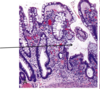
What are the three characteristic symptoms associated with the attached finding of a 40 y/o white male?

A

Whipple disease - note the distended foamy macrophages in lamina propria that contain T. whippelii.

  1. weight loss
  2. diarrhea
  3. polyarthritis
8
Q

On inspection you note the distended foamy macrophages in the lamina propria of this image, and you know that this, combined with the patients weight loss, diarrhea and polyarthritis means the patient has?

Which means we are looking at what part of the GI tract that this organism commonly infects?

A

Whipple disease

small intestine (usually proximal)

9
Q

What stain have you used to deted these T. whippleii?

A

PAS-D

10
Q

Celiac disease is an inflammatory disease of small bowel in genetically susceptible individuals. What are the genetic predispositions identified?

A

strong association with certain HLA types

  • HLA-DQ2
  • DQ8
11
Q

In addition to a genetic predisposition to celiac disease, there must also be an inciting agent exposure. What types of exposures are we talking about here?

A
  1. gluten (technically the etoh-soluble gliadin)
    1. from wheat, barley, oat, rye
12
Q

What mediates the inflammation in celiac disease?

A

T-cells: Altered peptides are presented to CD4+ T cells in mucosa, leading to an increase in CD8+ intraepithelial lymphocytes that produce proinflammatory cytokines

13
Q

Celiac is most common in the following age ranges…

  • Adults present usually between the ages of 30-60
  • Children present usually between the ages of 6 and 24 months.

What are two conditions in which a patient can have it but not see symptoms?

A
  1. Silent
  2. Latent
14
Q

What defines a silent celiac disease?

A
  • Positive serology
  • Positive villous atrophy
  • No symptoms
15
Q

What defines a latent celiac disease?

A
  • positive serology
  • no villous atrophy
  • may have symptoms
16
Q

Celiac disease presents typically with systemic signs of malapsorbtion. What will 10% of these patients also have?

A

dermatitis herpetiformis

17
Q

The vague abdominal discomfort and abdominal bloating can lead to a mistaken diagnosis of?

A

Irritable bowel syndrome

18
Q

What are two things that celiac disease increases the risk for?

A
  • enteropathy-associated T-cell lymphoma
  • small intestine adenocarcinoma
19
Q

In the past, we referred to a clinical presentation with weight loss, steatorrhea, diarrhea and nutritional deficiency as?

A

Typical celiac disease

20
Q

In contrast to typical celiac disease, now atypical celiac disease is more common. What are the ssx associated with atypical celiac disease?

A
  • anemia
  • fatigue
  • abdominal bloating/discomfort
  • osteoporosis
  • infertility
21
Q

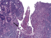
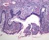
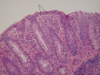
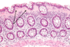
What is shown in each image?

A

Left: normal mucosa with villi

Right: atrophic mucosa without villi, d/t celiac disease

22
Q

Your patient presents with the attached skin lesions, your tissue sample is shown also. What is this called?

What is causing the lesions?

What condition does this person likely also have?

A
  1. Dermatitis herpetiformis is a chronic blistering intensely itchy skin condition, characterised by blisters filled with a watery fluid.
  2. Deposition of immunoglobulin A (IgA) in upper papillary dermis
  3. Celiac disease - 15-25% develop this
23
Q

Tropical sprue is of an unknown etiology and typically affects adults. What is the typical presentation?

A
  1. Diarrhea
  2. soreness of tongue (B deficiency)
  3. Weight loss
  4. Steatorrhea
24
Q

Autoimmune enteropathy is rare, and presents with what two main things?

A

•Intractable diarrhea and malabsorption associated with circulating gut autoantibodies and a predisposition to autoimmunity

25
Autoimmune enteropathy affects infants, young children and (rarely) adults evidence suggests a hyperactive immune state due to a defect in?
Regulatory T-cells.
26
toimmune enteropathy diagnostic criteria includes the presence of chronic diarrhea and malabsorption along with what five other criteria?
1. Partial or complete villous blunting 2. minimal intraepithelial lymphocytosis 3. deep crypt lymphocytosis and apoptotic bodies 4. Antienterocyte antibodies 5. anti-goblet cell antibodies
27
Does the absence of antibodies rule out autoimmune enteropathy?
No
28
Dissacharidase (lactase) deficiency can be either _______________ or \_\_\_\_\_\_\_\_\_\_\_\_\_\_\_\_\_\_.
Congenital or acquired
29
What is the inheritance pattern of congenital lactase deficiency?
AR
30
Acquired lactase defiiency is common, particularly among what populations?
Native americans african americans
31
In dissacharidase deficiency, incomplete breakdown of lactose leads to what?
Osmotic diarrhea from unabsorbed lactose
32
•Abetalipoproteinemia is a rare, _______________ disorder, characterized by fat malabsorption.
autosomal recessive
33
Abetalipoproteinemia is a rare, autosomal recessive disorder, characterized by fat malabsorption, and involves a mutation in what? What is the consequence of this mutation?
•Mutation in the microsomal triglyceride transfer protein (MTP) -Failure to assemble and export lipoproteins
34
What steps should be taken in the treatment of abetalipoproteinemia?
Dietary modification replacement of fat soluble vitamins
35
Based on the findings shown here, what is the likely mutation involved in this disease process affecting the small intestine?
Abetalipoproteinemia - microsomal trigylceride transfer protein (MTP) is mutated.
36
IBS is characterized by the presence of chronic abdominal discomfort or pain associated with changes in bowel habits. In GI practices one third of patients have functional GI disorders, IBS being the most common diagnosis. What is the cause?
Not known
37
Peak prevalence of IBS is between 20 and 40 years of age and demonstrates a significant female preponderance. What must you do to make the dx?
Rule out other causes such as enteric infection or IBD
38
What are the criteria that need to be met for IBS?
At least 3 days/month in the last 3 months associated with 2 or more of the following: * Improvement with defecation * Onset associated with a change in frequency of stool * Onset associated with a change in form (appearance) of stool
39
What are the two types of IBD?
Chron disease Ulcerative colitis
40
Inflammatory bowel disease (IBD) is a chronic condition resulting from inappropriate?
Mucosal Immune activation
41
Chron disease may involve any area of the GI tract and is frequently?
Transmural
42
Ulcerative colitis is limited to what two areas of the GI tract? What layers of the GI tract?
Colon and rectum mucosa and submucosa
43
Chron Disease is diagnosed most often among persons 15 to 30 years of age who complain of what three symptoms?
_Intermittent attacks of:_ 1. Diarrhea 2. Fever 3. Abdominal pain
44
What does Crohn disease most commonly affect? give 3
1. **Distal Ileum** 2. Colon 3. Can involve mouth to anus (dear lord)
45
What does Chron disease put a patient at increased risk of?
Adenocarcinoma risk increased 5-6 fold
46
Crohn disease primarily affects western nations, whites more than blacks and jews more than non-jews. Is more common in females than males and typically peaks in what decade(s)?
•Disease peaks in 2nd to 3rd decade; minor peak 6th to 7th
47
What percent of the time does Chron disease affect: Small bowel only (Ileum) Small bowel and colon Colon only ????
–40% small bowel only (Ileum) –30% small bowel and colon –30% colon only
48
What are some characteristic gross pathological features of Crohn Disease? 7
1. Non-contiguous mucosal inflammation 2. Fibrosing Strictures 3. Perforation and abscess 4. Adhesions 5. Thickened intestinal wall 6. fistula 7. creeping fat
49
What are some characteristics of the skip leasions associated with Crohn disease? 4
1. aphthous ulcers 2. serpinginous ulcers 3. fissures 4. cobblestone mucosa
50
What is shown in this image? What is it characteristic of?
Creeping fat Crohns disease
51
What gross pathological feature of Crohns disease are we looking at?
Aphthous ulcer
52
How would you describe the gross finding here? What is it associated with?
Cobblestone appearance Assoc: Crohn disease
53
What is this characteristic feature of Crohns?
Terminal ileum stricture with thick fibrotic walls.
54
What are six hisopathological features of Crohn disease?
1. Non-necrotizing **granuloma** 2. **transmural** lympoid aggregates 3. **Fissures** - transmural inflammation with deep ulcers 4. Crypt destruction/distortion 5. submucosal fibrosis 6. Mucosal ulcers
55
What is indicated by the lesions in this tissue sample taken from a patient with a likely diagnosis of Crohns?
Double arrow - Transmural inflammation Solo right arrow - Granuloma
56
What classic histopathological finding that is associated with Chrons is seen here?
Crypt distortion (resembling animals... here an antelope)
57
What is the characteristic histopathological feature of Crohns shown here?
Non-necrotizing granuloma
58
This image depicts one of the hallmarks of Crohn's disease. Which one?
Transmural lymphoid aggregates
59
What does this histology sample from a patient with Crohn disease show?
Aphthous lesions are small erosions that are associated with a neutrophilic infiltrate
60
What hallmark histopathological feature of Crohns is depicted here?
Fissuring ulcer extending into the submucosa
61
Ulcerative colitis affects approximately 800,000 people in the United States. Common symptoms include diarrhea, rectal bleeding, passage of mucus, urgency, and abdominal pain. Following the initial flare up what are the two typical courses?
40-65% have intermittent course 5-10% have chronic continuous course
62
Onset of disease peaks from ages 20-25 yrs ulcerative colitis is more common among nonsmokers than among current smokers. This is a systemic disorder associated with what 4 other conditions?
1. Migratory polyarthritis 2. ankylosing sponylitis 3. uveitis 4. primary sclerosing cholangitis
63
What antibody is present in 41 to 73% of UC patients?
pANCA
64
Ulcerative colitis increases the risk of what cancer? In what patient grou in particular?
Adenocarcinoma Risk is greatest in those with pancolitis of more than 10 years (20-30 fold risk)
65
UC tends to be more severe distally and progressively less severe more proximally. The initial presentation can vary. What are three common initial locations that this presents?
**–45% of patients with UC have disease limited to the rectosigmoid** –35% have disease extending beyond the sigmoid **–20% of patients have pancolitis.**
66
In UC there is a sharp transition between diseased and uninvolved segments of the colon. What does it always involve? What can occur in 5% of severe UC flares?
Always involves rectum Toxic megacolon in ~5% of cases
67
What is this?
Ulcerative Colitis
68
What do the arrows indicate? What condition is this?
_Ulcerative Colitis_ Top - Pseudopolyps Left four - Sharp demarcation from ulcerated tissue to normal Bottom three - Coalescence of mucosal erosion/ulcer
69
What is depicted by the arrows in this patient's case of UC?
Psuedopolyps
70
What is shown here, that was taken from an unlucky UC patient?
Toxic megacolon
71
Inflammation in UC is typically confined to the mucosa. What are some findings expected in UC hisopathology?
1. Cryptitis/crypt abscesses 2. gland destruction/distortion 3. Ulcers 4. pseudopolyps 5. Mucosa uniformly involved 6. Absence of granulomas
72
What is shown here? What is this indicative of?
Flask shaped ulceration of the mucosa - Ulcerative Colitis
73
What finding common to UC is depicted here?
Flask shaped ulcer again
74
What characteristic histopath feature of UC is shown here?
Crypt Abscess
75
What characteristic histopath finding for UC is shown here?
Crypt architectural distortion
76
What is happening in this sample from a UC patient?
Red arrows= non-dysplastic crypts White arrows = high grade dysplasia
77
What type of bowel movements are seen in crohn's?
Watery diarrhea Constipation
78
What kind of pattern of damage do you see in chrons?
Segmental with rectal sparing
79
What kind of damage pattern do you see in UC?
Extends proximally from the rectum, no skip lesions
80
Which involves the Ileum, Crohns or UC?
Crohns
81
Do you see strictures in UC and Crohns?
In crohns, yes commonly In Ulcerative colitis - rarely ~5%
82
Describe the inflammation for both: Ulcerative colitis Crohns
* UC: Diffuse mucosal inflammation/ulceration * Crohns: Focal inflammation and ulceration
83
In which are granulomas common, UC or Crohns?
Crohns
84
In which condition do you see sersitis/adhesions, UC or Crohns?
Crohns
85
D/t the extensive pathologic and clinical overlap between ulcerative colitis and Crohn disease definitive diagnosis is not possible in approximately what percent of IBD patients? What are these cases called?
Indeterminate colitis - 10% of IBD patients.
86
Lymphocytic colitis affects primarily female middle aged patients and appears normal on endoscopy. What is the typical compaint? tx?
Chronic watery diarrhea for months to years Corticosteroids
87
What is indicated by the arrows? Condition?
Intraepithelial lymphocytes Lymphocytic colitis
88
This colon was stained for CD3, what does the patient have?
Lymphocytic colitis
89
This patient complains of chronic watery diarrhea for months to years, is middle aged/old female more than male, and endoscopy was normal. What is this?
Collagenous Colitis
90
Diverticular disease refers to acquired pseudo-diverticular outpouchings. Where are they most common?
Most common in the sigmoid colon
91
\>80% of diverticular disease patients are asymptomatic. Inflammation of diverticula is called diverticulitis (pain, leukocytosis and fever). While uncommon, perforation can happen and results in? 3
1. **pericolonic abscesses** 2. sinus tracts 3. peritonitis
92
What is shown here?
Diverticular disease on endoscopy
93
What is shown on this barium enema?
Diverticular disease
94
What does this barium enema reveal?
Diverticular Disease
95
Where is the abnormality in this image? What is it?
Diverticular Disease See arrows in image
96
What is shown here?
Diverticulum - protrusion of mucosa and submucosa through the muscle wall.
97