Mechanics Flashcards
(11 cards)
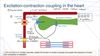
Cariac Myocytes ( explain the function and the sequence of events which lead to contraction and relaxation)
Cardiac Myocytes are Ventricular cells;
- 100μm long and 15μm wide
- they have a strained structure because they are invaginatated by T-tubules –> spaced approximatelly every z-line and carry out surface depolirazation
Sarcoplasmic reticulumn (SR);
- main store for Ca2+
- overlies microfilamnet and is close to T-tubules
- 4% of the cell volume
Excitation- Contraction
Excitation;
- L-type Ca2+ channel: senses depolarization and opens up–> allowing Ca2+ to enter the SR
- Some of the calcium will go to the microfilaments but MOST of it will bind to the SR Ca2+ release channel causing conformational change and allow calcium that is stored INSIDE the SR to efflux into the cytoplasm. ( calcium induced calcium release since Ca2+ is needed for calcium release )
- Ca2+ binds to troponin, actin, myosin etc —> contraction
DDifferenences with SKELETAL muscle; because there is no need for Ca2+ environment there is a mechanical linkage betweem the L-type Ca2+ channel and SR Ca2+ release channel
So,
- depolarization
- L-type Ca2+ channel
- activation of SR Ca2+ release channel
- Ca2+ effluxes to cytoplasm
Removal of Ca2+ from the cytoplasm –> allow cell to relax
- Ca2+ is pumped back to the cytoplasm ( against the concentration gradient) using Ca2+ ATPase (ATP is used)
- Na+/ Ca2+ exchanger; the calcium that triggers the SR is removed using the Na+ concentration
Define Isometric, Isotonic, Preload, Afterload
Isotonic; muschle fibers DO NOT change in lenght
Isotonic; there is SHORTENING of the muscle fiber
Preload; the weight by which the muscle is stretched before its stimulated to contract –> the more you stretch the produced force increases
Afterload; weight which is not apparent to muscle during resting stase –> increased afterload results in decreased shortening
Law of Laplace (cardiac): recall and explain the relationship of the law of Laplace to cardiac mechanics
Stork; is the work performed by the heart to eject blood into the aorta and the pulmonary artery
Law of Laplace; If pressure within a cylinder is held constant while radius increases TENSION is also increased.
T=PxR, wall thickness can be considered ( in this case we divide with n )
Starling’s Law of the Heart: explain the mechanisms of Starling’s Law of the heart
This law is the result of the observation that as filling of the heart was increased, the force contraction also increased.
Definition; Increased diastolic fibre lenght increases ventricular contraction
factors that might be causing this observation
- changes in microfilaments cross- bridges that interact as you stretch –> more cross bridges –> decrease lenght–> actin filaments overlap
- Changes to Ca2+ sensitivity of the myofilaments ( the longer, the more sensitivity)
Hyp 1.
- longer filaments; more force ( more sensitive)
- shorter filaments; less force
–> conformation to troponin C
Hyp 2.
- with stretch the spacing between myosin and actin filaments ( lattice spacing) decreases
- with lattice spacing decreasing the propability of cross bridges increases and there is therefore more force for the same Ca2+
Phases of the cardiac cycle; electrical and mechanical events, valve movements and points where this occurs on pressure-volume loops
Phases; two main phases
- Diastole;
- ventricular relaxation ( filled with blood )
- lasts approximately 2/3 of each beat
- split into 4 phases
- Systole;
- Ventricular contraction ( ventricles generate pressure to eject blood into the arteries)
- Lasts approximately 1/3 of each beat
- split into 3 phases
- Atrial systole;
- P-wave on ECG
- Atria fills up with bood due to passive filling ( driven by pressure gradient)
- Atria contract to fill up the volume of the ventricles
- Pacemaker ( at the top right ) stimulates the electrical impulse —> depolirazation of the atria
- 4th abnormal heart soud can be heard here
- Isovolumentric contraction
- QRS complex on the ECG
- In this interval the AV valves close
- contraction of ventricles with NO change in volumes (contracring against closed valves)
- It’s the 1st sound ( lub) made by the closing of the valves
- depolirization of ventricular cells
- Rapid ejection
- Defined by the opening of the aortic and pulmonary valves
- When ventricles contrac their inner pressure exceeds the aortic pressure and with the opening of the semi-lunar valves blood is pumped out and the volumes of the ventricles decrease
- There is no heart sound since no valves are closed ( just opened)
- Example of isotonic contraction since there is a decrease in volume
- Reduced ejection;
- This pahse marks the end of systole ( ventricles beging to repolarize—> T-wave on ECG)
- Reduced pressure gradient results to the closure of the semi-lunar valves
- Blood flow from ventricles decrease and ventricular volume decreases more slowly
- As pressure in ventricles decreases it falls bellow of that in arteries resulting in blood flowing backwards and semilunar valves to close
- Isovolumetric relaxation;
- Causes the 2nd sound (dub) due to the closure of the semilunar valves
- There is no change in volume
- Even though the semi-lunar valves shut the AV valves remain closed until ventricular pressure drops bellow atrial pressure
- There is a slight rise in atrial pressure
- Rapid passive filling
- Isoelectric ( flat) phase on ECG
- Once AV valves are open blood falls rapidly from the atria to the ventricles ( passivly)
- This is usually the time when a 3rd abnormal sound can be heard due to tuburlent ventricular filling ( severe hypertention or mitral incompetence)
- Reduced passive filling;
- Can be called the diastasis phase
- Venrticles are filled more slowly without the contracton of the atria
- Depinding on how much they fill this will determine the pre-load and therefore the after-load ( important for the ventricular pressure to be higher than the aortic)

Define; End-systolic volume, End-diastolic volume, Stroke volume, ejection fraction ( state normal values)
End-diastolic volume; the volume of blood into the ventricle just before they are about to contract ( normal= 108ml)
End-systolic volume; the blood which is left in the ventricles after their contraction (normal =36ml)
Stroke volume; the total amount of blood which is pushed out in one beat. Can be calculated by Stroke(ml)= End Diastolic- End Systolic ( normal= 72ml)
Ejection fraction(%); Amount of blood pushed out of the heart in relation to the amount of blood in the ventricles. (normal= 67%)
Can be calculated; EF= 100x ( stroke volume/ End diastolic volume)
Clinical sign of how the heart is contracting ( ventricles)
Pressure volume loops: draw cardiac pressure-volume loops

Pressure volume loops: draw cardiac pressure-volume loops (giving more information to preload and afterload)

Pressure volume loops: draw cardiac pressure-volume loops ( how preload and afterload influence stoke vvolume)
increase in preload= increases stroke volume
increase in afterload= decreases stroke volume

Cardiac output
Cardiac output= Heart rate x Stroke volume
Stroke volume can be indluenced by
- Pre-load
- After-load
- Contactility; strenght of contraction of the heart ( increased by sympathetic stimulatio–> adrenaline, nor-adrenaline), a simple way to measure it is ejection fraction
increase in contractility results in increased contraction
decrease in contraction results in decreased contraction

Pressures and volumes: define and state normal values for intra-cardiac pressures and volumes
The patterns of pressure changes in the right heart are essentially the same to those of the left
—> The pressure in the right heart and pulmonary circulation are much lower ( peak of systole 25mmHg in pulmonary artery)
Hoewver, they eject the same amount of bloof as the left since they pump it to a lower pressure circuit ( lower afterload)

