Microbial structure Flashcards
• Recognise the structural differences between eukaryotic and prokaryotic cells • Identify the main structural features of bacteria, viruses, protozoa, fungi, helminths, and yeasts. • Describe how bacteria and viruses replicate and how genetic information is transferred between bacterial cells. (31 cards)
What are the 2 basic types of cells?
What do eukaryotic and prokaryotic cells have in common?

Differences in Genetic Material for prokaryotes and eukaryotes
Eukaryotes
Has true nucleus; bound by double membrane
Differences in strucutre between prokaryotes and eukaryotes
Eukaryotes
Cytoplasm filled with large complex collection of organelles
Mitochondria with cristae are “energy centres”
Transcription requires formation of mRNA and movement of mRNA from nucleus to cytoplasm for translation
Prokaryotes
- No membrane bound organelles independent of plasma membrane
- Mesosomes are used in aerobic respiration
- Transcription and translation occur simultaneously
Bacteria - structural components
- Capsule
- Pili (fimbriae) • Flagellae
- Spores
- Slime
- Cell wall

Bacteria - capsule
- Loose polysaccharide structure
- Protectscellfrom phagocytosis
- Protectscellfrom dessication

Bacteria - Pili/Fimbriae
Singular = pilus “hair”
Composed of oligomeric pilin proteins
Appendage used for bacterial conjugation
Forms tube / bridge to enable transfer of plasmids between bacteria
Highly antigenic
Plays role in attachment
- Singular = fimbria (“thread”)
- Not on all bacteria
- May contain lectins which recognise oligosaccharide units on host cells
- Facilitates bacterial attachment to host surfaces
Bacterial adherence to host cell

Bacteria - Flagellae
Organs of locomotion
Single / multiple
Composed of flagellin protein
20nm-thick helical hollow tube
Driven by rotary engine at anchor point on inner cell membrane
Singular = flagellum (“whip”)

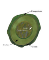
Bacteria - spores
Metabolicallyinertform triggered by adverse environmental conditions
Adapted for long-term survival allowing regrowth under suitable conditions
Hard,multi-layered coats making spore difficult to kill
Common diseases caused by sporing bacteria
Bacteria - slime
Polysaccharide material
secreted by some bacteria growing in biofilms
Protects against immune attack
Protects against eradication by antibiotics

Cell walls - gram staining created by christian gram
G-/G+ differences
The four steps of Gram staining
• Differentiates bacterial species into 2 groups:
–Gram positive (+)
–Gram negative (-)
• Based on chemical and physical properties of the cell walls
Primary stain (crystal violet dye)
– Stains all the bacterial cells purple
Trapping agent (Gram’s iodine) (Mordent)
– Forms CVI complexes in the cell wall (larger than CV so not to be easily washed out of the PGN layer)
Decolourisation (alcohol / acetone)
– Interacts with lipids in cell wall
– Gram negative: loses outer LPS layer; exposes thin inner PGN layer; coloured complexes mainly wash away
– Gram positive: becomes dehydrated and traps the complexes in thicker PGN layer of cell wall
Counterstain (safranin)
– Gram negative: pink / reddish – Gram positive: purple

Cell wall components
Bacterial replication of genome
• Reproduce by binary fission (asexual)
– 1 cell reproduces to give 2 daughter cells
Genetic information found in circular DNA
– Distributed equally between each daughter cell
DNA is a self-replicating molecule
– Can make an exact copy of itself before cell division
• Circular DNA – replication starts at “origin”
– Replicates in 2 directions (bi-directional replication)
– 2 replication forks split off from origin and meet at bottom

What is the bacterial growth cycle?
What are the four stages of the bacterial growth cycle?
- During active growth, the number of cells continuously doubles at specific time intervals
- Each binary fission takes a specific duration of time (depends on bacterium)
1 - 21 = 22 = 23 = 24 = 25 = 2n
n = number of generations
1 - lag phase
- Represents the period of active growth (ie. in size, not number)
- Bacteria prepare for reproduction ie. Synthesising DNA and enzymes for cell division
2 - log/ exponential phase
• Cells divide at maximum rate
- Uniform replication
- Graph line is almost straight
3 - stationary phase
- Cessation of growth
- Exhaustion of nutrients
- Accumulation of inhibitory end products of metabolism or oxygen availability
- Number of cells dying balances the number of new cells, so population stabilises
4 - death phase

Bacterial recombination
• Conjugation
– One bacterium connects itself to another through the pilus
– Genes are transferred from one bacterium to the other through this tube
• Transformation
– some bacteria are capable of taking up DNA from their
environment • Transduction
– involves the exchanging of bacterial DNA through bacteriophages.
How to we identify bacteria?
• Bacteria are identified using a series of physical, immunological or molecular characteristics
– Gram stain: Gram positive / negative
– Cell shape: cocci; bacilli; helical / spiral
– Atmospheric preference: aerobic, anaerobic, microaerophilic
– Key enzymes – Fastidiousness
What are some important bacteria?
Gram Stain
Gram pos - cocci - staphylococcus aureus
Gram negn - cocci - neisseria meningitidis
Gram neg - coccibaccili - haemophilus influenzae
Gram pos - bacilli - listeria monocytogenes
Gram neg - bacilli - escherichia coli
Gram neg - spiral - helicopter pylori
Cocci - spherical
Bacilli - rod- shaped
Spiral - helical rod

Viruses
Viral structural compnents
Nucleic acid
- Double stranded or single stranded
- Deoxyribonucleic acid or ribonucleic acid
ds dna, ss dna, ds rna, ss rna
Capsid
- Protein coat / shell
- Composed of protein subunits – capsomeres
- Capsomeres consist of aggregated protomeres
• Various shapes of capsids – Rod-like – Polyhedral – Complex
Envelope
- Amorphous structure surrounding some viruses
- Composed of lipid, protein & carbohydrate
Spikes
• Glycoprotein projections arising from envelope
- Highly antigenic
- May have enzymatic, adsorption or haemagglutin activity
Viral replication
- Intracellular obligate parasites
- Uses host’s cellular machinery to replicate
- Produces many progeny which leave host to infect other cells in the organism
- May infect any type of cell – Animal
– Plant
– Bacterial
Viral replication 6 steps
Step 1 - Adsorption
- Virus binds to host cell
- Highly specific
Step 2 - Penetration
• Virus injects its genome into host cell
• Occurs by
– Fusion
– Binding
– Ingestion
Step 3 - Replication
• Capsid digested by proteolytic enzymes
• Viral genome replicates using the host’scellular machinery
Step 4 - Assembly
• Viral components and enzymes are produced and begin to assemble
Step 5 - Maturation • Virus fully develops
Step 6 – Release of Naked Viruses
- Occurs at site of nucleic acid replication
- Viral enzymes break down bacterial cell wall
• RNA viruses released as they are produced
• DNA viruses expelled from the host cell: – as cells autolyse
– in inclusion bodies
Step 6 – Release of Enveloped Viruses
- Viruses migrate to either plasma membrane or nuclear membrane
- Envelopes formed around nucleocapsids by“budding” of cell membrane
- Slow continuous release of mature viral particles • No inclusion bodies
protozoa


