Microbiology Immunology Flashcards

(51 cards)

1
Q

Name and describe the three lines of defense

A

Lines of Defense

  • Skin and mucous membranes
  • Innate (natural) immunity
    • Functions immediately after microbial infiltration
    • Nonspecific targeting of antigens
    • No memory: Does NOT arise from previous infection or vaccination
      • Natural killer (NK) cells
      • Polymorphonuclear neutrophils (PMNs)
      • Macrophages
      • Complement system
      • Nonspecific enzymes (cytokines, lysozyme, etc)
  • Acquired (adaptive) immunity
    • Functions days after microbial infiltration
    • Specific targeting of antigens
    • Exhibits diversity: Responds to millions of unique antigens
    • Memory: improves on multiple exposure to microorganism
    • Two types of acquired immunity
      • Cell-mediated: T cells
      • Antibody-mediated (humoral): B cells, antibodies
2
Q

Name and describe the two classifications of acquired immunity

A

Classification of Acquired Immunity

  1. Active
    • Mediators: Antibodies and T cells
    • Occurs after exposure to foreign antigens
    • Slow onset (days)
    • Lasts a long time (years)
    • Ex: Previous microbial infection, Vaccination with live attenuated or killed antigens
  2. Passive
    • Mediators: Antibodies
    • Occurs after exposure to preformed antibodies from another host
    • Immediate onset
    • Short duration (months)
    • Ex: Pregnancy (IgG), Breast feeding (IgA), Vaccination with antibodies
3
Q

Name and describe what an antigen is and examples/characteristics listed below:

Immunogen

Hapten

Superantigen

Epitope

Adjuvant

A

Antigens

  • Most are proteins, but many are also polysaccharides, lipoproteins, and nucleoproteins
  • Immunogens: Molecules that react with antibodies to induce an immune response. All immunogens are antigens, but not all antigens are immunogens
  • Hapten: An antigen that cannot elicit an immune response on its own (Can’t activate Th cells); it must be bound to a carrier protein
    • Many drugs are haptens. ie. Penicillin
  • Superantigen: Activates a large number of Th cells at one time. (Eg. TSST)
  • Epitopes: The specific antibody-binding site on an antigen
  • Adjuvant: A molecule that enhances the immune response to an antigen
    • Added to a vaccine to decrease absorption and increase the effectiveness
    • Elicits stronger T and B cell response
    • Eliminates the need for repeated boosters
4
Q

Explain the difference between cell-mediated and antibody-mediated immunity

A
  • Cell-Mediated Immunity
    • Host defense:
      • Viruses,
      • Bacteria (intracellular),
      • fungi
      • Protozoa
    • Mediators:
      • T cells
      • NK cells
      • Macrophages
    • Ex: Intracellular infections, Granulomatous infections, Tumor suppression, Organ transplant rejection, Graft vs. host reactions, Type IV (delayed) hypersensitivity
  • Antibody-mediated (humoral)
    • Hoste defense:
      • Bacteria
      • Some viruses
      • Helminths
    • Mediators:
      • B cells
      • Antibodies
    • Examples: Bacterial toxin-induced infections, Autoimmune reactions, Type I, II, III hypersensitivity
5
Q

Explain Freund’s adjuvant

A

Freund’s adjuvant:

  • Inactivated M. tuberculosis suspended in lanolin and mineral oil
  • Functions as an immunopotentiator (booster)
  • Used for research as it is toxic in humans
6
Q

State the cellular components of the immune system

A

T cells

B cells

Natural killer (NK) cells

Monocytes and Macrophages

Dendritic Cells

Polymorphonuclear Neutrophils (PMNs)

Eosinophils

Basophils and Mast Cells

7
Q

Explain a T-cell and its differentiation pathway

A

T Cells

  • Differentiate in the thymus
  • Long lifespan, ranging from months to years
  • Have a CD3 associated T-cell receptor (TCR) , which recognizes a unique antigen only in conjunction with MHC proteins

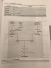
T-cell differentiation:

  1. Made in bone marrow
  2. Thymus cortex = positive selection
    • CD4+ CD8+ T cell
  3. Thymus Medula = Negative selection
    • CD8+ T cell -> Cytotoxic T cell (lymph node)
    • CD4+ T cell -> Helper T cell (lymph node)
  4. Lymph node
    • CD8 T cell -> Cytotoxic T cell (lymph node)
      • Kills virus-infected, neoplastic, and donor graft cells
        • Endogenous MHC I
    • CD4 T cell -> Helper T cell (lymph node)
      • Th1 cell (cell-mediated response)
        • Makes IL-2, IFN-GAMMA, AND ACTIVATES MACROPHAGES AND CD8+ T cells
          • ​​Inhibited by IL-10
      • Th2 cells (humoral response)
        • Makes IL-4, IL-5, IL-10, and help B cells make antibody (IgE > IgG)
          • Inhibited by INF-gamma
      • Exogenous MHC II
8
Q

Explain the following about the identified T Cell:

CD4 lymphocytes, helper T cells (Th Cells)

Th1 cells

Th 2 cells

  • Function
  • Characterization
A

CD4 lymphocytes, helper T cells (Th Cells)

  • Function: none listed
  • Characterization: Responds to antigen associated with Class II MHC proteins

Th1 cells

  • Function:
    • Signal CD8 cells to differentiate into cytotoxic T cells
    • Signal macrophages in Type IV (delayed) hypersensitivity reactions
  • Characterization:
    • Secrete:
      • IL-2 (CD8+ cells)
      • INF-gamma (macrophages)

Th2 cells

  • Function:
    • Signal B cells to differentiate into plasma cells, producing antibodies
  • Characterization:
    • Secrete:
      • IL-4
      • IL-5
9
Q

Explain the following about the identified T Cell:

CD4 lymphocytes, cytotoxic T cells (Tc cells)

  • Function
  • Characterization
A

CD4 lymphocytes, cytotoxic T cells (Tc cells)

  • Function:
    • Kill virus-infected, tumor, and allograft cells
    • Two ways:
      • Release perforins (disrupt cell membranes)
      • Induce apoptosis (programmed cell death)
  • Characterization
    • Respond to antigen associated with Class I MHC proteins
10
Q

Explain the following about the identified T Cell:

Memory T Cells

  • Function
  • Characterization
A

Memory T Cells

  • Function:
    • Activated in response to re-exposure to antigen
  • Characterization
    • Exist for years after initial exposure
11
Q

Explain clonal selection

A

The process by which an antigen binds to a specific TCR (T cell) or Ig (B cell), activating that immune cell to clonally expand into cells of the same specificity is called clonal selection

12
Q

Explain a B cell and state/describe its major types

Types (Plasma Cells, Mature B cells, Memory B cells)

  • Function
  • Characteristics
A

B cells

  • Differentiate in the bone marrow
  • Short life span, ranging from days to weeks

Major Types of B Cells:

Plasma Cells

  • Function:
    • Synthesize immunoglobulins (antibodies)
  • Characteristics:
    • Only monomeric IgM and IgD are expressed on their surface as antigen receptors

Mature B Cells

  • Function:
    • Antigen presentation
  • Characteristics:
    • Express class II MHC proteins
      • APC that presents to CD4 Th cells

Memory B Cells

  • Function:
    • Activated in response to re-exposure to antigen
  • Characteristics:
    • Exist for years after initial exposure

Recall:

  • Class I MHC surface proteins: on all nucleated cells. Recognition of self vs. non-self.
  • Class II MHC surface proteins: ONLY on ANTIGEN PRESENTING CELLS (APCs) Present antigen to Th cells
13
Q

Explain Natural Killer (NK) Cells

A

Natural Killer (NK) Cells

  • Lack a CD3-associated TCR and surface IgM or IgD
    • IgG antibodies enhance NK cell effectiveness via antibody-dependent cellular cytotoxicity (ADCC)
  • Are NOT specific to any antigen and do not need to recognize MHC proteins
  • No memory : Do not require previous exposure to antigen
  • Activated by IL-12 and INF-Gamma
  • Functions:
    • Kill Virus-infected cells and tumor cells (induce apoptosis via perforins and granzymes)
14
Q

Explain Monocytes and Macrophages

A

Monocytes and Macrophages

  • Agranular leukocytes
  • Derived from bone marrow histiocytes
  • Exist in plasma (monocytes) and in tissues (macrophages)
  • Activated by bacterial LPS, peptidoglycan, and DNA, as well as TH1 cell-mediated INF-Gamma
  • Functions:
    • Phagocytosis: Via Fc and C3b receptors
    • Antigen presentation: Express Class II MHC proteins
    • Cytokine Production: IL-1, IL-6, IL-8, INF and TNF

Monocytes and macrophages are major components of the reticuloendothelial system, which includes all phagocytic cells except for granulocytes (PMNs)

15
Q

What are other phagocytes besides monocytes and macrophages?

A

Other Phagocytes:

  • Histocytes: CT
  • Microglia: CNS
  • Dust cells: Lungs
  • Kupffer cells: Liver
16
Q

Explain Dendritic cells

What are Langerhans cells?

A

Dendritic cells

  • Agranular leukocytes
  • Located primarily in the skin and mucous membranes
  • Functions:
    • Antigen presentation express Class II MHC proteins

Langerhans cells: Are the major dendritic cells of the gingival epithelium

17
Q

Explain Polymorphonuclear Neutrophils (PMNs)

What are the major contents of PMN cytoplasmic granules?

A

Polymorphonuclear Neutrophils (PMNs)

  • Granular leukocytes.
  • Cytoplasmic granules (lysosomes) contain several bacteriocidal enzymes
  • Functions:
    • Phagocytosis
    • Cytokine production

Major contents of PMN Cytoplasmic Granules

Granule Type/ Enzymes

Primary (azurophilic)

  • Hydrolase
  • Myeloperoxidase
  • Neuraminidase

Secondary

  • Collagenase
  • Lysozyme
  • Lactoferrin
18
Q

Explain Eosinophils

A

Eosinophils

  • Granular leukocytes
  • Blind antigen-bound IgG or IgE, subsequently releasing cytoplasmic granules
  • Do not present antigen to T cells
  • Functions:
    • Defense against parasitic infections (especially nematodes)
    • Mediate hypersensitivity diseases: Release histaminase, leukotrienes, and peroxidase
    • Phagocytosis
19
Q

Explain Basophils and Mast Cells

A

Basophils and Mast Cells

  • Granular leukocytes
  • Exist in plasma (basophils) and in tissues (mast cells)
  • Bind antigen-bound IgE, subsequently releasing cytoplasmic granules (histamine, heparin, peroxidase, and hydrolase) and inflammatory cytokines.
  • Functions:
    • Mediate immediate hypersensitivity reactions such as anaphylaxis
20
Q

Explain what Opsonization is

What are the two major opsonins?

A

Opsonization

  • Enhances phagocytosis of encapsulated microorganisms
  • Antibody (IgG) or complement protein (C3b) coat the outer surface of microorganisms, allowing phagocytes to bind and engulf them more efficiently

THE TWO MAJOR OPSONINS ARE IgG AND C3b

21
Q

What are Antigen presenting cells and what do they express?

A

Antigen-presenting cells (APCs) express class II MHC proteins and present antigen to CD4 T cells. The predominant APCs of the immune system are monocytes and macrophages, dendritic cells (langerhans cells) and B cells

22
Q

What are Chemokines (give examples)

A

Chemokines (IL-8, C5a, LT-B, FMLP) are chemotactic cytokines for PMNs and macrophages

23
Q

What is phagocytosis and what are the stages

Stage

event

characteristic

A

Phagocytosis

  • The process by which microorganisms, cell debris, dead or damaged host cells, and other insoluble particles are taken up and broken down by phagocytes

Stages of Phagocytosis

Adhesion

  • Plasma phagocytes (PMNs, monocytes) bind to vascular endothelium
  • Mediated by selectin and cellular adhesion molecules (CAMs)

Migration

  • Phagocytes migrate toward the microorganisms
  • Diapedesis is the movement of the phagocyte through the vascular endothelium
  • Mediated by chemokines (IL-8, C5a, LT-B4, FMLP)

Ingestion

  • The phagocyte cell membrane forms pseudopods, which surround and engulf the microorganisms
  • Phagosome formation occurs when the internalized endosome fuses with lysosomes
  • Mediated by opsonization (C3B, IgG)

Lysosomal degranulation

  • The lysosome empties its hydrolytic enzymes into the phagosome, killing the microorganism
  • Mediated by lysosomal enzymes
24
Q

State the lysosomal contents and what makes them up

A

Lysosomal Contents

  • Superoxide radicals (O2-)
  • Superoxide dismutase
    • Produces hydrogen peroxide (H2O2)
  • Myeloperoxidase
    • Produces hypochlorite ion, which damages cell walls
  • Lactoferrin
    • Chelates iron from bacteria
  • Lysozyme
    • Degrades bacterial cell wall peptidoglycan
  • Proteases
  • Nucleases
  • Lipases
25
What are Lysosomes
**Lysosomes** membrane-bound vesicles that contain hydrolytic enzymes necessary for intracellular digestion
26
What are catalase and other peroxidases
Catalase and other peroxidases (enzymes that break down H2O2) are located in membrane-bound organelles called **peroxisomes (microbodies).** *_Bacteria that contain **catalase** (staphylococci) are able to resist*_ _*cidal*_ _*effects of H2O2_*
27
What are immunoglobulins and what are the types Type * Location of action * Function * Characteristics
***_Immunoglobulins (antibodies)_*** * Y-shaped glycoproteins secreted by plasma cells * Contain two identical **light polypeptide** chains and two identical **heavy polypeptide** chains linked by **disulfide bonds** ***_IgA_*** * Location of action * Blood plasma (monomer) * Exocrine secretion (dimer) * Function: prevents microbial attachment to mucous membranes * 2nd most abundant antibody ***_IgD_*** * Location of action: * B cells * Function: Uncertain * Least abundant antibody ***_IgE_*** * Location of action: * Mast cells * Basophils * Eosinophils * Function: Mediates **type I** hypersensitivity rxn. (**anaphylaxis)** * Main host defense against **parasites** (especially _helminths_) ***_IgM_*** * Location of Action * B cells (monomer) * Plasma (pentamer * Function: Main antimicrobial defense of **primary response** , _Activates complement, opsonizes B cell_ * **Largest** antibody, Most potent activator of complement, Has highest avidity of all antibodies ***_IgG_*** * Location of Action: Plasma * Function: Maintain antimicrobial defense of **secondary response** , _Opsonized bacteria,_ Activates complement, Neutralizes bacterial toxins and viruses * **_Most_** **_abundant antibody, Crosses the placenta_**, Has four subclasses
28
Explain the Immunoglubulin structure
* The **constant regions** **of the two heavy chains** form the **_Fc site,_** which binds to **APCs or C3b.** They define the immunoglobulin class (**_isotype_**) * The **two variable regions** of the heavy and light chains form the **_Fab sites_**, which are **specific for binding antigen** and determine the **_idiotype_** * Can be bound to plasma membrane of B cells, or free in extracellular fluid * Functions: * Neutralize bacterial toxins and viruses * Opsonization (enhances phagocytosis) * Activates complement via the ***_classical pathway_*** * Inhibit microbial attachment to mucosal surfaces
29
Explain the specialness of IgA and IgM
**IgA and IgM** are the only antibodies that can exist as polymers, as a dimer, and a pentamer, respectively. Only the polymeric forms contain a **J chain**, which initiates the polymerization process
30
Explain the difference between secretory IgA (sIgA) and serum IgA
**Secretory IgA (sIgA)** differs from serum IgA in that it is **_more resistant to proteolytic degradation._** It always exists as a **dimer**
31
What are the only two antibodies that can activate complement?
IgM and IgG are the only two antibodies that can activate complement
32
What is complement and what are the pathways of complement activation
***_Complement_***: * Consists of about 20 plasma proteins * Mostly synthesized in the **_liver_** * Augment the humoral immune system (B cell) and inflammation * All modes of activation lead to the production of C3 **_Pathways of Complement Activation_** Pathway/Characteristics **_Classic_**: * Primarily activated by **antigen-antibody complexes with IgG (1,2,3) or IgM** **_Alternate_**: * Primarily activated by bacterial **LPS (endotoxin)** **_Lectin_** * Primarily activated by microorganisms containing cell-surface **mannan** (a polymer of mannose)
33
State the Major functions of complement and its mediators
***_Complement_*** Viral neutralization * Mediator: C1, C2, C3, C4 Opsonization * **C3b** Chemotaxis * **C5a** Anaphylaxis * C3a, C5a (most potent) Cell lysis (cytolysis) * **Membrane attack complex (MAC)** disrupts cell membrane permeability (composed of **C5b and C6-9)**
34
Explain the types of Grafts
Types of Grafts * **_Autograft_**: Transplantation of tissue from one site to another within the same individual * **_Isograft_**: Transplant of tissue between two genetically identical individuals in the same species * **_Allograft_**: Transplant of tissue between two genetically different individuals in the same species * **_Xenograft_**: Transplant of tissue between two different species
35
Explain Graft Rejection Process
***_Graft Rejection_*** * **_T-cell-mediated_** (mostly ***_CD8 Tc cells_***) immune response against donor alloantigens * The severity and rapidity of graft rejection is determined by the degree of differences between donor and recipient class I and II MHC proteins * If a *second* graft from the same donor is given to a sensitized recipient, an accelerated rejection response occurs due to the presence of presensitized Tc cells * Allografts are the most common grafts used for organ transplantation, blood transfusions, and other tissue grafts * Graft rejection can occur at different time intervals
36
State and explain the three types of Graft Rejections - time after transplant - common reason for rejection
***_Types of Graft Rejection_*** * **_Hyperacute_** * Minutes after transplant * Preformed antibody-mediated immune response to graft antigens * **_Acute_** * Weeks after transplant * T=cell=mediated immune response to foreign class I and II MHC proteins * **_Chronic_** * Months to years * Antibody-mediated necrosis of graft vasculature NOTE: The most common type of hyperacute rejection are _ABO blood mismatches_
37
What are Graft-Versus-Host (GVH) Reaction
* Immunocompetent T cells from the graft recognize the recipient's cells as foreign, eliciting their destruction * Host cells are targeted because the recipient generally undergoes radiation therapy, inducing severe immunocompromise * Occurs most commonly after bone marrow transplants and **can be fatal**
38
What is a Hypersensitivity? Name and describe each type
Hypersensitivity * Hypersensitivity reactions elicit exaggerated immune responses, which are damaging and destructive to the host Remember ACID ***_Type_*** ***_I :_*** ***_immediate_*** (***anaphylactic***) * Mediator: **IgE** * RxN: Antigen-bound IgE activates the release **histamine** and other mediators from **mast cells** and basophils * Ex: Atopic allergy, Angioedema, Anaphylaxis ***_Type II: Cytotoxic_*** (Binds with Ab) * Mediators: **IgM and IgG** * RxN: IgM or IgG bind to host cell surface antigens, activating **complement** and producing MAC-mediated **cell destruction** * (antigen on cell membrane -\> antibody + complement = cell death) * Ex: Hemolytic anemia ***_Type III: Immune-complex (Binds soluble Ag)_*** * Mediator: Antigen-antibody complexes * RxN: **Antigen-antibody complexes** (IgG, IgM, and IgA) are **deposited** in various tissues, activating **complement** and eliciting PMN/macrophage-mediated tissue **destruction** * (antigen+antibody complex in blood -\> deposited on vessel walls, complement activated -\> cell death) * Ex: Arthritis reaction, Glomerulonephritis, Serum sickness ***_Type IV: Delayed (cell-mediated)_*** * Mediator: T cell * RxN: Macrophage present antigen (MHC Class II), activating T cells and producing **lymphokine-mediated tissue destruction** * (macrophage produces IL-1, IL-12 -\> Th and Helper T cell produces INF-Gamma -\> macrophage) * Starts hours - days after contact with antigen * Ex: Contact dermatitis, Tuberculin (PPD) tests, Tuberculosis, Sarcoidosis, Leprosy
39
What are Atopic allergies
Common type I hypersensitivity reactions that have a strong genetic predisposition for excessive IgE production. Clinical manifestations include asthma, edema, and erythema ("wheal and fire") and urticaria (hives). Common allergens include pollen, animal danders, food (shellfish and peanuts) drugs (penicillin), bee venom, and latex
40
What is Angioedema
***_Angioedema_*** = a more generalized version of type I hypersensitivity. It involves larger areas and deeper tissues beneath the skin and underlying tissues, causing a more diffuse swelling. May involve the hands, feet, lips, eyelids, genital, oral mucosa, and airway **Rapid(immediate) onset**
41
What is anaphylaxis?
**_Anaphylaxis_** The most severe form of _type I hypersensitivity,_ leading to bronchoconstriction and hypotension (shock); can be life-threatening without treatment. Its treatment is **epinephrine** IV. Once vital signs are stabilized, antihistamines or corticosteroids can be administered
42
Name the 5 common antigen-antibody laboratory tests - Function - Common use
***_Common Antigen-Antibody Laboratory Tests_*** **_Agglutination_** * Antibody cross-links with a **particulate** antigen, creating visible clumping is positive * Common use: ABO blood typing **_Precipitation_** * Antibody cross-links with a **soluble** antigen, creating visible precipitates if positive * Common use: Detection of serum antigen or antibody **_Radioimmunoassay (RIA)_** * Radio-labeled antibodies cross-link with unlabeled (unknown)antigen, creating measurable radioactive complexes is positive * Common use: Detection of serum antigen or hapten **_Enzyme-linked immunosorbent assay (ELISA)_** * Enzyme-labeled antibody binding to serum antibody-antigen complexes. A substrate is then added, activating the enzyme and eliciting a color reaction (determined by spectrophotometry) is positive * Common use: Detection of antigen or antibody in patient specimens **_Immunofluorescence_**: * Fluorescent-labeled antibodies bind to unlabeled (unknown) antigen, creating visible fluorescence in UV light if positive * Common use: Detection of antigen in histologic sections or tissue specimens
43
What is the universal donor for blood? What is the universal recipient of blood?
**_Universal donor: Type O blood_** (no A or B antigens) **_Universal Recipient: Type AB blood_** (Neither anti-A or anti-B antibody)
44
What is an autoimmune disease and what are examples
***_Autoimmune Diseases_*** * Caused by immune reactions versus self (host tissue) * Usually involves auto-antibodies * HLA antigen association * Mechanism: * Hypersensitivity reactions * Disordered immunoregulation * Increased Th cell function * Decreased Ts cell function * Nonspecific B-cell activation * Examples: * Graves' disease * Hashimoto's thyroiditis * Pernicious anemia * Sjogren's syndrome * Systemic lupus erythematosus * Scleroderma * Polyarteritis nodosa * Rheumatoid arthritis * Reiter's syndrome * Ankylosing spondylitis * Myastheria gravis * Multiple sclerosis
45
What is the Thymus gland important for?
Thymus gland * Important for development of the immune system, beginning prenatally * Involved in T cell development and differentiation * Located behind the sternum * Functions in childhood then gradually atrophies with age
46
Name the 4 autoimmune diseases affecting oral mucosa
1. Lichen planus 2. Mucous membrane pemphigoid 3. Pemphigus vulgaris 4. Erythema multiforme
47
Explain Lichen planus * Autoantibody * Histopathology * IF * Clinical signs
***_Lichen planus_*** * **_Autoantibody_**: Unknown * **_Histopathology_**: * Epithelial acanthosis with "sawtooth" rete pegs * Dense accumulation of T cells in underlying CT and into epithelium * **_IF_**: Fibrinogen in a "shaggy" linear pattern along BM * **_Clinical signs:_** * **Oral lesion on buccal mucosa, tongue, and gingiva** * Types: **Reticular** (with Wickham's striae), **erosive, plaque** * Certain medications can cause lichenoid reaction * Skin lesions appear as clusters of **p**ruritic **p**urplish **papules** with a white keratotic "cap"
48
Explain Mucous membrane pemphigoid * Autoantibody * Histopathology * IF * Clinical signs
**_Mucous membrane pemphigoid_** * **_Autoantibody_**: Anti-BP-1 (of hemidesmosomes) * **_Histopathology_**: * Separation of **epithelial basal cells from BM** * **_IF_**: * IgG and C3 in linear pattern along BM * **_Clinical signs:_** * Erythematous and erosive lesions on gingiva precede other oral areas: buccal mucosa, palate, FOM * Can extend to other mucosa: nasopharynx, esophagus, vaginal, eye conjunctiva (**symblepharon formation**) and skin
49
Explain **Pemphigus** **vulgaris** * Autoantibody * Histopathology * IF * Clinical signs
***_Pemphigus_*** ***_vulgaris_*** * **_Autoantibody_**: Anti-desmoglein (of desmosomes) * **_Histopathology_**: * **Suprabasilar acantholysis with Tzanck cells** * **_IF_**: * IgG antibody in "fishnet" pattern within spinous layer * **_Clinical signs:_** * Oral lesions often precede skin lesions * Erosive lesions on soft palate, buccal mucosa, gingiva, lateral tongue * High **mortality** rate
50
Explain **Erythema Multiforme** * Autoantibody * Histopathology * IF * Clinical signs
**Erythema Multiforme** * **_Autoantibody_**: Immune complexes * **_Histopathology_**: * Intraepithelial pooling of eosinophilic amorphous coagulum * Perivascular infiltration of mononuclear cells with lamina propria * **_IF_**: * IgM and C3 in perivascular pattern with lamina propria * **_Clinical signs:_** * Causative agents include infections (HSV), medications, GI diseases (Crohn's, UC) * **Bullous "Target" lesions** on skin and oral mucosa (25% prevalence), which may collapse and crust over (**stevens-johnson syndrome**) * Severe form is toxic epidermal necrolysis (TEN)
51