Midterm images Flashcards
(110 cards)
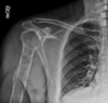
how do you describe this?
what is it?

ground glass
fibrous dysplasia

what benign 1˚ bone neoplasm has a 1% chance of malignant transformation?
what benign 1˚ bone neoplasm has a 25% chance of malignant transformation?
1% chance of malig = osteochondroma >30yo w/ Pain
25% chance of malig = Hereditary Multiple Exostosis (HME)
what is this?

aneurysmal bone cyst (ABC)
highly expansile
lytic, septated
eccentric
markedly thinned cortex
metaphyseal, can extend to epiphysis ITS THE ONLY BENIGN TUMOR TO CROSS GROWTH PLATE
periosteal response common
what is this?
malignant transformation rate? what does it turn into if it does transform?

ollier disease
- multiple enchondromas: usually unilateral, monomelic
25-50%; chondrosarcoma
autosomal dominant diseases?
gardner syndrome
fibrous dysplasia
neurofibromatosis
what is this?
enlarged IVF

neurofibromatosis
how do you describe this?
what does it point to?

blade of grass
paget disease
what is this?

osteosarcoma
- MC location is distal femur/proximal tib
- cumulus cloud appearance
- cortical destruction, aggressive periosteal response
MC location for Ewing’s sarcoma?
diaphysis of humerus and femur

what is this?
typical age?

osteoid osteoma
- reactive sclerosis
- nidus (black arrow on viewing right)
- painful: not relieved by rest, worse at night, relieved by aspirin
10-25 yo
epiphysis / apophyseal region is key for…?
chondroblastoma

what is this?

acral mets
what is this?
what is the triad associated with it?

Gardner Syndrome
well efined
opaque
clean borders
- multiple osteomas
- colonic polyps (considered pre-malig)
- soft tissue fibromas
what is this?

multiple myeloma
- punched out lesions: multiple, well-defined, round, osteolytic defects
what is this?
how do you describe it?

paget disease
cotton wool appearance
basilar impression (tip of the odontoid process projects above the foramen magnum)
Local P and swelling.
Systemic signs: fever, anemia, increased ESR.

Ewing sarcoma
- 10-25 yo
- laminated onion skin
- periosteum getting eaten
- endosteal scalloping
- long zone of transition
what is the next step?

for multiple myeloma: get MRI
painless, lumpy joints?
what % chance of malignancy?

HME
malignant degeneration in 25% of cases
what is the ddx list for this?

- osteoblastic mets
- multiple ivory vertebra
-
osteoid osteoma** most likely this
- pain
- has nidus
- osteoma
- located in sinuses
- smooth edges
what is this?

polyostotic fibrous dysplasia (FD)
what is this?
MC age group?

enchondroma
10-30 yo
what is this phenomenon called?
and what are the 3 most common causes of it?

ivory vertebra
osteoblastic mets (multiple ivory vertebrae), paget disease (cortical thickening, expansion), lymphoma (anterior body scalloping)
what is this?

ABC
describe this. what is this?

distal radius
extends into subchondral region
diminished bone density
appears aggressive
malignant GCT