Morphology Flashcards
(42 cards)
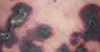
identify the morphology of this lesion

MACULE
examples of macules
freckles, flat moles, the rashes of rickettsial infections, rubella, measles
describe a macule
flat, circumscribed
<1cm in diameter
hypo or hyper pigmented
can be other colors (pink, red, violet)
identify the morphology of this lesion

patch
describe a patch
flat, circumscribed
>1cm in diameter
hypo or hyper pigmented
also other colors
a bigger macule
examples of a patch
tattoos, port-wine stains, cafe-au-lait spots
identify the morphology of this lesion
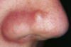
papule
describe a papule
elevated lesions
<1cm in diameter
palpable
thickening of epidermis and/or cells or deposits within dermis
may have secondary changes
distinguish from vesicle or pustule
what are examples of papules
nevi, warts, lichen planus, insect bites, seborrheic keratoses, actinic keratoses, some lesions of acne, and skin cancers
identify the morphology of this lesion

plaque
describe a plaque
elevated, circumscribed
>1 cm in diameter
elevation due to increased thickness of the epidermis and/or cells or deposits within the dermis
may have secondary changes (e.g. scale, crust)
bigger papule
what are examples of plaques?
Lesions of psoriasis and granuloma annulare
identify the morphology of this lesion
nodule
describe a nodule
elevated, circumscribed
larger volume than papule, often >2cm in diameter
involves the dermis, may extend to subcutis
greatest mass may be beneath the skin surface
what are some examples of nodules?
cysts, lipomas, and fibromas
identify the morphology of this lesion

vesicles
what are examples of vesicles?
herpes infections, acute allergic contact dermatitis
describe a vesicle
elevated, circumscribed
<1cm in diameter
primarily filled with clear fluid
may become pustular, umbilicated or an erosion
identify the morphology of this lesion

bulla
describe a bulla
elevated, circumscribed
>1cm in diameter
filled with clear fluid
what are some examples of bulla?
burns, bites, irritant contact dermatitis or allergic contact dermatitis, and drug reactions
identify the morphology of this lesion

pustule
describe a pustule
elevated, circumscrived
usually <1cm in diameter
filled primarily with purulent fluid
describe the morphology of this lesion
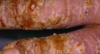
crust
dried serum, blood or pus