MSK Flashcards
(176 cards)
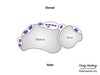
Rice bodies
TB and RA
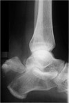
Diagnosis?
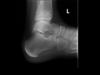
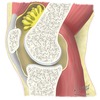
Sever’s
- calcaneal apophysis
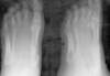
Diagnosis?

Bizarre parosteal osteochondromatous proliferation (BPOP)
- also known as Nora lesions
- benign osteochondral lesions which have the appearance similar to an osteochondroma
- typically seen in hands and feet
- continuous with underlying cortext without continuation of the medulla
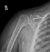
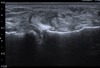
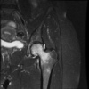
Concern for septic joint. Sign and indication?

Pneumoarthrogram sign
- Presence of air in the joint excludes a joint effusion and is a normal finding
- No septic joint
T score vs Z score for DEXA
T score = Density relative to young adults
> - 1.0 normal, -1.0 to -2.5 osteopenia, < -2.5 osteoporosis
- False positive - absent normal structures such as laminectomy
- False negative - too much osteophytces, dermal calcifications, metal
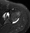
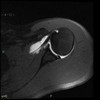
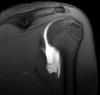
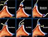
Name 4 types of impingments of the shoulder
- External Primary: abnormal coracoacromial arch
- External Secondary: multidirectional instability “increased glenohumeral volume” with injection
- Internal Posterior Superior: Throwers, posterior superior labrum torn
- Internal Anterior Superior: associated with subscapular damage
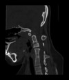
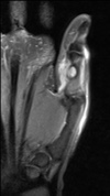
Diagnosis?

Os trigonum syndrome
Diagnosis?

Partial Ulnar Collateral Ligament Tear
- Throbwers, anterior bundle of ulnar collateral ligament torn (medial and poster bundles can also be involved)
- T sign
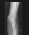
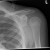
Diagnosis?

Monteggia Fracture
Best radiotracer for osteomyelitis in the spine?
Gallium > WBC scan (In-111)
Diagnosis?

Cortical Desmoid
- misnomer, not a true desmoid
- typically ages 10-15
- repetitive stress injury
- typically seen posterior medial femur at site of medial gastroc attachment or distal adductors
Grading of stress fracture on MRI
Grade 0: Normal
Grade 1: Subtle periosteal edema
Grade 2: Periosteal edema with marrow edema on T2
Grade 3: More edema with changes on T2 and T1
Grade 4: True stress fracture with visible fracture line on MRI or radiograph
Sign and diagnosis?

Double PCL
- Medial meniscus bucket handle tear
- Proves ACL is intact
Pincer type FAI keywords and associations
- Coxa profunda
- Acetabular protusion
- Prominent ischial spine
Asscoiated with os acetabuli, labral tears and early arthritis
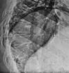
Sign and associations?

Looser zones
- wide lucent bands that transverse bone at right anles to the cortex
- Classic locations are femoral neck and pubic rami and have surrounding sclerosis
- Think of osteomalacia and rickets, less common OI
- They are a type of insufficiency fracture
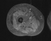
Diagnosis?

Medial epicondylitis
- less common than lateral
- seen in golfers
- common flexor tendon and ulnar nerve may enlarge from chronic injury
Name these 3 wrist factures

- Colles’ fracture - Dorsal angulation
- Smith fracture - Volar angulation
- Barton fracture - involving radial rum, volar is more common
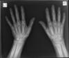
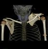
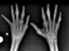
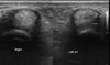
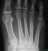
Diagnosis?

Thyroid acropachy (drumstick fingers)
Sign and association?

Arcuate sign
- avulsion of proximal fibula (insertion of arcuate ligament complex)
- 90% are associated with cruciate ligament injury PCL > ACL
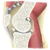
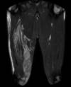
Diagnosis?
Transient osteoporosis of the hip
- Ddx for MRI - AVN (has serpigenous dark lines and involves more the subchondral femoral head)
- Bones is lucent on X-ray which looks very different than AVN (sclerotic)
- Regional migratory osteoporosis is similar however affects different joints
DDx Vertebra Plana
MELT
Mets/myeloma
EG
Lymphoma
Trauma/Tb
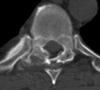
Diagnosis?

Trevor Disease
- multiple osteochondromas develop at the epiphysis resulting in joint deformity
- most common in ankle and knee
- these point toward the joint
- aka dysplasia epiphysealis hemimelica (DEH)
Diagnosis?

Nail-patella syndrome
- AKA Fong disease and bunch of other names
- Absent/hypoplastic nails from birth, flexion conractures, recurrent knee disloctions, hypoplastic patellas
- Posterior iliac horns (Fong prongs) is pathognomic
- In elbow can see dysplastic radial head, hypoplastic capitellum and lateral epicondyle and prominent medial epicondyle

Has the appearance of a chondroblastoma, but is in adult
Think clear cell chondrosarcoma