Muscle Physiology COPY Flashcards
(248 cards)
1
Q
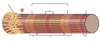
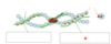
Name the three varieties of muscle in the body
A
- Skeletal
- Cardiac
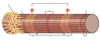
- Smooth
2
Q
The contractile cells of muscle tissue
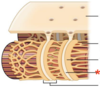
A
Myocytes/Myofibres
3
Q
Another name for the skeletal muscle
A
Striated muscle
4
Q
A
Skeletal muscle
5
Q
A
Epimysium
6
Q

A
Muscle fascicles
7
Q
A
Muscle fascicle
8
Q

A
Perimysium
9
Q

A
Endomysium
10
Q

A
Muscle Fibres
11
Q

A
Muscle fibre
12
Q

A
Sarcolemma
13
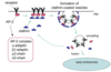
Q
Fibril diameter
A
100 - 1000 µm
(1mm)
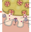
14
Q
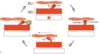
Myocyte diameter
A
10-100µm
15
Q
Myofibril diameter
A
1µm
16
Q
What is located between two Z-bands?
A
1 sarcomere
17
Q

A
A-band
18
Q

A
H-zone
19
Q

A
1/2 I-bands
20
Q
A
Z-bands
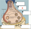
21
Q

A
M-line
22
Q

A
Sarcomere
23
Q

A
Sarcoplasmic reticulum
24
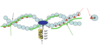
Q
A
Actin filament
25

H-zone
26
Z-disc
27

Myosin filament
28

I-band
29

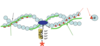
A-band
30

M-line
31

Sarcolemma
32

Sarcoplasmic reticulum
33

Terminal cister
34
T-tubule
35

Triad
36
What permits the conduction of electrical impulses in the muscle fibre?
Narrow T-tubules
37
What regulates the intracellular levels of calcium?
Sarcoplasmic reticulum
38
The membrane triad of myocytes is composed of...
* 2 x terminal cisternae
* 1 x T-tubule
39
List the 3 additional proteins in the sarcomere
* Titin
* Nebulin
* Alpha-actinin
40
Titin
* Largest protein of the body
* From Z-lines → Myosin bundles
* Ensures precise return of actin and myosin bundles to original position
41
Nebulin
* Determines the direction and placement of actin polymerisation (during development)
* Protects actin fibres from rearranging
42
Alpha-actinin
* Creates the Z-band
* Net-like
* Provides a binding site for actin complexes
43

Nebulin
44

Titin
45

Alpha-actinin
46
A motor unit
* Motor neuron
* Skeletal muscle fibres innervated by the neurone's axonal terminals
47
Summarise the pathway from neural activation to muscle contraction
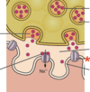
1. Generated AP→ Myoneural junction
2. ACh-containing vesicles open at synaptic knobs
3. ACh attach to the sarcolemma ACh-R
4. ACh channel opens
5. Na+ enters inner surface → Local end plate potential generated
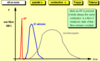
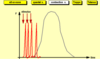
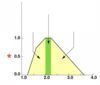
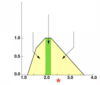
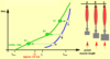
6. AP generated → Activates SR though T-system
7. Ca2+ release into sarcoplasm → Actin-myosin contraction
8. Ca2+ repumping into:
* SR
* Mitochondrium
* EC
48
AP on the myolemma is generated only if...
The AP is stimulated through a nerve
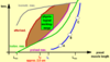
49
Transmission of neural AP to the muscle takes place in the...
Myoneural junction
50
Give the summary of the processes that occur at the neuromuscular junction
1. AP reaches nerve terminal → ACh release
2. ACh → Nicotinic receptors on muscle membrane
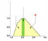
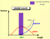
3. Ligand-activated cationic channels open
4. EPP produced
5. Voltage-gated Na+ channels open
6. AP formed in myolemma
51

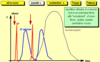
Synaptic Vesicle
52

Synaptic Cleft
53
ACh receptor
54

* ACh binding to its receptor
* Ligand-activated cationic channel opens
55
ACh released by synaptic vesicle
## Footnote
*Exocytosis*
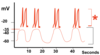
56
Lifecycle of neurotransmitter

Neurotransmitter synthesis
* In cell body (cytosol)
* In the terminal
57
Lifecycle of neurotransmitter
Neurotransmitter packaged into vesicles
58
Lifecycle of neurotransmitter

Neurotransmitter released
59
Lifecycle of neurotransmitter

Neurotransmitter binding
60
Lifecycle of neurotransmitter

Neurotransmitter diffused away
* Catabolysed or transported back into the terminal
61
Give the actions when AP reaches the NMJ
* Voltage-gated Ca2+ channels open, influx from EC space
* [Ca2+] increases 100x → ACh exocytosis initiated
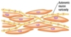
62
Clathrin
* Protein on inner membrane
* Stimulates endocytosis
63
What is shown?
Clathrin-dependent endocytosis
64
Composition of the nicotinic acetylcholine receptor
What does this allow?
* Two alpha subunits
* Two beta subunits
* One delta subunit
Allows blocking effect of _curare_ and _bungarotixin_
65
Give the 3 possible conductance states of the nicotinic acetylcholine receptor
* Closed
* Open
* Inactivated
66
Which potential is amplitude-coded?
EPP
67
Which potential is frequency-coded?
AP
68
Decremental conduction
Decrease of signal strength with distance travelled
69
Role of Mg2+ in muscle contraction
* Antagonises ACh receptor
* Blocking function of sarcomere
70
Give the importance of Mg2+ in cattle
* High Ca2+ secretion after calving → Low plasma Ca2+ levels
* [Mg2+] becomes relatively high
* Muscles relax: _Parturient paresis_
71

* AP from axon
* Ca2+ enters from EC, ACh vesicle release
72
Filling up with ACh
73

* ACh binds to the receptor
* EPP generated
* AP generated, Ca2+ influx, contraction
74
Effect of nicotine on the neuromuscular junction
* Same effect as ACh
* Cannot be degraded by cholinesterase
* Conc. therefore increases → Permanent depolarisation
* Intensive spasm
75
Effect of cholinesterase inactivators on the neuromuscular junction
* ACh not hydrolysed
* High ACh accumulation
* Repetitive stimulation of muscle fibres
* Spasm/laryngeal spasm
76
Effect of curariform drugs on the neuromuscular junction
* ACh receptor blocked
* No depolarisation → No contraction
* Paresis
77
Effect of botulin toxin on the neuromuscular junction
* Blocking of ACh release
* Pareisis
78
Effect of myasthenia gravis (autoimmune disease) on the neuromuscular junction
* ACh receptor blocked by antibodies
* No ACh binding
* Paresis
79
Depending on the task of the given muscle, there can be variations in...
Nerve:muscle fibre ratio
* Occular muscles (1:1)
* Skeletal muscles (1:100)
80
The fusimotor system
* Intrafusal fibres
* Modified muscle fibres → Stretch detection
* Also located in tendons → Golgi tendon receptor organs
81
Static fibres
Sensitive to static changes of tension (Length)
82
Dynamic fibres
Sensitive to dynamic changes of tension (length & velocity)
83
Intrafusal fibres
84

Extrafusal fibres
85

Sensory fibres
86
Myotatic reflex
* (Contraction of stretched muscle)
* Efferentation returns to the same muscle where afferentation occurs
* Monosynaptic
87
Give the responses of Myotactic reflex
1. Increased stretching causes increased tension (Servo-mechanism)
2. Fusimotor activation
88
Give the steps of the servo-mechanism
1. Muscle stretching → Muscle spindles stimulated
2. Sensory neuron activated
3. Information processing at motor neurone
4. Motor neurone activation
5. Muscle contraction
89
Co-activation
CNS participation (+servo-mechanism) in fusimotor system activation
90
Describe Co-Activation mechanism
* α- + γ- motorneurones stimulated by cerebral centre _(Co-__acitivation__)_
* Intrafusal & extrafusal fibres contract with the _same rate and strength_
91
Describe Co-Activation in the case of sudden increased load
* Extrafusal fibre tension is stronger than intrafusal
* AP frequencies accelerate in Ia and II afferents
* Locally adjust the tension of extrafusal fibres (fine tuning)
92
1

AP → Myolemma
93
2

* AP reaches L-type Ca2+ channels in the T-tubuli
* L-type channels open
94
3

Ryanoid-Ca2+ open
95
4

Ca2+ enters the IC from the SR
96
5
* Ca2+ channels open on myolemma
* Ca2+ influx from the EC
97
6

* IC Ca2+ high in and around the sarcomere
* Contraction
98
Contraction and relaxation of muscle require...
ATP
99

The triad
100
Cardiac equivalent to the triad
Diad
101
Triad
* Basis of excitation-contraction coupling
* Where t-tubule is closest to the SR IC
102
What occurs at the triad?
* AP → Change in L-type Ca2+ receptors
* T-type ryanodin Ca2+ channels open
* High amount of Ca2+ released from SR to IC space
* Positive feedback:
* Ca2+ opens SR Ca2+ channels
* **Increase in [Ca2+] → Cross-bridge cycle triggered**
* Calcium signal stimulates its own inactivation
103

Potential dependent DHP proteins
104
T-type (Ryanodin) calcium channel
105
Briefly describe the figure

* Receptors in the T-tubule changing from their closed state to their opened state
* Influx of Ca2+ ions into the triad
106
What is the main component of the actin-complex?
G-actin
107
Give the function of tropomyosin
Used in stimulation of ATPase activity of myosin
108
What is shown?

F-actin α-helix
109

G-actin subunit
110
Myosin binding site
111

Tropomyosin (Inactive)
* Blocks 7 binding sites
112
Tropomyosin (Active)
* Slides into the groove of α-helix, leaving binding sites free
113
What is shown?

Troponin-complex
114

Troponin-C
115

Troponin-T
116

Troponin-I
117
Describe activation of tropomyosin
* Ca2+ binding → Removes tropomyosin to grove (Active)
* Tn-complex binds to the tropomyosin attached to actin
* Tropomyosin-troponin complex kept on the helix surface (inactive)
118
Describe the structure of myosin
1 bundle = 6 myosin molecules
* 2 heavy chains (HC)
* 2 light chains (LC)
* Globular part (head/cross bridge)
119
Angle of myosin head with alpha helices
90°
120
Maximum bend angle of myosin heads
45°
121
Give the three types of ATPases
How do they differ in function?
* LC-1
* LC-2
* LC-3
Difference determines the speed of ATPase activity
122
LC-2 ATPases are found in...
Fast twitch muscle
123
LC-3 ATPases are found in...
Slow-twitch muscle
124
* Tail / Heavy chain
* Formed by α-helix
125

Head Cross-bridge
126
Actin binding site
127
ATP binding site
128
Myosin filament composition
200 miosin units
129
Calcium transient
1. IC [Ca2+] increased x1000
2. Calcium (re)pumping mechanisms
3. Calcium elimination from cytoplasm
4. Ca2+ levels decrease near the sarcomere
130
Give the steps of the Cross-bridge cycle
1. Relaxation/Resting
2. Ca2+ release from AP
3. Tropomyosin removed → myosin binding sites exposed
4. Cross bridge binds to actin
5. Contraction (head tilts, ADP released)
6. Ca2+ removed from outside → Myosin detaches
131
ATP in the cross bridge cycle
* Myosin head binds to ATP
* Energy deliberated → ADP + P
* Head tilts back to 90° (Cocked head)
132
* Myosin head detached
* ATP hydrolysed
133

ADP + P bound to myosin as myosin head attaches to actin
134

* ADP + P release
* Head changes position
* Actin filament moves
135

* ATP binding to head
* Head returns to resting position
136
When ATP isn't present in the muscle
* Myosin cannot dissociate from actin
* Muscle becomes contracted and inactive
Rigor mortis → Autolysis follows after
137
When Ca2+ isn't present in the muscle
* Tropomyosin slides over myosin
* Activating part of the actin
* Muscle relaxes
* Myosin heads can't bind
138
The ratchet mechanism
Myosin filament cannot fall back
* Myosin heads need to work asynchronously to contract

139
How is calcium removed from the cytosol
All are ATP dependent:
* Na+/Ca2+ ion antiporter
* Secondary active transport
* Ca2+ repumping into the SR
* Other
* Cell organelles
* Mitochondria
140
Composition of muscle tissue
* 75% water
* 20% protein
141
Describe the importance of the macroscopic structure of muscles
* Most sarcomere orientations aren't parallel to the direction of macroscopic contraction
* Skeletal muscle is wasteful but provides extreme spatial flexibility
142
Which metabolism is expressed in red/slow twitch muscle?
Oxidative
143
Which metabolism is expressed in white muscle?
Anaerobic
144
Which metabolism is expressed in pink muscle?
Mixed
145
Fast twitch muscle types
Pink and White
146
Atrophy
* Decrease in skeletal muscle size
* Myonuclear loss
* Decreased myofibrillar proteins
* Decreased CSA
147
Muscle Hypertrophy
* Increase in skeletal muscle size
* Myonuclear addition
* Increased myofibrillar proteins
* Increased CSA
*
148
The fibre spectrum of an individual is determined by...
* Genetic factors
* Usage of specified muscle
149
Which two factors contribute to hypertrophy
* Sarcoplasmic hypertrophy - Increased glycogen storage
* Myofibrillar hypertrophy - Increased myofibril size
150
Remodelling of 'slow' muscles
Mass of fibres increase slower than nutrient/energy storage of myocytes
151
Myocyte ATP concentration
5 mmol/l
152
List the energy sources of muscle contraction
* Creatin-phosphate
* Anaerobic glycolysis
* Oxidative phosphorylation
153
Creatin phosphate
ADP + CrP → **ATP** + Cr
154
Creatin phosphate conc. in myocytes
20 mmol/l
155
Anaerobic Glycolysis as muscle energy source
In cases of outstanding load
* Glycogen
* Glucose
4 ATP produced
* If more ATP is used than produced → Oxygen debt
* Produced lactic acid inhibits contraction
156
Oxidative phosphorylation as a muscle energy source
Pyruvate → Acetyl-Coenzyme A
36 ATP produced
Process and _contraction are slow_
No oxygen debt
157

Working in an oxygen-free environment
158

Oxygen debt
159
Muscle can replenish glycogen and creatine phosphate by...
Oxygen consumption
160
Give the types of muscle contraction
* Isotonic
* Isometric
* Mixed
* Auxotonic
* Preload
* Afterload
161
Isotonic contraction
Contraction with constant tension
162
Isometric contraction
Contraction when only tension is changed, no length changes
## Footnote
*e.g lifting an unliftable load*
163
Auxotonic contraction
Working against increasing tension and resistance
## Footnote
*e.g against a spring*
164
Preload contraction
* Muscle length is adjusted until equilibrium
* Isotonic contraction follows
## Footnote
*e.g locomotion related muscle work*
165
Afterload contraction
* Isotonic contraction until the contraction is blocked
* Isometric contraction follows
## Footnote
*e.g. m. masseter*
166
The sum of observable and biological latency
Virtual latency
167
Which muscle elements reach equilibrium with the load first, why?
SEC elements, dues to the contraction of the contractile components
(only tension is increased at this stage)
168
Summation of muscle contraction
Addition of muscle contraction forms
* Increase the contractile capacity of individual fibres
* Recruits more fibres
169
Give the types of Summation
* All or none law
* AP Frequency
* Quantal summation
* Contraction summation
* (Staircase effect)
* Tetanus
170
All or none law
* Applies for a single fibre
* Adequate stimulus causes maximal contraction
* Stimulus strength can't influence amplitude of contraction
171
AP frequency summation
* Increased frequency
* Prolonged Ca2+ release
* Stronger contraction
172
Quantal summation
* Increased AP frequency
* More fibres contract
173
Contraction summation
* Additional Ca2+ release before the end of Ca2+ transient
* Increased amplitude of contraction
174
Staircase effect summation
* Warmup phenomenon - not graded contraction
* If new stimuli arrive after the end of the first twitch
* Increased efficiency of ion channels
* Ca2+ accumulation
* Increased amplitude of contraction

175
Tetanus summation
* Stimuli applied with increasing frequency
* Muscle eventually reaches max contraction state (tetanus0

176
How is the length-tension curve obtained?
* Stimulation of a muscle which is passively stretched with varying loads
* Isometric, isotonic, preload and afterload experiments carried out
177
What does the length and tension diagram show?
The area where muscles execute normal work
178
Maximal tension value
3kg/cm2 muscle cross section
179
Tension (g) generated upon stimulation
180
Sarcomere length (µm) before stimulation
181

Overly contracted
182

Optimum resting length
183
Overly stretched
184
Length x Tension =
Work
185
Describe obtaining length-tension diagram in _isotonic conditions_
* Muscle stretched to A, B, C distances (Above L0)
* Muscle is stimulated with max single stimuli
* The isotonic maximum (It) curve can be obtained
186
Describe obtaining length-tension diagram in _isometric conditions_
* Shortening isn't possible
* Only changes of tension can be measured
* Isometric maximum (Im) curve achieved

187
Describe obtaining length-tension diagram in the _preload experiment_
* Tension increase is followed by contraction
* Preload maximum (Pm) curve achieved

188
Length-tension diagram in the _afterload conditions_
Afterload-maximum (Am) curve achieved

189
How is working range achieved from the 4 length-tension experiments?
Area is constructed on a graph
190
Compare 'normal working range' and 'length measured under max power' in _skeletal muscle_
Both are identical to eachother
191
Compare 'normal working range' and 'length measured under max power' in _cardiac muscle_
* The normal working range is much below the length
* Ensuring maximal tension

192
Velocity x Tension =
Power
193
As tension is...velocity becomes...during muscle work
Low; high
194
Unloaded muscle contracts with...velocity
Maximal
195
Overloaded muscle contracts with...velocity
Zero
196
Velocity related to an actual tension is determined by...
The type of muscle
* Phasic (Fast)
* Tonic (Slow)
197
Using the velocity-tension diagram rather than the length-tension diagram gives a better indication of...
The power of the muscle
198
Intermediate tension and intermediate velocity result in...
Maximal power
199
Describe the figure
Grey rectangles:
* Small tension = High velocity
* High tension = Small velocity
Red rectangle:
* The optimal position where maximum power can be achieved
200
Max speed of muscle contraction
7 m/sec
201
Total force of skeletal muscle
200N
## Footnote
*relative to 100kg mass*
202
Efficiency of skeletal muscle
20%
203
Power maximum of skeletal muscle
* Short term: 3-5000 W
* Long term: 1200 W
204
How do muscles produce heat?
* Contraction: ATP breakdown
* After contraction: Synthetic processes create heat
205
When do phasic/fast/white fibres produce heat most?
During restoration/recovery
206
When do tonic/slow/red fibres produce heat most?
207
Give the phases of heat production
* In resting (Muscle maintenance/BMR)
* Initial heat production
* Activation heat
* Ca2+ release
* Myosin-activation
* Contraction heat
* Sliding filament
* Ca2+ repumping
* Restitution heat
208
Muscle fatigue is dependent on...
Ratio of phasic and tonic fibres
209
Signs of fatigue on a mechanogram

210
In Vitro fatigue
* Lack of O2
* Lack of Transmitter
211
In Vivo fatigue
* Peripheral
* Decreased energy sources
* Lactic acid
* Central fatigue
* Exhaustion of motor-unit
* Exhaustion of myoneural junction
212
Subjective feelings of fatigue can be caused by...
* Increased heat production
* Decreased pH
* Lactic acid
* Dehydration
* Hypoglycaemia
213
Fatigue develops earlier in...
Phasic fibres

214
Smooth muscle
Used in maintenance and form of visceral organs
* Single-unit smooth muscles
* Multi-unit smooth muscles
215
Multi-unit smooth muscle
* Individual fibres not connected with gap junctions
* Fibres under direct neural control (not by AP)
* Capable of fast and accurate movements
* Transmitters cause local depolarisation
## Footnote
*Located in the eye*
216
Single unit smooth muscle
* Many hundreds of fibres
* Form a _functional syncytium_
* Fibres are innervated by varicosities
## Footnote
*e.g muscle of vessels*

217
What causes contraction of smooth muscle
* Dense bodies and intermediate filaments
* Networked through the sarcoplasm
218
Proportion of myosin:actin in smooth muscle
1:15
219
Give the steps of contraction in smooth muscle
* If IC Ca2+ is high, _MLCK enzyme_ is used
* Actomyosin complex formation
* Contraction stays continuous until MP enzyme triggers relaxation
## Footnote
*Most muscles are in weak but continuous contraction*
220
Summarise the structure of smooth muscle
* _No transverse tubular system_
* Poor blood supply
* Non-striated
* Small SR
221
What is shown?
Smooth muscle sarcomere
222
Calmodulin
223

Caldesmon
224
Describe the role of caldesmon in smooth muscle contraction
* Ca2+ binds to the Calmodulin-Ca2+ complex
* Removes tropomyosin from binding sites
225
Describe smooth muscle myosin
Heads contain a unique MLC subunit:
_P-LCh_
* Phosphorylated = Actin binding → Contraction
* Non-phosphorylated = No actin binding
226
In smooth muscle:
If there is no Ca2+
MLCK is inactive → P-LCh is not phosphorylated
227
Elimination of Ca2+ activates...
Myosin phosphatase (MP)
228
Activation of MP
Dephosphorylation of P-LCh → Relaxation
229
Characteristics of smooth muscle contraction
* Prolonged tonic contraction (hours/days)
* Energetically economic - low energy use
* Length of contraction 30x longer than skeletal
230
Max contraction length of smooth muscle
66% of resting length
231
Varicosities
* Series of axon-like swellings
* From autonomic neurons
* Form motor units in the smooth muscle

232
Example of multi-unit smooth muscle
m. ciliaris
233
Example of single unit smooth muscles
Gastrointestinal muscles
234
What is shown?

Special structure of smooth muscle SR
235
Sources of Ca2+ in the smooth muscle
* Minor: SR
* Major: EC-Space
236
Which channels can be found in the smooth muscle myolemma?
* Voltage-gated Ca2+ channels
* _Ligand-gated_ _Ca2+ channels_
237
Describe AP in smooth muscle
* Single-unit only
* RMP = _-50mV_
* Forms of AP:
* Typical peak potential
* AP with 'plato'
238
Importance of extremely prolonged repolarisation of smooth muscle
Prolonged contraction:
* Myocardium
* Uterus
239
Factors causing contraction of smooth muscle
* AP
* Binding of chemical ligands
* IC IP3 release
* G-Protein or phospholipase C (PLC) mediated Ca2+ influx

240
What stimulates relaxation of smooth muscle?
All stimuli which can increase IC cAMP or cGMP levels
* _Sympathetic beta2 receptor agonists_

241
Chemical factors influencing smooth muscle contraction
* Lack of O2
* Excess of CO2
* Increased H+
* Increased K+
242
Bayliss effect
Extension of smooth muscles results in contraction
## Footnote
*Not related to neural or hormonal influences*
243
What is the mechanism of the Bayliss effect?
* Stretching opens the _mechano-sensitive cation-channels_
* Depolarisation
244
Where would the spontaneous generation of smooth muscle AP be observed?
In the gut
245
Spontaneous generation of smooth muscle AP is associated with...
The 'slow wave' rhythm

246
AP (Spike potential)
247

Slow wave potential
* Not AP
* Local potential
* If above -35mV, AP is initiated
248
Each slow wave initiates...
More than one AP
| (Also called pacemaker waves)
