Myeloid Neoplasms Flashcards

(43 cards)

1
Q

Characterization of acute myelogenous (myeloid) leukemia? (AML)

A

Accumulation of immature myeloid precursors in the BM and suppression of normal hematapoiesis

2
Q

Characteristics of Myelodysplastic syndromes?

A

Ineffective hematopoiesis with cytopenias

3
Q

Characterization of chronic myeloproliferative disorders (CML)?

A

Increased production of terminally differentiated myeloid cells

4
Q

What is found in the BM in AML?

A

Undifferentiated blasts

(results in anemia, neutropenia, and thrombocytopenia)

5
Q

Diagnosis of AML?

A

Myeloid blasts >20% of marrow cells

6
Q

M1 myeloblast characteristics in AML?

A
  • Delicate chromatin
  • 2-4 faint nucleoli
  • Cytoplasm with azurophilic peroxidase positive granules
7
Q

M3 acute promyelocytic leukemia cell characteristics?

A
  • Auer rods (definitive evidence)
  • Peroxidase positive
8
Q

M5 monoblast cell characteristics in AML?

A
  • Folded/lobulated nuclei
  • Esterase positive
  • NO auer rods or peroxidase
9
Q

AML cell markers?

A
  • +CD34 (multipotent stem cell marker)
  • +CD33 (immature myeloid marker)
  • -CD64 (mature myeloid)
10
Q

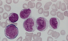
What is this? What can you distinctively see that indicates that?

A

Acute promyelocytic leukemia, M3

Can clearly see the Auer Rods

11
Q

What is this?

A

AML, M1

Can see the faint nucleoli

Dispersed chromatin

12
Q

What is this?

A

AML M5

The nuclei are lobulated/folded.

13
Q

AML signs and symptoms?

A
  • Pancytopenia
  • Weakness, fatigue, infections, and bleeding
  • Less extramedullary tissue infiltration in comparison to ALL
  • M4/M5 differentiation:
    • Infiltration of the skin and gingiva may occur

(60% remission with therapy; only 15-30% disease free after a year)

14
Q

Bone marrow in Myelodysplastic Syndromes? (MDS)

What are the two forms?

A

Marrow is partly or completely replaced by the clonal progeny of a defective stem cell

  • 2 forms
    • Idiopathic
    • Therapy related (2-8 yrs after cancer therapy)
      • More rapid transformation to AML
15
Q

MDS clinical presentation? Survival?

A
  • Weakness
  • Infections
  • Hemorrhages
  • Survival = 1-2 years
16
Q

MDS:

Describe A?

B?

C?

D?

A
  • A
    • nucleated RBC progenitors
  • B
    • Ringed sideroblasts
    • Iron in mitochondria (stains blue)
  • C
    • Neutrophils with only 2 nuclear lobes
  • D
    • Megakaryocytes with multiple nuclei
17
Q

4 most common syndromes of chronic myeloproliferative disorders?

A
  • Chronic myelogenous leukemia
  • Polycythemia vera
  • Essential thrombocytosis
  • Primary myelofibrosis
18
Q

Simmilar characteristics in the chronic myeloproliferative disorders?

A
  • Stem cells circulate and initiate extramedullar hematopoiesis
    • Organomegaly
  • All can progress to acute leukemia
19
Q

Chronic myelogenous leukemia (CML):

Cause?

A
  • Philadelphia chromosome 9:22 translocation
    • c-abl proto-oncogene (9)
    • bcr 22
    • transposition yields tyrosine kinase activity
20
Q

CML leukocytosis cells?

A

Neutrophils

Metamyelocytes

Myelocytes

21
Q

CML presentation?

Treatment?

A
  • Presentation
    • anemia, fatigue, weight loss, anorexia
    • splenomegaly
  • Treat
    • imatinib
22
Q

CML types of blast crises?

A
  • 70% myeloblast
    • AML-like
  • ALL-like
    • Tdt
    • CD10
    • CD19
23
Q

Polycythemia vera:

Cells produced?

A

Increased production of erythroid granulocytic, and megakaryocytic progenitors.

24
Q

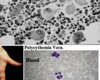
Presentation of polycythemia vera?

A
  • Erythrocytosis, granulocytosis, and thrombocytosis in peripheral blood
  • Erythromelalgia
    • burning pain in feet/hands with erythema, pallor, or cyanosis
  • Abnormal blood flow: bleeding and thrombosis
  • Marrow fibrosis
    • Extramedullary hematopoiesis –> hepatosplenomegaly
25
What do progenitor cells proliferate so quickly/much?
Decreased requirements for EPO and other growth factors (serum EPO levels are very low)
26
A FYI CARD IF YOU WANT TO LEARN IT: Hemoglobin levels of PV? Hematocrit levels? WBC levels? Platelet count?
Hb: 14-28 (normal 12-16) Hct: \>60% (normal 31-52%) WBC: 12-50,000 (normal 4-11,000) Platelets: \>500,000 (normal 140-450,000)
27
Essential thrombocytosis: Proliferation of?
Megakaryocytic progenitors (with reduced requirement for growth factors)
28
Platelets of essential thrombocytosis? Marrow? Peripheral blood?
* Platelets * dysfunctional --\> thrombosis and bleeding * Marrow * megakaryocytes * Peripheral blood * thrombocytosis with giant platelets
29
Characteristic symptom of essential thrombocytosis?
Erythromelalgia
30
Primary myelofibrosis: What is this disease characterized by?
Obliterative marrow fibrosis that then results in suppression of hematopoiesis and causes cytopenias (this leads to more extramedullary hematopoiesis; yet, is inefficient and cytopenia persists)
31
What occurs to the bone marrow in primary myelofibrosis?
* Collagen deposition (by normal cells) * Megakaryocytes are large and clustered * Later: fibrotic marrow may be converted to bone (osteosclerosis)
32
Cell characteristics in primary myelofibrosis?
* Nucleated erythroid progenitors in peripheral blood * Granulocytic progenitors in the peripheral blood * RBCs are teardrop shaped from being pushed through fibrotic marrow * Dacryocytes
33
Signs and symptoms of primary myelofibrosis?
* Anemia * Splenomegaly * Splenic infarcts * Infections * Thrombosis and bleeding * 5-20% transform to AML
34
Langerhans cell histiocytosis: Cell involved? Categories?
Langerhans cell histiocytosis: * Involves immature dendritic cells * Letterer-Siwe syndrome * Hand-Schuller-Christian disease * Eosinophilic granuloma
35
Cell markers in Langerhans cell histiocytosis?
* Cell markers * CD1 * S-100 * HLA-DR * Birbeck granules * characteristic areas of dilation in the cytoplasm
36
Multifocal multisystem Langerhans cell histiocytosis (letterer-siwe syndrome): Tumor masses are composed of? Organs involved? Presentation?
* Tumor masses * CD1+ langerhans cells * Organs * Spleen, liver, lung, and eventually BM * Presentation * Anemia, thrombocytopenia, and infections
37
Unifocal and multifocal unisystem Langerhans cell histiocytosis (eosinophilic granuloma): Characterized by?
* Characterized by erosive accumulations of langerhans cells * Often in medullary cavities of bone * Eosinophil is the dominant infiltrate * Also plasma cells, lymphocytes, and neutrophils
38
Unifocal lesion of Langerhans cell histiocytosis (eosinophilic granuloma) Characterization?
* Asymptomatic or local pain * Usually indolent
39
Multifocal unisystem lesions of Langerhans cell histiocytosis (still eosinophilic granuloma): Gross findings? Signs/symptoms?
* Gross findings * erosive growths in bone that may expand into soft tissue * Signs/symptoms * 50% have invasion of the posterior pituitary stalk * leads to diabetes insipidus
40
What is the Hand-schuller-christian traid in Langerhans cell histiocytosis?
* Involvement of: * The calvaria * Diabetes insipidus * Exopthalmos
41
What is this?
Essential thrombocytosis: large platelets
42
What is this? What is the black arrow pointing at?
This is primary myelofibrosis Black arrow: teardrop shaped RBC
43
What disease is this found in? What in this image is distinctive?
This is in Langerhans cell histiocytosis The birbeck granules with characteristic areas of dilation in the cytoplasm