Chapter 13 - Blood vessels Flashcards

(130 cards)

1
Q

Review:

What is the intima?

What is the media?

What is the adventitia?

How are they supplied?

A
  • Intima
    • Single layer of endothelial cells with little subendothelial CT
    • Supplied by the lumen
    • Separated from media by the internal elastic lamina
  • Media
    • Smooth muscle
    • Inner layers: lumen
    • Outer layers: small arterioles (vasa vasorum)
  • Adventitia
    • CT, Nerves, and Vasa Vasorum
2
Q

Review

Difference between large, medium, and small arteries?

A
  • Large (elastic arteries)
    • Media = rich in elastic fibers that alternate with layers of smooth muscle
  • Medium (muscular arteries)
    • Media = mostly smooth muscle
  • Small (less than 2mm)
    • Structurally similar to medium arteries
    • Found within tissue and organs
3
Q

Review still:

Arterioles:

Composition?

Importance?

Capillaries:

Composition?

Importance?

A
  • Arterioles
    • Small amount of smooth muscle
    • NO elastic lamina
    • *Principle sites of BF resistance*
  • Capillaries
    • Endotheilal lining but NO media
    • BF slows dramatically here obbbbbviously for nutrient exchange
4
Q

Review:

Where does blood flow after capillaries? What is important about these?

Veins in comparison to arteries?

Composition of lymphatics?

A
  • Postcapillary venules (after cap)
    • Site of inflammation induced leakage
  • Veins
    • Large lumens and walls (less organized than art.)
    • Contains 2/3 of all blood
  • Lymphatics
    • Thin-walled, endotheium lined channels
    • Drain intersitial fluid and inflammatory cells
5
Q

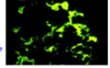
How are endothelial cells identified immunohistochemically?

A

Endothelial cells identified by:

  • PECAM-1 (CD31)
  • CD34
  • vWF
6
Q

Functions of endothelial cells?

A

Endothelial cell function:

  • Maintenance of permeability barrier
  • Elaboration of compounds that control coagulation
  • Produce ECM proteins for healing/angiogenesis
  • Production of vasoconstrictors (endothelin/angiotensin)
  • Production of vasodilators (NO/prostacyclin)
  • Important mediators of inflammation
    • Produce IL-1/6/8
    • Express VCAM-1, ICAMS, E/P selectin
  • Produce growth factors
7
Q

What is endothelial dysfunction associated with?

A

Endothelial dysfunction:

  • Abnormal thrombus formation
  • Atherosclerosis
  • Vascular lesions of hypertension
8
Q

Functions of vascular smooth muscle cells?

A

Vascular smooth muscle cells:

  • Vasodilation/constriction in response to mediators
  • Synthesis extracellular basement membrane proteins
    • Collagen, elastin, and proteoglycan
  • They can also proliferate and be migratory
    • In response to cytokines (IL-1, IFN)
    • In response to GF (PDGF, endothelin-1, and FGF)
9
Q

What layer of the artery thickens in response to vessel injury?

A

The intima

10
Q

What is the pathology of intimal thickening in response to vessel injury?

A

Pathology

  • Endothelial cells lose the ability to contract but they proliferate and produce ECM proteins, creating what is known as the neointima*
  • If chronic - associated with atherosclerosis
11
Q

Arteriosclerosis:

Generic term for?

What are the 3 variations?

A

Arteriosclerosis:

  • Arterial wall thickening and the loss of wall elasticity

3 variations:

  • Monckeberg medial calcific sclerosis
  • Arteriolosclerosis
  • Atherosclerosis
12
Q

What is monckeberg medial calcific sclerosis?

A

Characterized by excessive Ca++ deposits in muscular arteries (50+ yo) that are usually benign because the deposits do not cause obstruction

13
Q

What is arteriolosclerosis?

A

Involves arterioles and small arteries

Hyaline and hyperplastic forms occur and are associated with thickening of the vessel walls and obstruction of the lumen

14
Q

What is atherosclerosis characterized by?

A

Characterized by intimal lesions known as atheromas or atheromatous fibrofatty plaques

(they may obstruct the lumen and weaken the media)

15
Q

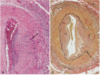
What is this an example of?

A

This is Monckeberg medial calcific sclerosis with the collection of calcium deposits (arrows) in the media of small muscular arteries

16
Q

Where do atherosclerotic lesions primarily develop?

Atherosclerotic symptomatic disease most often involves the arteries supplying what organs?

A
  • Lesions develop in:
    • Elastic arteries (aorta, carotid, and iliac)
    • Large/medium muscular arteries (coronary/popliteal)
  • Symptomatic disease involves arteries supplying:
    • Heart
    • Brain
    • Kidneys
    • Lower extremities
17
Q

Major outcomes of atherosclerosis?

A

Atherosclerosis major outcomes:

  • MI
  • Stroke
  • Aortic aneurysms
  • Peripheral vascular disease
18
Q

Earliest lesion of atherosclerosis

A

Earliest lesion:

  • Fatty streaks in the intima
    • Lipid filled macrophages (foam cells)
    • Not raised so no disturbance in BF
19
Q

Atherosclerotic plaques typically have what 3 principal components?

A
  • Cells
    • Smooth muscle, macrophages, and lymphocytes
  • ECM proteins
    • Collagen, elastic fibers, and proteoglycan
  • Intra/Extracellular Lipid
    • Cholesterol clefts are common
20
Q

Top risk factors for CVD?

A
  • Hyperlipidemia
  • Hypertension
  • Cig smoking (oops)
  • Diabetes
  • Advanced age
21
Q

Response to injury hypothesis implies?

A

Implies that atherosclerosis is a chronic inflammatory response of the artery wall initiated by injury to the endothelium

22
Q

Lesion progression of atherosclerosis is sustained by?

A

Sustained by interactions between

  • lipoproteins
  • Macrophages
  • T cells
  • Smooth muscle cells
23
Q

Steps in lesion development?

A
  • Chronic injury
  • Accumulation of lipoproteins (mostly LDL)
  • Increased adhesion of monocytes/leukocytes to endo.
  • Adhesion of platelets
  • Release of factors that induce smooth muscle migration
  • Smooth muscle proliferation
  • Elaboration of extracellular matrix proteins
24
Q

What is C-reactive protein?

A

CRP = one of the cheapest and most sensitive predictors of the risk of MI, stroke, peripheral arterial disease, and sudden cardiac death

25
How can the endothelium become damaged to create plaques?
Endothelial damage: * May be associated with infection * Initiated by normal hemodynamic disturbances * Eg: at branch points * Hypercholesterolemia
26
Inflammatory role associated with plaque formation?
* Increases expression of adhesion molecules by endothelial cells * Allows for infiltration of inflammatory cells to intima * This is initially protective until T cells become activated * Secrete proinflammatory cytokines that induce smooth muscle migration/proliferation
27
What are the major lipids in atheromatous plaques? Correlation between the severity of atherosclerosis and?
* Major lipids * Plasma derived cholesterol and cholesterol esters * Correlation of severity with: * High levels of plasma cholesterol or LDL
28
Smooth muscle cells convert the fatty streak into what? Catastrophic evens are associated with what aggregation?
* Smooth muscle cells convert fatty streak to * Fibrofatty atheroma * Catastrophic events are associated with platelet aggregation over the plaque * This is followed by eventual disruption of the fibrous cap
29
When does atherosclerotic coronary artery disease begin?
Childhood (modification should begin then)
30
Hyptertensive vascular disease BP: Normal? Prehypertensive? Stage 1 hypertension? Stage 2 hypertension?
* Normal * \<120/80 * Prehypertension * 120-129/80-89 * Stage 1 * 140-159/90-99 * Stage 2 * \>160/100
31
Types of hypertension?
* Essential hypertension * idiopathic and some develop potentially fatal BP * Secondary * Underlying renal or adrenal disease
32
What relationship is altered in arterial hypertension?
The relationship between _cardiac output_ and _TPR_ ( BP = CO x TPR )
33
What is cardiac output dependent upon? Where is TPR determined?
* CO * Blood Vol influenced by Na+ homeostasis * TPR * Determined at the arterioles
34
Role of the kidney in BP regulation?
* Renin converts A to AI which is then converted to _AII_ bye ACE * AII increases BP by increasing _peripheral resistance_ and increasing the _blood volume_ by increasing Na+ resorption * Kidney also produces _antihypertensive_ substances (PG/NO) * Counterbalances AII * _Natriuretic Factors_ inhibit Na+ resorption and induce vasodilation
35
What three pathological dysfunctions alter Renin secretion?
* ( - ) Fibromuscular dysplasia * Genetic disorder * ( + ) Renal artery stenosis * Acquired Disorder * ( + ) Renin-secreting tumor * Acquired Disorder
36
What two things alter angiotensin (A) secretion?
* ( + ) Oral contraceptives * Acquired * ( - ) Angiotensinogen Variants * Genetic disorder
37
What three pathological dysfunctions alter Na+ reabsorption?
* ALL ( - ) Genetic Disorders * Liddle Syndrome * Pseudohypoaldosteronism * Gitelman syndrome
38
Hyptertension is associated with what two forms of arteriolosclerosis?
Hyaline and Hyperplastic
39
What is _hyaline arteriolosclerosis_?
* Lesion consists of a homogeneous hyaline thickening of the wall with eventual narrowing of the lumen * These reflect the leakage of plasma components across the endothelium and excessive ECM production by smooth muscle secondary to the hemodynamic stress associated with hypertension * Contribute to hypertension related renal failure
40
What is _hyperplastic ateriolosclerosis_?
* Associated with severe hypertension * Concentric, laminated thickening of the arterioles composed of smooth muscle and basement membrane components * May become necrotic and infiltrated with fibrin * Also common in the kidney
41
What is this?
Hyperplastic arteriolosclerosis (Concentric - looks like an onion )
42
What is this?
Hyaline Arteriolosclerosis
43
What is a TIA? Characterization of a TIA?
* TIA * Mini stroke that lasts less than 24 hours * Characterized by focal neurological defects that resolve within 24 hours
44
Difference between an aneurysm and a false aneurysm? What is a Dissection?
* Aneurysm * Abnormal dilation of a vascular wall * False aneurysm * Breach in the vascular wall leading to an extravascular hematoma * Dissection * Blood enters the artery wall and travels for a distance Note\* aortic aneurysm/dissections are clinically the most important
45
Despite that the picture kindddda tells you.. what are these from left to right (1-4)?
1. True aneurysm (saccular) 2. True aneurysm (fusiform) 3. False aneurysm 4. Dissection
46
Saccular versus fusiform aneurysms?
* Saccular * Spherical and involve only a portion of the vessel wall * Usually contain a thrombus * 5-20 cm in diameter * Fusiform * Vary in diameter/length * May involve the entire segment
47
Major cause and location of abdominal aortic aneurysms?
Atherosclerosis usually right below the renal arteries and above the bifurcation
48
What predisposes to aneurysms and dissections?
* Genetic defects in CT structure and/or assembly * Example: Marfan syndrome * Overactive metalloproteinases (w/ chronic inflammation) may degrade CT and weaken the vessel wall
49
Clinical course of the abdominal aortic aneurysm?
* May rupture with massive hemorrhage, obstruction of lumen, obstruction of neighboring vessels/organs, and/or emolism * May present as a mass simulating a tumor * Most expand until they rupture
50
What occurs in syphilitic aneurysms?
* This occurs in tertiary syphilis as an aortic obliterative endarteritis * Inflammation and fibrosis weaken the media
51
What is aortic dissection characterized by?
* Dissection of blood between and along the laminar planes of the media * Often rupture outward causing a massive hemorrhage (90% in men btwn 40-60yo with HT, others due to CT abnormalities or as a result of arterial cannulation)
52
Most common pathologic abnormality of aortic dissection
* Cystic medial degeneration * Characterized by fragmentation of the elastic tissue and separation of the elastic/fibromuscular element by clefts * Frequently occurs in Marfan syndrome
53
Major risk factor for dissection? Pathology?
Hypertension Path = varying degrees of elastic tissue fragmentation
54
Classic symptoms of aortic dissection?
* Sudden onset of pain * Begins in chest * Radiates to the back * Moves downward as the dissection progresses
55
Vasculitis (inflammation) typically affects what vessels? Two most common causes?
* Typically affects arterioles, venules, and capillaries * _Small vessel Vasculitis_ * May have systemic symptoms - _Systemic necrotizing vasculitides_ * Causes * Infection * Immune mediated reactions
56
3 most common pathologies of non-infectious vasculitis?
1. Immune complexes 2. Antineutrophil cytoplasmic antibodies 3. Anti-endothelial antibodies
57
How do Immune complexes create vasculitis?
* ICs and Complement components deposit in vessel walls * ICs induce neutrophil accumulation/activation/degran. * Results in tissue necrosis * IC formation may be drug-related, associated with infection or autoimmune disease
58
Role of Antineurophil cytoplasmic antibodies (ANCA) in vasculitis?
Abs form to mostly granule enzymes in neutrophils (also monocytes and endothelial cells)
59
Anti-endothelial antibodies are seen in what 2 diseases?
SLE and Kawasaki disease
60
Most common form of systemic vasculitis in adults? What does it involve?
_Giant cell (temporal) arteritis_ * Ganulomatous inflammation principally affecting the arteries of the head (esp _temporal arteries_) * May also involve vertebral/ophthalmic arteries and the aorta (_giant cell aortitis_)
61
Morphology of Giant cell arteritis? Where does granulomatous inflammation occur?
Nodular intimal thickening (varying lumen occlusion) Inflammation occurs in the media with a _mononuclear infiltrate (T cells/macrophages) with both foreign body and Langhans giant cells common_ Heals with fibrosis
62
Clinical manifestations of giant cell arteritis?
Either: * Vague and systemic * Fever/fatigue/wt. loss * Or Localized * facial pain/headache * Most serious when ophthalmic artery involved (blindness)
63
Diagnosis?
ESR is nearly always elevated Biopsy
64
What is _Takayasu arteritis?_
Ganulomatous vasculitis of medium and large arteries Predominantly the aortic arch and its branches with irregular thickening of the intima
65
Inflammatory infiltrate of Takayasu arteritis? Clinical Manifestation?
Similar to giant cell arteritis and is composed of mononuclear cells and giant cells Clinical: * Weak pulses in upper extremities * May also involve eyes/CNS
66
How to diagnose the difference between Takayasu and Giant cell arteritis in the aorta?
Same infiltrate thus _AGE_: _Takayasu_: under 50 _Giant cell_: over 50
67
What is _polyarteritis nodosa (PAN)_?
Systemic vasculitis involving small/medium muscular arteries Often involve the renal/visceral vessels but _SPARE_ the pulmonary circulation
68
What is PAN characteristically associated with?
Necrotizing inflammation with a mixed infiltrate (neutro/eosinophils and mononuclear cells)
69
Hallmark of PAN?
All stages of inflammation coexist in different or the same vessels
70
Clinical manifestations of PAN?
* Fever and weight loss * Abdominal pain (mesenteric arteritis) * Peripheral neuritis * Myalgia and muscle weakness * Renal involvement (doesnt EVERYTHING involve the kidneys???!)
71
What is _polymyalgia rheumatica (PMR)_?
Systemic inflammatory disease that occurs in 50% of patients with Giant cell arteritis (GCA)
72
Differences between GCA (giant cell ateritis) and PMR (polymyalgia rheumatica) symptoms?
_GCA_: systemic inflammation accompanies the vascular manifestations _PMR_:same systemic symptoms but the _vascular lesions are subclinical_
73
Similarities between GCA (giant cell) and PMR (polymyalgia rheumatica)?
* Both have an _increased ESR_ * Both present with anorexia/wt. loss, fever, depression, and maliase
74
Additional presentation of PMR?
* Normochromic normocytic anemia * Pain/stiffness in the shoulders, upper arms, hips, thighs, and/or neck (associated with synovitis of the affected joints)
75
What is the most common form of systemic vasculitis in kids?
_Henoch-Schonlein purpura_
76
Classical manifestation of Henoch-Schonlein purpura?
* _Palpable purpura_ * _Arthralgia/arthritis_ * _Abdominal pain_ * _Renal disease_: mild porteinuria to insufficiency
77
Pathological finding in Henoch-Schonlein purpura?
_IgA immune complex deposition_
78
What is this?
Giant cell arteritis: Granulomas in the media (black arrows) Lumen is almost entirely occluded (yellow arrow)
79
What is this? What is the black arrow pointing at in figure A? Figure B?
Giant cell arteritis _A_: degenerated internal elastic lamina in active arteritis _B_: focal destruction of internal elastic lamina and intimal thickening of long-standing/healed arteritis
80
What is this?
PAN: Polyarteritis Nodosa Shows inflammation, necrosis, and fibrosis (and karyorrhexis as a bonus if you remember nuclear fragmentation)
81
What disease is this?
Takayasu arteritis A: narrowing of aortic arch branches C: destruction of the media w/ mononuclear infiltrates, giant cells, and fibrosis
82
What disease are you likely to find this in? (THIS ONES A TOUGHY!)
Henoch-Schonlein purpura: IgA deposits limited to the mesangium and thus the capillary walls are not outlined
83
Leading cause of acquired heart disease in children?
Kawasaki disease
84
Kawasaki disease: AKA? Acute symptoms?
Kawasaki AKA mucocutaneous lymph node syndrome: * Fever * cervical adenopathy * buccal erythema * strawberry tongue * erythematous rash involving the palms and soles (resembles TS/Scarlet fever)
85
Associated with what mediators?
T cell and macrophage activation Anti-endothial/smooth muscle Antibodies
86
The vasculitis of Kawasaki resembles? What can this progress to?
Vasculitis resemples that of PAN Can progress to _vasculitis of the coronary vessels_ (can cause aneurysms/thrombosis/MI)
87
Diagnosis of Kawasaki?
Fever at least 5 days + 4 of the 5: * Conjunctival injection * Oral mucous membrane changes * Peripheral extremity changes (erythema of palms/soles) * Polymorphous rash * Cervical lymphadenopathy
88
What two diseases demonstrate large globular mesangial IgA-IC deposits?
IgA nephropathy Henoch-Schonlein purpura (just makin sure youre learning something over derr)
89
What is _Wegener's granulomatosis_?
Necrotizing vasculitis characterized by: 1. Acute necrotizing granulomas of the U/L Respiratory tract 2. Necrotizing or granulomatous vasculitis of small/med vessels 3. Focal necrotizing glomerulonephritis (pathogenesis resembles PAN)
90
Wegener's URT lesions? Cells?
URT lesions range from inflammatory sinusitis to ulcerations Macrophages, giant cells, lymphocytes, and plasma cells
91
What could happen to the Wegener's lesions in the lungs?
The lesions may coalesce and undergo cavitation
92
Early vs. Late kidney lesions in wegeners?
Early * Focal necrotizing glomerulonephritis w/ acute focal proliferation, necrosis, and thrombosis (isolated) Late * Diffuse necrosis, proliferation, and crescent formation
93
Wegener's granulomatosis presentation?
* Persistent pneumonitis * Chronic sinusitis * Mucosal ulceration * Palpable purpura * Myalgia/arthralgia * Evidence of renal disease
94
What is _microscopic polyangitis_?
Necrotizing vasculitis of arterioles, capillaries, and venules
95
How do lesions in microscopic polyangiitis compare to those in PAN? Where do they occur?
They all are the same age and commonly occur in the skin, lungs, and kidney
96
Characteristic infiltrate of microscopic polyangiitis?
Mononuclear and neutrophil infiltrate w/ fibrinoid necrosis -neutrophils migrate into the wall and disintegrate releasing cytoplasmic components (_leukocytoclastic vasculitis_)
97
Differences in the morphology between microscopic polyangiitis and Wegeners?
MP has _NO_ granulomas in the lesions and wegeners does
98
Differences between P-ANCA and C-ANCA? (ANCA = antineutrophil cytoplasmic antibodies)
* P-ANCA * Stain is limited to perinuclear region and cyto is nonreactive * Abs directed at myeloperoxidase * C-ANCA * Heavy stain in cyto w/ nonreactive nuclei * Abs directed against proteinase 3
99
Which is P-ANCA? C-ANCA? What Diseases are each associated with?
_P-ANCA_: on the left; associated with Microscopic Polyangiitis _C-ANCA_: center and right; associated with Wegener's
100
What is _Thromboangiitis obliterans_?
Buerger's Disease: Occurs in conjunction with cig. smoking and affects the distal medium sized A/V of the extremities
101
Characteristics of Thromboangiitis obliterans?
Acute/Chronic inflammation with thrombosis (organization/recanalization) The thrombi often contain abscesses
102
What happens in thromboangiitis obliterans without treatment? What is the treatment?
Without treatment it spreads into other vessels/nerves and all structures become fibrotic (looks gangrenous and will require amputation) Treatment = quit smoking
103
Review (will have more ?? per slide) What is _Raynaud's phenomena_? Primary vs secondary?
Paroxysmal pallor of the digits of hands/feet (white), progressing to cyanosis with pain/numbness (blue), followed by hyperemia (red) _Primary_: exaggeration of vasomotor responses to cold/stress _Secondary_: associated with CT diseases (slceroderma/SLE/RA), occlusive diseases, drugs, neurologic disorders, and trauma. Arterial insufficiency may result in ulceration or gangrene
104
Most common manifestation of chronic venous disease?
Varicose veins
105
Varicose veins are most common where? How do they occur? What do they look like?
Most common in superficial veins of legs often due to long periods of standing (pressure increases leading to stasis/edema) and they are dilated, tortorous veins.
106
Morphology of varicose veins? What patients may have esophageal varicosities?
Veins with varicosities have thing walls, imcompetent valves, with thus stasis, congestion, pain, and edema Patients with cirrhosis and portal hypertension may have esophageal varicosities
107
What is _thrombophlebitis/phlebothrombosis_? Predisposing conditions?
DVT that can result in life threatening venous thromboembolism Predisposing conditions: * HF * Neoplasia * Pregnancy * Obesity/prolonged immobilization * Surgery/Previous DVT
108
Clinical manifestations of DVT?
Leg pain, tenderness, swelling, dilation of the superficial veins, and cyanosis
109
What is lymphangitis?
Bacterial infections that spread through lymphatics causing inflammation
110
What is a Hemangioma?
Localized superficial cutaneous lesion of childhood
111
Capillary vs. Cavernous hemangioma?
* Capillary hemangioma * Skin, subcutaneous tissue, and oral/lip mucous memb. * Covered with an intact epithelium * Unencapsulated aggregates of thin capillaries filled with blood and separated by scant CT * Cavernous * Larger than capillary, well defined, unencapsulated * Thrombosis and dystrophic calcification is common
112
What is a pyogenic granuloma? What is this during pregnancy?
Rapidly growin exophytic red nodule attached to the skin or oral mucosa that bleeds easily and often ulcerates? _Pregnancy = granuloma gravidarum_
113
Histological presentation of a pyogenic granuloma?
Proliferating capillaries Edema with neutrophils
114
What is a Lymphangioma? Difference between capillary and cavernous lymphangiomas?
Lymphangioma = benign lymphatic analog of hemangioma * Capillary: * Lesion = small channels beneath the epidermis * Resemble hemangioma but w/o blood cells * Cavernous * Occur mostly in children in the neck/axilla * Resemble cavernous hemangioma but w/o blood cells
115
What is a glomus tumor?
A painful benign tumor that arises from the modified SMC of the glomus body (specialized arteriovenous anastomosis involved in thermoregulation/BF)
116
Histology and common locations of glomus tumors?
* Histology * vascular channels separated by CT w/ masses of glomus cells * Commonly occurs: * Distal digits (esp under fingernails)
117
What is Vascular ectasias? 3 major examples?
Localized dilation of pre-existing channels Nevus Flammeus Spider Telangeictasia Hereditary hemorrhagic telangiectasia
118
What is nevus flammeus?
"Birthmark" on head or neck that ranges from pink-deep purple with dilation of vessels in the dermis. Most regress, others (like port wine stains) may grow proportionately with the child
119
What is spider telangiectasia and where is it likely to occur?
Form an array of dilated subcutaneous arteries/arterioles that blanch with pressure Common on face, neck, or chest in pregnancy/pts w/ cirrhosis
120
What is Hereditary hemorrhagic telangiectasia?
The most common genetic (AD) cause of vascular bleeding. The most frequent symptom is epistaxis Capillaries are dilated with a loss of subendothelial structures
121
What is _Bacillary angiomatosis_?
Opportunistic infection caused by Bartonella Henselae Associated with cat bites/scratches
122
Two main histologic forms of bacillary angiomatosis?
Bacillary angiomatosis Cat-scratch disease (granulomatous)
123
Pathology of bacillary angiomatosis? Characteristics of the Lesions?
Proliferation of capillaries, acute inflammatory infiltrate, and bacteria Lesions = cutaneous or mucocutaneous and tender --\> may ulcerate and crust (disseminated in AIDS)
124
Pathology of Cat-scratch disease?
Granulomas in the lymph nodes | (treat with azithromycin)
125
Virus of Kaposi sarcoma? Clinical and histological manifestations?
Kaposi sarcoma = HHV-8 * _Clinical_ * _​_cutaneous lesions that are red-purple macules that become raised plaques * _Histological_ * Spindle cells and inflammatory infiltrate that line vascular channels
126
Different forms of Kaposi sarcoma?
* Chronic/Classic/European * Older men * Indolent and confined to lower extremities * Lymphadenopathic/African/Endemic * Immunosuppressive-associated KS * Transplants and AIDS
127
What is _angiosarcoma_?
Malignant endothelial neoplasms Occurs in older adults Initially involve the skin, heart, breast, or liver
128
Angiosarcoma is the most common sarcoma arising in what two organs? Morphology and clinical course?
Most common arising in the liver and heart All degrees of endotheial differentiation occurs These neoplasms are locally invasive, prone to metastases, and yield a generally poor prognosis
129
What is percutaneous transluminal coronary angioplasty?
Placement of a balloon, catheter, or stent to open a stenotic vessel. Use of this + thrombolytic therapy is best for an MI High success rate (lower with complete occlusion)
130
Restenosis issues with PTCA? How to make better?
_Elastic recoil_: occurs w/o placement of a stent and is a mechanical renarrowing caused by adventitial constriction _Neointimal hyperplasia_: proliferation of SMC and production of ECM proteins in response to the injury caused by the balloon and/or stent Use a stent that elutes antiproliferative agents (sirolimus/paclitaxel) however, may increase risk for stent-associated thrombosis