Neoplasia Flashcards

(55 cards)

1
Q

Define Neoplasia

A
  • NOT the same as malignant
  • Neoplasia is a disorder of cell growth (from an array of causes) that is triggered by one or more mutations affecting a single cell and its clonal progeny
  • Monoclonal - almost always malignant
  • Polyclonal - not always malignant
  • There are benign and malignant form of neoplasia
2
Q

Define Benign tumors

A
  • tumor is considered benign when its gross & microscopic appearances are considered relatively innocent, implying that it will remain localized, will not spread to other sites and is amenable to local surgical resections
3
Q

Define malignant tumors

A
  • latin = crab
  • Tumor is considered malignant when its gross & microscopic appearances (and locations) indicates that it will not remain localized, and has or will spread to other sites, making cure from local excision likely
4
Q

Nomenclature - Carcinoma

A
  • Modifiers may refine the epithelial source
  • Adenocarcinoma means glandular epithelium (ex. breast cancer)
  • Bronchogenic carcinoma - epithelium of airway passages
  • Tumors from connective & muscle tissue - hard (bone), soft (stromas)
5
Q

Nomenclature - Sarcoma

A
  • More deadly then carcinoma
  • Modifiers may refine source
  • Rhabdomyosarcoma - striated muscle
  • Leimyosarcoma - smooth muscle
6
Q

Nomenclature - Tumors from neural cells

A
  • Glioma (benign)
    • Oligodendroglioma
    • Astrocytoma
  • Glioblastoma
    • Glioblastoma multiforme
  • Retinoblastoma
7
Q

Nomenclature - Tumors from brain coverings

A
  • Meningioma - brain covering
    • same term used for benign & malignant with addition of invasive, if malignant
8
Q

Nomenclature - Tumors from blood vessles and blood cells

A
  • Leukemia - derived from granulocytic, nucleated blood cells
  • Lymphomas - derived from lymphocytes or their precursors
  • Multiple myeloma - malignancy of plasma cells
  • Hemangioma/Lymphangioma - benign tumor of blood/lymphatic vessels (image)
  • Hemangioblastoma/Lymphangiosarcoma - malignant tumor of blood/lymphatic vessels
9
Q

Nomenclature of benign vs malignant CT tumors

A
  • Benign tumors from CT usually end in -oma
    • Leiomymoa (smooth muscle)
    • Rhabdomyoma (skeletal m)
    • Chondroma (cartilage)
    • Fibroma (Soft CT - fibroblasts)
    • Osteoma (bone)
  • Malignant tumors from connective & muscle tissues usually end in -sarcoma
    • Leiomysarcoma
    • Rhobdomysarcoma
    • Chondrosarcoma
    • Fibrosarcoma
    • Osteosarcoma
  • Exception is malignant form melanoma (CT melanocytes). - think of melanosarcoma, b/c that is how it behaves
    • Benign form is nevus
10
Q

What does this image show?

A
  • Iris leiomyoma
  • The only smooth muscle in the iris is the sphincter (NOT Dilator, myoepithelium)
  • Mass at the pupil
11
Q

What does this image show?

A
  • Diabetic lacy vacuolization of the iris that increases deposition of glycogen in epithelia but NOT smooth muscle.
  • Since dilator muscle is the myo component of epithelium, it is affected. The sphincter, being true smooth muscle, is not. Hence diabetic do not dilate well but constrict just fine
12
Q

Epithelial Dysplasia

A
  • consists of an expansion of immature cells (such as ectoderm), with a corresponding decrease in the number and location of mature cells
  • Dysplasia is often indicative of early neoplastic process.
  • The term dysplasia is typically used when the cellular abnormality is restricted to the originating tissue, as in the case of an early, in-situ neoplasma that occupies less than the full thickness of the epithelium
  • Nothing to do with hip displasia (development anomalies)
13
Q

Conjunctival Intraepithelial Neoplasia (CIN)

A
  • Carcinoma in-situ - full replacement of epithelium
  • If the process has invaded the basement membrane, it is considered to be squamous cell carcinoma
14
Q

Differentiation vs Anaplasia

A
  • Differentiation describes the degree to which neoplastic tissues resemble the full differentiated cells from which they derive and retain their differentiated function (well differentiated, looks like normal tissue)
  • Anaplasia poor cellular differentiation
  • The more differentiated the tissue = less malignant potential it has
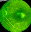
  • Primary retinal tumor caused by mutation of a tumor suppressor gene.
    • Avg 18 mo but as late as 12 yrs
    • Retinoblastoma
15
Q

Name the degrees of differentiation of retinoblastoma mimic photoreceptor differentiation

A
16
Q

What does this image show?

A
  • Poorly-differentiated (anaplastic) Retinoblastoma
17
Q

Degree of De-differentiated Ortho vs. Parakeratosis

A
  • In several neoplastic skin conditions (most of which are not malignant), the epithelium is turning over too rapidly (cell balance is loss)
  • As a result, certain differentiative stages are sacrificed
  • Perakeratosis - absence granular layer (stratum granulosum) and retention of nuclei in the stratum corneum.
  • Hyperkeratosis - thicker keratin on surface, but the granular cell layer is retained and the keratinized cells have lost their nuclei
18
Q

What are the cellular characteristics of Malignancy

A
  • Pleomorphism
  • Prominent nucleoli
  • Coaruse clumping of chromatin
  • Increased mitotic index/Tripolar mitoses
  • Loss of polarity & loss of contact inhibition
19
Q

What does this image show?

A

Cellular characteristic of malignancy - pleomorphism

20
Q
A

Cellular Characteristics of malignancy - Prominent nucleoli

21
Q

What does this image show?

A

Cellular characteristics of malignancy - coarse clumping of chromatin

aka hyperchromaticity

22
Q

What does this image show?

A
  • Cellular characteristic malignancy
    • Left increased mitotic index
    • Tripolar mitoses
23
Q

What does this image show?

24
Q
A
  • Note pleomorphism, prominent nucleoili, increased mitotic index, loss of of polarity and loss of contact inhibition, but minimal hyperchomaticity
25
What are the 4 steps of invasion?
* **Loosening of cell-cell contacts** - inactivation of E-cadherin through a variety of pathways * **Degradation of ECM -** basement membranes and interstitial matrix degradation is mediated by proteolytic enzymes secreted by tumor cells & stromal cells, such as matrix metalloproteases and cathepsins * **Attachment to novel ECM components** - proteolytic enzymes also release growth factors sequestered in the ECM & generate chemotactic and angiogenic fragments from cleavage of ECM glycoproteins * **Migration of tumor cells**
26
What do these images show?
27
Metastasis
* **once in the blood stream, cancer cells are still vulnerable** * Mechanical shear stress from blood flow * Apoptosis stimulated by loss of adhesion (anoikis) * And the innate and acquired immune system, if the cell can be identified as foreign * **Once they reach target site they extravastate** * Within the blood, tumor cells tend to cluster via homotypic adhesions between tumor cells or heterotypic between tumor cells & WBC/platelets. These may create **emboli** * Many tumors arrest in the first capillary bed they encounter (lung, liver, most commonly) * Solid tumor cells often display **CD44** adhesion molecule, which is the same vehicle used by T cells to extravastate in inflammation * **Tropism** * **​**Certain tumors tend to target tissues for metastatic spread * Prostate prefers spread to bone * Bronchogenic CA spread to adrenal brain * Neuroblastoma spread to liver bone * Skeletal muscle and spleen rarely sites of metastasis
28
Pathways of Spread
* **Hematogenous** * **​**This is typical route of metastasis for **sarcomas**, but it is also the favored route for certain types of carcinoma, such as those originating in kideny renal cell carcinoma. Because of their thinner walls, veins are more frequently invaded than are arteries, and metastasis tends to follow the pattern of venous flow * Remember that uveal melanoma is a sarcoma and behaves as one * **Lymphatic** * **​**Lymphatic can transport tumor cells to lymph nodes and then to other parts of the body. This is the **most common route of metasis for carcinomas.** * Uncommon for a sarcoma to metastasize via this route. * Lymphatic system does eventually drain to systemic venous via the **azygous vein**, and therefore these metastatic cells can eventually spread through the hematogenous route
29
Seeding
Common in retinoblastoma
30
TNM system - Staging
* The **TNM Classification of Malignant Tumors (TNM)** is a cancer staging notation system that describes the stage of a solid tumor with alphanumeric codes * **T** desribes the size of orginial (primary) **tumor** and whether it has invaded nearby tissue * size or direct extent of the primary tumor * **Tx**: tumor cannot be evaluated * **Tis**: carcinoma in situ * **To**: no signs of tumor * **T1, T2, T3, T4**: size and/or extension of the primary tumor * **N** describes nearby (regional) lymph **nodes** that are involved * Degree of spread to regional lymph nodes * **Nx**: lymph nodes cannot be evaluated * **No**: tumor cells absent from regional lymph nodes * **N1**: regional lymph node metastasis present; at some sites, tumor spread to closest or small number of regional lymph nodes * **N2**: tumor spread to an extent betwen N1 & N3 (N2 not used at all sites) * **N3**: tumor spread to more distant or numerous regional lymph nodes (N3 is not used at all sites) * **M** describes distant **metastases** * **​**Presence of distant metastasis * **Mo:** no distant metasis * **M1:** metastasis to distant organs (beyond regional lymph nodes) * **Mx:** used variable to mean mutliple sites in different organs
31
TNM - system - other parameters
* **G** (1-4): the grade of the cancer cells (ex. they are "low grade" to "high grade". High grade = poorly differentiated * **S** (0-3): elevation of serum tumor markers * **R** (0-2): the completeness of the operation (resection-boundaries free of cancer cells or not) * **L** (0-1): invasion into lymphatic vessels * **V** (0-2): invastion into vein (no, microscopic, macroscopic) * **C** (1-5): a modifier of the **certainty** (quality) of the last mentioned paramter
32
TNM system - Prefix Modifiers
* **c:** stage given by clinical examination of the pt. The c-prefix is implicit in absence of the p-prefix * **p:** stage given by pathologic examination of surgical specimen * **y:** stage assessed after chemotherapy and/or radiation therapy; in other words, the individual neoadjuvant therapy of some kind (ex. tamoxifen) * **r:** stage for a recurrent tumor in an individual that had some period of time free from the disease * **a:** Stage determined at autopsy * **u:** stage determined ultrasonography or endosonography
33
TNM system - Stage 1 grouping
* small, low-grade cancer, no metastasis, no spread to regional lymph nodes, cancer completely removed, resection material seen by pathologist: pT1 pNo Mo Ro G1; this grouping of T,N, and M, would be considered stage 1 * **pT1** - smallest detectable tumor (seen by pathologist) * **pNo** - lymph nodes resected and pathologist found no tumor cells * **Mo** - no metastases * **Ro** - resection complete with no tumor at surgical margins * **G1** - abnormal but minimal anaplasia
34
If a pt develop mets from primary tumor (lung cancer) which spread to liver or brain, which one do they die from? Lung tumor or liver/brain tumor?
* If a pt develops mets from their primary tumor (ex. lung cancer) to a vital (ex. liver or brain) and it is the mets that lead to their demise, they did not die of liver cancer or brain cancer. They died of lung cancer metastatic to the liver or brain.
35
What does this image show?
* Benign does not mean inconsequential (unimportant) * **Spenoid ridge meningioma** - most common primary brain tumor * Potentially blinding * Reduced acuity, afferent defect and acquired red-green color vision defect
36
What are the Hallmarks of Cancer cells?
* **Self suficiency in growth signals -** cells can proliferate without external stimuli - **oncogenes** * **Insensitivity to growth inhibition signals** - usually due to inactivation of **tumor supressor genes** * **The Warburg Effect -** cancer cells switch from oxidative phosphorylation (26 ATPs) to aerobic glycolysis (2 ATPs). Why? b/c the cell wishes to use most of the available metabolic intermediates to fabricate cell components for building new cells, rather than burn them up for just ATP * **Evasion of apoptosis -** malignant cells evade apoptosis (which is worrisome thing as we try to develop ways for retinal ganglion cells to avoid apoptosis in glaucoma * **Limitless Replicative potential -** essentially de-differentiated (which is worrisome thign as we try to develop ways for retinal ganglion cells to avoid apoptosis in glaucoma) * **Sustained angiogenesis** - anti-VEGF, currently used to inhibit neovascularization in macular degeneration, was first explored to as avenue to kill tumors * **Ability to invade and or metastasize** - ability to evade host immune response
37
Self-sufficiency in growth signals - oncogenes, protooncogenes
* **Oncogenes -** mutated genes that promote autonomous cell growth * Their un-matured counterparts are called **proto-onocogenes** - products of proto-oncogenes promote cell growth but are regulated * Oncogenes encode **oncroproteins** that can promote cell growth in the absence of the usually required growth-promoting cell signals * It is the main signaling pathways that are altered including: * Receptor tyrosine kinase pathway, G-protein receptor pathway, the WNT pathway, the Notch pathway, the Hedgehog pathway, the TFG-beta/SMAD pathway and the NF-kB pathway
38
Insensitivity to growth inhibition signals - Tumor Suppressor genes
39
TP53- Gaurdian of the genome
* TP53, a tumor suppressor gene, located on chromosome 17p, regulates cell cycle progression, DNA repair, cellular senescence and apoptosis * It is the most frequently mutated gene underlying human cancers
40
Rb
* the first quintessential tumor suppressor gene and knudson's 2-hit hypothesis
41
Carcinogens
* Chemical * Radiation * Inflammation * Microbial (Bacteria & viral - both RNA & DNA)
42
Chemical Carcinogens
* Chemical carcinogens have highly reactive electrophile groups that directly damage DNA, leading to mutation & eventually cancer * Direct-acting agents do not require metabolic conversion to become carcinogenic, while indirect-acting agents are not active until converted to an ultimate carcinogen by endogenous metabolic pathways * After exposure of a cell to a mutagen or an initiator, tumorigenesis can be enhanced by exposure to promoters, which stimulate proliferation of the mutated cells * Ex. of human carcinogens are direct-acting agents (ex. alkylating agents used for chemotherapy), indirect acting agents (eg. benzo[a]pyrene, azo dyes, aflatoxin) and promotor or agents that cause pathologic hyperplasia of the endometrium or regenerative activity in the liver
43
Radiation Carcinogenesis
* Ionizing radiation causes chromosome breakage, translocations, and, less frequently, point mutations, leading to genetic damge and carcinogenesis * UV rays induce formation of pyrimidine dimers within DNA, leading to mutations. Therefore, UV rays can give rise to squamous cell carcinomas & melanomas of the skin. Individuals with defects in the repair of pyrimidine dimers suffer from xerodema pigmentosa and are particularly high risk. * Exposure to radiation during imaging procedures such as CT scans, pan-dental x-rays is linked to a very small, but measurable,increase in cancer risk in children
44
Inflamed-Associated
* Acquired conditions that predisopose to cancer can be divided into chronic inflammations, precursor lesions, and immunodeficiency states. Chronic inflammation disorders and precursor lesions span a diverse set of conditions that are all associated with increased cellular replication, which appears to create a "fertile" soil for the development of malignant tumors. * Indeed, repeated rounds of cell division may be required for neoplastic transformation, in that proliferating cells are the most at risk for accumulating the genetic lesions that lead to carcinogenesis * Tumors arising in the context of chronic inflammation are most carcinomas, but also include mesothelioma and several kinds of lymphomas * Immunodeficiency states predispose to virus-induced cancers
45
Immunodeficiency & cancer
* Pt who are immunodeficient & those who have deficits in T-cell immunity (ex. AIDS), are at increased risk for cancers, especially those caused by **oncogenic viruses.** * These virally associated tumors include mainly lymphomas, but also certain carcinomas & even some sarcomas and sarcoma-like proliferations
46
Why does cancer increase with age?
* Compare the size of 25 year old thymus (left) to that of an 80 year old (right). Each is normal for pt age and is outlined with red arrows * B/c our immune system weakens & fail to target precancerous cells
47
Microbial Carcinogenesis
* **HPV:** an important cause of benign warts, cervical cancer, and oropharyngeal cancer * Oncogenic types of HPV encode 2 viral oncoproteins, E6 & E7, that bind to p53 and Rb, respectively, with high affinity and neutralize their function * HPV cancers can be prevented by vaccination against high-risk HPV types * **Hepatitis B virus and hepatitis C virus:** cause of between 70% and 85% of liver CA * **Oncogenic effects** are multifactorial dominant effect seems to be immunologically mediated chronic inflammation, hepatocellular injury, and reparative hepatocyte proliferation * **HBx protein** of HBV and the HCV core protein can activate signal transduction pathways that also may contribute to carcinogenesis. New medications can cure Hep-C * **H. pylori** (cause of stomach ulcers) implicated in gastric adenocarcinoma and MALT lymphoma * Pathogenesis of H. pylori-induced gastric cancers is multifactorial, including chronic inflammation and reparative gastric cell proliferation * H. pylori pathogenecity genes, such as CagA, also may contribute by stimulating growth factor pathway * **Chronic H. pylori infection** leads to polyclonal B-cell proliferations that may give rise to a monoclonal B-cell tumor (MALT lymphoma) of the stomach as a result of accumulation of mutations
48
What are the principles of management?
* Surgery * Radiation (general or focused) * Adjuvant ( general chemo or receptor-targeted therapy)
49
Principles of mangement - Hormone receptors in breast CA
* Estrogen receptors * Progesterone receptors * **HER2** - human epidermal growth factor receptor 2. It causes these cells to grow and spread faster than the one with normal levels of the protein * **Triple negative breast cancer** - If all 3 receptors are negative. Absent targetable receptors, general chemo is usually required. (All 3 receptor fuel the growth of breast cancer) * Those with **BRCA1 or BRCA2** mutations are at increased risk of triple negative breast cancer
50
How are fruit flies used to help with cancer research?
* Researcher using fruitflies, bred to mimic genetic mutations in cancer pt. * He sequences DNA from pt tumor to look for errors and then replicates those errors into fruit flies to create "fly avatars". The fly develops tumor and thats when the drug testing comes in. * By doing this he comes up with a cocktail of 2 to 3 drugs that work together against the tumor. He passes on his findings to pt & the pt oncologist
51
How are monoclonal antibodies used to help cure cancer?
* Researchers used monolconal antibodies to treat pt with **B cell lymphomas** * Each B cell have a unique receptor (BCR) on the surface which helps them respond to different types of infections * As lymphoma express the same type of BCR. By targeting this BCR we can eliminate lymphoma. * To test this, they produced monoclonal antibodies specific for unique BCR expressed by a pt lymphoma cells aka **anti-idiotype therapy.** * This was successful but technique is too difficult & costly for clinics.
52
What is paraneoplastic syndrome? (autoimmune, cancer associated retinopathy)
* **Cancer-associated retinopathy** is characterized by * painless and progressive visual loss * photopsia (Visual distortion) * ceco-central scotoma * loss of rod & cone function * demonstrated by **electroretinography** due to production of autoantibodies against retinal antigens stimulated by a systemic, non-ocular tumor. * Anti-retinal antibody testing is commercially available * **VF:** central, cecocentral, or equatorial scotomas * **Fundus angiography:** usually normal. Rarely show areas of leakAge if vasculitis or macular edema present * **OCT:** retinal thinning * **Electroretinogram (ERG):** Full field ERG almost always abnormal (attenuated or absent photopic and scotopic response). In CAR where mainly the cones are affected, full field ERG could be normal but multifocal ERG will be abnormal * **FAF:** Parafoveal ring with enhanced autofluorescence with normal autofluorescence within the ring and hypoautofluoresence outside the ring * **The malignancy most commonly associated with this disorder** * small-cell lung cancer * followed by gynecologic (uterine & cervical) & breast cancers. * Occasional cases have been associated with non-small lung cancer, hodgkin lymphoma, and pancreatic, prostate, bladder, laryngeal, and colon cancers
53
What does this image show?
Ceco-central VF loss
54
Which tumors are most commonly metastatic to the eye?
* **Cancer metastasis that appear in and around the eye are usually from** * Breast cancer (w) * Lung cancer (m) * **Other less common sites include** * the prostate * the kidney * the thyroid * gastrointestinal tract * lymphoma and luekemia can also metastasize to the eye and orbit
55
Within the eye, tumors most often metastasize to places with \_\_\_\_\_\_\_\_.
normally permeable vessels (episclera, choroid, ciliary body) If primary unknown,usually difficult to determine source from biops of a met in the eye